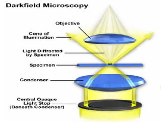
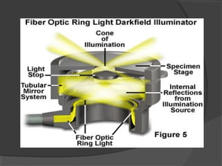
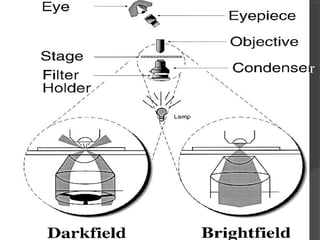
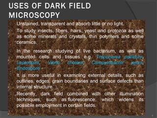
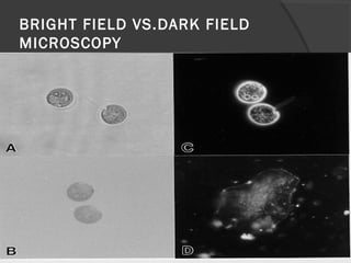
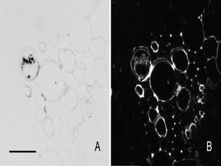
DARK FIELD
MICROSCOPY
 Dark field microscopy allows viewer to
observe living unstained cell and organisms
simply by changing the way in which they
illuminate the object.
 A hollow cone of light is focused on the
specimen in such a way that unreflected and
unrefracted rays do not enter the objective.
 Only light that has been reflected or
refracted by the specimen forms the image
 The field surrounding specimen appears
dark while the object brightly illuminated.
 The dark field microscope can reveal
considerable internal structure in larger
eukaryotic microorganism.
r
8
USES OF DARK FIELD
MICROSCOPY
 Unstained, transparent and absorb little or no light.
 To study insects, fibers, hairs, yeast and protozoa as well
as some minerals and crystals, thin polymers and some
ceramics.
 In the research studying of live bacterium, as well as
mounted cells and tissues e.g. Treponema pallidum,
Leptospira, Vibrio cholera, Campylobacter jejuni,
Endospore
 It is more useful in examining external details, such as
outlines, edges, grain boundaries and surface defects than
internal structure.
 Recently, dark field combined with other illumination
techniques, such as fluorescence, which widens its
possible employment in certain fields.
BRIGHT FIELD VS.DARK FIELD
MICROSCOPY
Advantages
 The advantage of darkfield microscopy also
becomes its disadvantage: not only the
specimen, but dust and other particles
scatter the light and are easily observed
 For example, not only the cheek cells but the
bacteria in saliva are evident.
 The dark field microscopes divert illumination
and light rays thus, making the details of the
specimen appear luminous.
 Dark field light microscopes provide good
results, especially through the examination of
live blood samples.
 It can yield high magnifications of living
bacteria and low magnifications of the
tissues and cells of certain organisms.
 Certain bacteria and fungi can be studied
with the use of dark field microscopes.
THANK

Dark field microscopy (1) By Ehtisham Ul Haq

  • 1.
  • 2.
     Dark fieldmicroscopy allows viewer to observe living unstained cell and organisms simply by changing the way in which they illuminate the object.  A hollow cone of light is focused on the specimen in such a way that unreflected and unrefracted rays do not enter the objective.
  • 3.
     Only lightthat has been reflected or refracted by the specimen forms the image  The field surrounding specimen appears dark while the object brightly illuminated.  The dark field microscope can reveal considerable internal structure in larger eukaryotic microorganism.
  • 7.
  • 8.
    8 USES OF DARKFIELD MICROSCOPY  Unstained, transparent and absorb little or no light.  To study insects, fibers, hairs, yeast and protozoa as well as some minerals and crystals, thin polymers and some ceramics.  In the research studying of live bacterium, as well as mounted cells and tissues e.g. Treponema pallidum, Leptospira, Vibrio cholera, Campylobacter jejuni, Endospore  It is more useful in examining external details, such as outlines, edges, grain boundaries and surface defects than internal structure.  Recently, dark field combined with other illumination techniques, such as fluorescence, which widens its possible employment in certain fields.
  • 9.
    BRIGHT FIELD VS.DARKFIELD MICROSCOPY
  • 11.
    Advantages  The advantageof darkfield microscopy also becomes its disadvantage: not only the specimen, but dust and other particles scatter the light and are easily observed  For example, not only the cheek cells but the bacteria in saliva are evident.  The dark field microscopes divert illumination and light rays thus, making the details of the specimen appear luminous.
  • 12.
     Dark fieldlight microscopes provide good results, especially through the examination of live blood samples.  It can yield high magnifications of living bacteria and low magnifications of the tissues and cells of certain organisms.  Certain bacteria and fungi can be studied with the use of dark field microscopes.
  • 13.