Downloaded 1,667 times
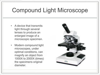


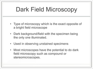
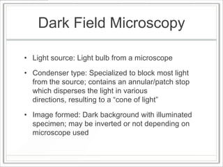


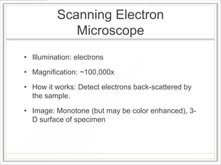


The document discusses different types of microscopy techniques. It provides details on: 1) Compound light microscopes which can magnify objects up to 2000x and transmit light through lenses to produce enlarged images. 2) Bright field microscopy which uses visible light and staining to produce contrasting images at magnifications up to 1000x. 3) Dark field microscopy which illuminates specimens on a dark background without staining, allowing observation of unstained samples. 4) Phase contrast microscopy which converts phase shifts in light waves into brightness contrasts, enhancing detail in living cells without staining at magnifications up to 400x. 5) Scanning electron microscopes which use electron beams at 100,000x magnification to


