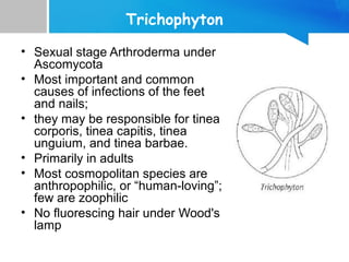
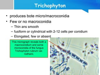
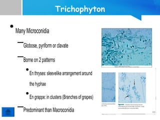
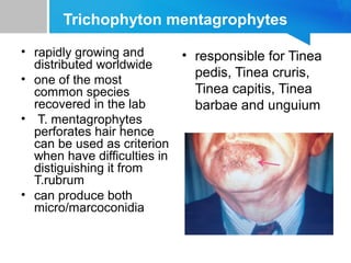
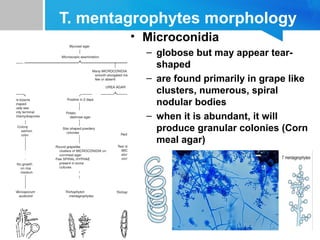
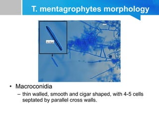
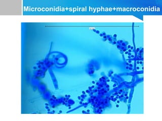
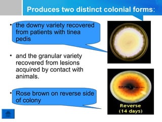
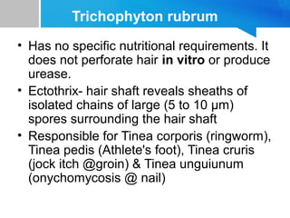
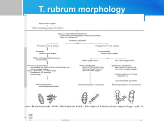
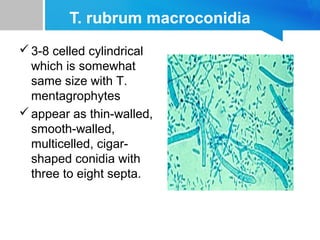
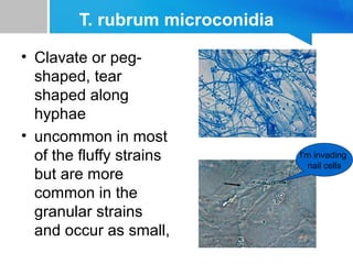
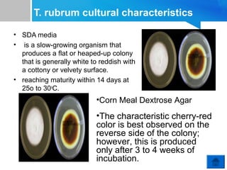
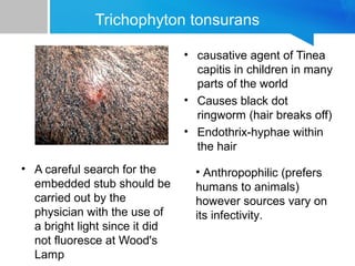
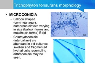
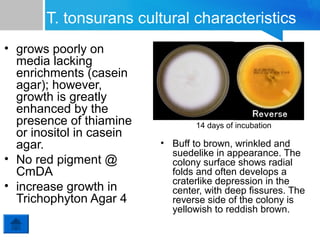
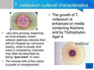
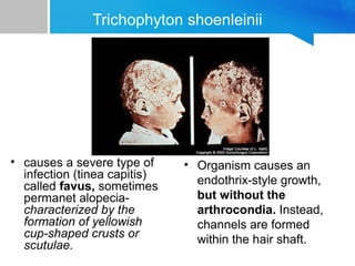
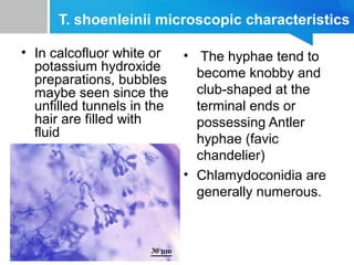
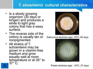
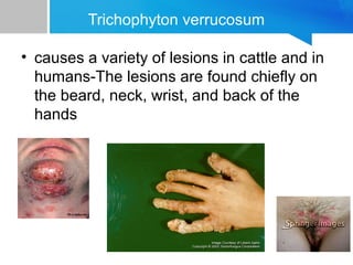
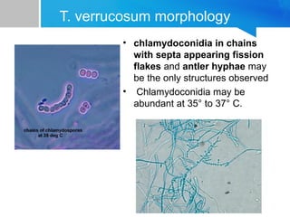
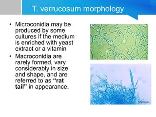
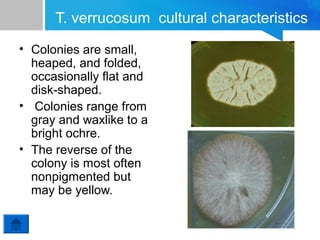
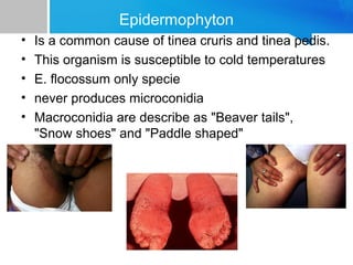
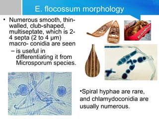
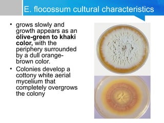
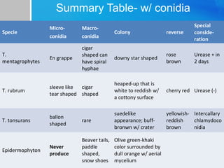
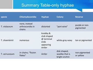
Trichophyton &
Epidermophyton
• PRINCESS ALEN AGUILAR
CONTENTS
• Trichophyton

– With Conidia

• T. mentagrophytes
• T.rubrum
• T. tonsurans

– Only Hyphae

• T. violaceum
• T.shoenleinii
• T. verrucosum

• Epidermophyton floccosum
Trichophyton
• Sexual stage Arthroderma under
Ascomycota
• Most important and common
causes of infections of the feet
and nails;
• they may be responsible for tinea
corporis, tinea capitis, tinea
unguium, and tinea barbae.
• Primarily in adults
• Most cosmopolitan species are
anthropophilic, or “human-loving”;
few are zoophilic
• No fluorescing hair under Wood's
lamp
Trichophyton
• produces bote micro/macroconidia
• Few or no macroconidia
– Thin ans smooth
– fusiform or cylindrical with 2-12 cells per conidium
– Elongated, few or absent
This micrograph reveals both a
macroconidium and some
microconidia of the fungus
Trichophyton rubrum var.
rodhaini.
Trichophyton

• Many Microconidia
–Globose, pyriform or clavate
–Borne on 2 patterns
• En thryses: sleevelike arrangement around
the hyphae

• En grappe: in clusters (Branches of grapes)

–Predominant than Macroconidia
Trichophyton mentagrophytes
• rapidly growing and
distributed worldwide
• one of the most
common species
recovered in the lab
• T. mentagrophytes
perforates hair hence
can be used as criterion
when have difficulties in
distiguishing it from
T.rubrum
• can produce both
micro/marcoconidia

• responsible for Tinea
pedis, Tinea cruris,
Tinea capitis, Tinea
barbae and unguium
T. mentagrophytes morphology
• Microconidia
– globose but may appear tearshaped
– are found primarily in grape like
clusters, numerous, spiral
nodular bodies
– when it is abundant, it will
produce granular colonies (Corn
meal agar)
T. mentagrophytes morphology

• Macroconidia
– thin walled, smooth and cigar shaped, with 4-5 cells
septated by parallel cross walls.
Microconidia+spiral hyphae+macroconidia
Produces two distinct colonial forms:
• the downy variety recovered
from patients with tinea
pedis
• and the granular variety
recovered from lesions
acquired by contact with
animals.
• Rose brown on reverse side
of colony
Trichophyton rubrum
• Has no specific nutritional requirements. It
does not perforate hair in vitro or produce
urease.
• Ectothrix- hair shaft reveals sheaths of
isolated chains of large (5 to 10 μm)
spores surrounding the hair shaft
• Responsible for Tinea corporis (ringworm),
Tinea pedis (Athlete's foot), Tinea cruris
(jock itch @groin) & Tinea unguiunum
(onychomycosis @ nail)
T. rubrum morphology
T. rubrum macroconidia
 3-8 celled cylindrical
which is somewhat
same size with T.
mentagrophytes
 appear as thin-walled,
smooth-walled,
multicelled, cigarshaped conidia with
three to eight septa.
T. rubrum microconidia
• Clavate or pegshaped, tear
shaped along
hyphae
• uncommon in most
of the fluffy strains
but are more
common in the
granular strains
and occur as small,

I'm invading
nail cells
T. rubrum cultural characteristics
• SDA media
• is a slow-growing organism that
produces a flat or heaped-up colony
that is generally white to reddish with
a cottony or velvety surface.
• reaching maturity within 14 days at
25o to 30oC.

•Corn Meal Dextrose Agar

•The characteristic cherry-red
color is best observed on the
reverse side of the colony;
however, this is produced
only after 3 to 4 weeks of
incubation.
Trichophyton tonsurans
• causative agent of Tinea
capitis in children in many
parts of the world
• Causes black dot
ringworm (hair breaks off)
• Endothrix-hyphae within
the hair
• A careful search for the
embedded stub should be
carried out by the
physician with the use of
a bright light since it did
not fluoresce at Wood's
Lamp

• Anthropophilic (prefers
humans to animals)
however sources vary on
its infectivity.
Trichophyton tonsurans morphology
• MICROCONIDIA
– Balloon shaped
(cornmeal agar),
numerous clavate varying
in size (balloon forms and
matchstick forms) if old
– Chlamydoconidia
(intercallary) are
abundant in old cultures;
swollen and fragmented
hyphal cells resembling
arthroconidia may be
seen.
Trichophyton tonsurans morphology
• MACROCONIDIA

– rare

T.tonsurans' macroconidia often
show an irregular or wavy
(undulating/ S-shaped) structure.
T. tonsurans cultural characteristics
• grows poorly on
media lacking
enrichments (casein
agar); however,
growth is greatly
enhanced by the
presence of thiamine
or inositol in casein
agar.
• No red pigment @
CmDA
• increase growth in
Trichophyton Agar 4

14 days of incubation

• Buff to brown, wrinkled and
suedelike in appearance. The
colony surface shows radial
folds and often develops a
craterlike depression in the
center, with deep fissures. The
reverse side of the colony is
yellowish to reddish brown.
Conidia is Absent, only Hyphae seen:
Trichophyton violaceum
• Produces an infection of the scalp and
body and is seen primarily in persons
living in the Mediterranean region, the
Middle and Far East, and Africa.
• Hair invasion is of the endothrix type; the
typical “black dot” type of tinea capitis is
observed clinically
Trichophyton violaceum infection
• Direct microscopic
examination of the
calcofluor white or
potassium hydroxide
preparation of the
nonfluorescing hairs
shows dark, thick
hairs filled with
masses of
arthroconidia
arranged in chains.
T. violaceum Microscopic Characteristics
• Microconidia and
macroconidia are
generally not present;
only sterile, distorted
hyphae and
chlamydoconidia are
found.
• Young hyphae usually
stain well in lactophenol
cotton blue, whereas
older hyphae stain poorly
and show small central
fat globules and granules.
T. violaceum cultural characteristics

•

very slow growing, beginning
as cone-shaped, creamcolored, glabrous colonies then
will turn heaped up, verrucous
(warty), violet to purple, and
waxy in consistency. Colonies
may often be described as
being “port wine” in color.
• The reverse side of the colony
is purple or nonpigmented.

• The growth of T.
violaceum is
enhanced on media
containing thiamine
and by Trichophyton
Agar 4.
Trichophyton shoenleinii

• causes a severe type of
infection (tinea capitis)
called favus, sometimes
permanet alopeciacharacterized by the
formation of yellowish
cup-shaped crusts or
scutulae.

• Organism causes an
endothrix-style growth,
but without the
arthrocondia. Instead,
channels are formed
within the hair shaft.
T. shoenleinii microscopic characteristics
• In calcofluor white or
potassium hydroxide
preparations, bubbles
maybe seen since the
unfilled tunnels in the
hair are filled with
fluid

• The hyphae tend to
become knobby and
club-shaped at the
terminal ends or
possessing Antler
hyphae (favic
chandelier)
• Chlamydoconidia are
generally numerous.
T. shoenleinii cultural characteristics
• Is a slowly growing
organism (30 days or
longer) and produces a
white to light gray
colony that has a waxy
surface.
• The reverse side of the
colony is usually tan or
nonpigmented.
• All strains of T.
schoenleinii may be
grown in a vitamin-free
medium and grow
equally well at room
temperature or at 35° to
37° C.

Sabouraud dextrose agar, 25℃, 62 days

Potato dextrose agar , 25℃, 27 days.
Trichophyton verrucosum
• causes a variety of lesions in cattle and in
humans-The lesions are found chiefly on
the beard, neck, wrist, and back of the
hands
T. verrucosum morphology
• chlamydoconidia in chains
with septa appearing fission
flakes and antler hyphae may
be the only structures observed
• Chlamydoconidia may be
abundant at 35° to 37° C.
T. verrucosum morphology
• Microconidia may be
produced by some
cultures if the medium
is enriched with yeast
extract or a vitamin
• Macroconidia are
rarely formed, vary
considerably in size
and shape, and are
referred to as “rat
tail” in appearance.
T. verrucosum cultural characteristics
• grows slowly (14 to 30 days), and growth is
enhanced at 35° to 37° C and also on media
enriched with thiamine and inositol.
• Kane and Smitka described a medium for the
early detection and identification of T.
verrucosum- The ingredients for this medium are
4% casein and 0.5% yeast extract.
– The organism is recognized by its early hydrolysis of
casein and very slow growth rate. Chains of
chlamydoconidia are formed regularly at 37°C.
• hence can be differentiated into T. shoenleinii
T. verrucosum cultural characteristics
• Colonies are small,
heaped, and folded,
occasionally flat and
disk-shaped.
• Colonies range from
gray and waxlike to a
bright ochre.
• The reverse of the
colony is most often
nonpigmented but
may be yellow.
Epidermophyton
•
•
•
•
•

Is a common cause of tinea cruris and tinea pedis.
This organism is susceptible to cold temperatures
E. flocossum only specie
never produces microconidia
Macroconidia are describe as "Beaver tails",
"Snow shoes" and "Paddle shaped"
E. flocossum morphology
• Numerous smooth, thinwalled, club-shaped,
multiseptate, which is 24 septa (2 to 4 μm)
macro- conidia are seen
– is useful in
differentiating it from
Microsporum species.

•Spiral hyphae are rare,
and chlamydoconidia are
usually numerous.
E. flocossum cultural characteristics
• grows slowly and
growth appears as an
olive-green to khaki
color, with the
periphery surrounded
by a dull orangebrown color.
• Colonies develop a
cottony white aerial
mycelium that
completely overgrows
the colony
Summary Table- w/ conidia
Microconidia

Macroconidia

Colony

reverse

Special
consideration

T.
mentagrophytes

En grappe

cigar
shaped can
have spiral
hyphae

downy star shaped

rose
brown

Urease + in
2 days

T. rubrum

sleeve like
cigar
tear shaped shaped

heaped-up that is
white to reddish w/
a cottony surface

cherry red Urease (-)

T. tonsurans

ballon
shaped

rare

suedelike
appearance; buffbronwn w/ crater

yellowish- Intercallary
reddish
chlamydoco
brown
nidia

Never
produce

Beaver tails,
paddle
shaped,
snow shoes

Olive green-khaki
color surrounded by
dull orange w/ aerial
mycelium

Specie

Epidermophyton
Summary Table-only hyphae
specie

Chlamydoconidia

Hyphae

Colony

Reverse

T. violaceum

none, instead
arthroconidia in
chains

distorted

"port wine"

purple or nonpigmented

T. shoenleinii

numerous

knobby &
club shaped
@ terminal
ends
appearing
Antler

white-gray waxy

tan or pigmented

T. verrucosum

in chains; "fission
flakes"

Antler

disk shaped;
waxlike that is
bright onchre

non-pigmented
or yellow
Trichophyton and Epidermophyton

Trichophyton and Epidermophyton

  • 1.
  • 2.
    CONTENTS • Trichophyton – WithConidia • T. mentagrophytes • T.rubrum • T. tonsurans – Only Hyphae • T. violaceum • T.shoenleinii • T. verrucosum • Epidermophyton floccosum
  • 3.
    Trichophyton • Sexual stageArthroderma under Ascomycota • Most important and common causes of infections of the feet and nails; • they may be responsible for tinea corporis, tinea capitis, tinea unguium, and tinea barbae. • Primarily in adults • Most cosmopolitan species are anthropophilic, or “human-loving”; few are zoophilic • No fluorescing hair under Wood's lamp
  • 4.
    Trichophyton • produces botemicro/macroconidia • Few or no macroconidia – Thin ans smooth – fusiform or cylindrical with 2-12 cells per conidium – Elongated, few or absent This micrograph reveals both a macroconidium and some microconidia of the fungus Trichophyton rubrum var. rodhaini.
  • 5.
    Trichophyton • Many Microconidia –Globose,pyriform or clavate –Borne on 2 patterns • En thryses: sleevelike arrangement around the hyphae • En grappe: in clusters (Branches of grapes) –Predominant than Macroconidia
  • 6.
    Trichophyton mentagrophytes • rapidlygrowing and distributed worldwide • one of the most common species recovered in the lab • T. mentagrophytes perforates hair hence can be used as criterion when have difficulties in distiguishing it from T.rubrum • can produce both micro/marcoconidia • responsible for Tinea pedis, Tinea cruris, Tinea capitis, Tinea barbae and unguium
  • 7.
    T. mentagrophytes morphology •Microconidia – globose but may appear tearshaped – are found primarily in grape like clusters, numerous, spiral nodular bodies – when it is abundant, it will produce granular colonies (Corn meal agar)
  • 8.
    T. mentagrophytes morphology •Macroconidia – thin walled, smooth and cigar shaped, with 4-5 cells septated by parallel cross walls.
  • 9.
  • 10.
    Produces two distinctcolonial forms: • the downy variety recovered from patients with tinea pedis • and the granular variety recovered from lesions acquired by contact with animals. • Rose brown on reverse side of colony
  • 11.
    Trichophyton rubrum • Hasno specific nutritional requirements. It does not perforate hair in vitro or produce urease. • Ectothrix- hair shaft reveals sheaths of isolated chains of large (5 to 10 μm) spores surrounding the hair shaft • Responsible for Tinea corporis (ringworm), Tinea pedis (Athlete's foot), Tinea cruris (jock itch @groin) & Tinea unguiunum (onychomycosis @ nail)
  • 12.
  • 13.
    T. rubrum macroconidia 3-8 celled cylindrical which is somewhat same size with T. mentagrophytes  appear as thin-walled, smooth-walled, multicelled, cigarshaped conidia with three to eight septa.
  • 14.
    T. rubrum microconidia •Clavate or pegshaped, tear shaped along hyphae • uncommon in most of the fluffy strains but are more common in the granular strains and occur as small, I'm invading nail cells
  • 15.
    T. rubrum culturalcharacteristics • SDA media • is a slow-growing organism that produces a flat or heaped-up colony that is generally white to reddish with a cottony or velvety surface. • reaching maturity within 14 days at 25o to 30oC. •Corn Meal Dextrose Agar •The characteristic cherry-red color is best observed on the reverse side of the colony; however, this is produced only after 3 to 4 weeks of incubation.
  • 16.
    Trichophyton tonsurans • causativeagent of Tinea capitis in children in many parts of the world • Causes black dot ringworm (hair breaks off) • Endothrix-hyphae within the hair • A careful search for the embedded stub should be carried out by the physician with the use of a bright light since it did not fluoresce at Wood's Lamp • Anthropophilic (prefers humans to animals) however sources vary on its infectivity.
  • 17.
    Trichophyton tonsurans morphology •MICROCONIDIA – Balloon shaped (cornmeal agar), numerous clavate varying in size (balloon forms and matchstick forms) if old – Chlamydoconidia (intercallary) are abundant in old cultures; swollen and fragmented hyphal cells resembling arthroconidia may be seen.
  • 18.
    Trichophyton tonsurans morphology •MACROCONIDIA – rare T.tonsurans' macroconidia often show an irregular or wavy (undulating/ S-shaped) structure.
  • 19.
    T. tonsurans culturalcharacteristics • grows poorly on media lacking enrichments (casein agar); however, growth is greatly enhanced by the presence of thiamine or inositol in casein agar. • No red pigment @ CmDA • increase growth in Trichophyton Agar 4 14 days of incubation • Buff to brown, wrinkled and suedelike in appearance. The colony surface shows radial folds and often develops a craterlike depression in the center, with deep fissures. The reverse side of the colony is yellowish to reddish brown.
  • 20.
    Conidia is Absent,only Hyphae seen:
  • 21.
    Trichophyton violaceum • Producesan infection of the scalp and body and is seen primarily in persons living in the Mediterranean region, the Middle and Far East, and Africa. • Hair invasion is of the endothrix type; the typical “black dot” type of tinea capitis is observed clinically
  • 22.
    Trichophyton violaceum infection •Direct microscopic examination of the calcofluor white or potassium hydroxide preparation of the nonfluorescing hairs shows dark, thick hairs filled with masses of arthroconidia arranged in chains.
  • 23.
    T. violaceum MicroscopicCharacteristics • Microconidia and macroconidia are generally not present; only sterile, distorted hyphae and chlamydoconidia are found. • Young hyphae usually stain well in lactophenol cotton blue, whereas older hyphae stain poorly and show small central fat globules and granules.
  • 24.
    T. violaceum culturalcharacteristics • very slow growing, beginning as cone-shaped, creamcolored, glabrous colonies then will turn heaped up, verrucous (warty), violet to purple, and waxy in consistency. Colonies may often be described as being “port wine” in color. • The reverse side of the colony is purple or nonpigmented. • The growth of T. violaceum is enhanced on media containing thiamine and by Trichophyton Agar 4.
  • 25.
    Trichophyton shoenleinii • causesa severe type of infection (tinea capitis) called favus, sometimes permanet alopeciacharacterized by the formation of yellowish cup-shaped crusts or scutulae. • Organism causes an endothrix-style growth, but without the arthrocondia. Instead, channels are formed within the hair shaft.
  • 26.
    T. shoenleinii microscopiccharacteristics • In calcofluor white or potassium hydroxide preparations, bubbles maybe seen since the unfilled tunnels in the hair are filled with fluid • The hyphae tend to become knobby and club-shaped at the terminal ends or possessing Antler hyphae (favic chandelier) • Chlamydoconidia are generally numerous.
  • 27.
    T. shoenleinii culturalcharacteristics • Is a slowly growing organism (30 days or longer) and produces a white to light gray colony that has a waxy surface. • The reverse side of the colony is usually tan or nonpigmented. • All strains of T. schoenleinii may be grown in a vitamin-free medium and grow equally well at room temperature or at 35° to 37° C. Sabouraud dextrose agar, 25℃, 62 days Potato dextrose agar , 25℃, 27 days.
  • 28.
    Trichophyton verrucosum • causesa variety of lesions in cattle and in humans-The lesions are found chiefly on the beard, neck, wrist, and back of the hands
  • 29.
    T. verrucosum morphology •chlamydoconidia in chains with septa appearing fission flakes and antler hyphae may be the only structures observed • Chlamydoconidia may be abundant at 35° to 37° C.
  • 30.
    T. verrucosum morphology •Microconidia may be produced by some cultures if the medium is enriched with yeast extract or a vitamin • Macroconidia are rarely formed, vary considerably in size and shape, and are referred to as “rat tail” in appearance.
  • 31.
    T. verrucosum culturalcharacteristics • grows slowly (14 to 30 days), and growth is enhanced at 35° to 37° C and also on media enriched with thiamine and inositol. • Kane and Smitka described a medium for the early detection and identification of T. verrucosum- The ingredients for this medium are 4% casein and 0.5% yeast extract. – The organism is recognized by its early hydrolysis of casein and very slow growth rate. Chains of chlamydoconidia are formed regularly at 37°C. • hence can be differentiated into T. shoenleinii
  • 32.
    T. verrucosum culturalcharacteristics • Colonies are small, heaped, and folded, occasionally flat and disk-shaped. • Colonies range from gray and waxlike to a bright ochre. • The reverse of the colony is most often nonpigmented but may be yellow.
  • 33.
    Epidermophyton • • • • • Is a commoncause of tinea cruris and tinea pedis. This organism is susceptible to cold temperatures E. flocossum only specie never produces microconidia Macroconidia are describe as "Beaver tails", "Snow shoes" and "Paddle shaped"
  • 34.
    E. flocossum morphology •Numerous smooth, thinwalled, club-shaped, multiseptate, which is 24 septa (2 to 4 μm) macro- conidia are seen – is useful in differentiating it from Microsporum species. •Spiral hyphae are rare, and chlamydoconidia are usually numerous.
  • 35.
    E. flocossum culturalcharacteristics • grows slowly and growth appears as an olive-green to khaki color, with the periphery surrounded by a dull orangebrown color. • Colonies develop a cottony white aerial mycelium that completely overgrows the colony
  • 36.
    Summary Table- w/conidia Microconidia Macroconidia Colony reverse Special consideration T. mentagrophytes En grappe cigar shaped can have spiral hyphae downy star shaped rose brown Urease + in 2 days T. rubrum sleeve like cigar tear shaped shaped heaped-up that is white to reddish w/ a cottony surface cherry red Urease (-) T. tonsurans ballon shaped rare suedelike appearance; buffbronwn w/ crater yellowish- Intercallary reddish chlamydoco brown nidia Never produce Beaver tails, paddle shaped, snow shoes Olive green-khaki color surrounded by dull orange w/ aerial mycelium Specie Epidermophyton
  • 37.
    Summary Table-only hyphae specie Chlamydoconidia Hyphae Colony Reverse T.violaceum none, instead arthroconidia in chains distorted "port wine" purple or nonpigmented T. shoenleinii numerous knobby & club shaped @ terminal ends appearing Antler white-gray waxy tan or pigmented T. verrucosum in chains; "fission flakes" Antler disk shaped; waxlike that is bright onchre non-pigmented or yellow