General characteristics
• Phylum:Ascomycota
• Strict aerobes
• Septate hyphae
• Requires and use keratin for growth
– Invade superficial keratinized structures such as skin, hair, claws etc
• Recognized in 3 anamorphic state/genera
– Microsporum
– Trichophyton
– Epidermophyton
• Teleomorphic genus –
– Arthroderma
• Zoophilic and anthropophillic dermatophytes – obligate parasites
• Geophilic dermatophytes – Saprophytes in soil.
• Dermatophytes characteristically cause circular skin infection
termed’RINGWORM"
Affects animal species mainly
3.
Species Host PreferencesHosts Geographical distribution
M. canis Zoophilic Cats, dogs Worldwide
M. gallinae Zoophilic Chickens, Turkeys Worldwide
T. Equinum Zoophilic Horses Worldwide
T. mentagrophyte Zoophilic Rodents, dogs,
horses etc
Worldwide
T.verrucosum Zoophilic Cattle Worldwide
M. cookeii Geophilic
M. gypseum Geophilic Horses, dogs,
rodents
Worldwide
M.nanum Geophilic Pigs North and South America,
Europe and Australasia
T. simii Geophilic Monkeys, Poultry,
Dogs
India, Brazil, Guinea
E. floccosum Anthropophilic Humans
M. audouinii Anthropophilic Humans
M. ferrugineum Anthropophilic Humans
T. rubrum Anthropophilic Humans
T. schoenleinii Anthropophilic Humans
4.
Clinical Presentation
• Dermatophytosiscan be focal or generalized1,3
affecting
• Face
• Ears
• Legs
• Tail
• +/- trunk
• Most patients have little to no evidence of pruritus, although
chronic and extensive cases can be severely pruritic.
5.
On skin, clinicalpresentation most often involves1-5
• Partial to complete alopecia
• Scale/dry or greasy seborrhea
• Erythema
• Papules
• Pustules
• Crusts
• Epidermal collarettes (ringlike appearance)
• +/- feline miliary dermatitis (crusted papules)
• Nodules, draining tracts, feline chin acne, paronychia (inflammation of the
claw fold), onychomycosis (fungal infection of the claw), or onychodystrophy
(abnormal growth of the claw) occur less commonly
6.
Laboratory Diagnosis
• Identificationof individual species depends
mainly on
colonial morphology
Microscopic appearance of macroconidia
Chlamydospores etc
7.
KOH mount ofinfected skin scales (left) and nail material (right) showing typical
dermatophyte hyphae breaking up into ARTHROCONIDIA (arthrospores)
Note:
1. Arthrospores are the infectious form associated with tissue invasion
2. Released by fragmentation of hyphae in keratinized structures, and form a sheath
around the infected structures.
3. Resistant forms can remain viable for more than 12 months in a suitable environment.
DIRECT MICROSCOPY
8.
Tinea capitis
Tinea capitisrefers to dermatophytosis of the scalp. Three types of in vivo hair invasion are
recognised:
9.
1. KOH mountof infected hairs showing
"small spored" ectothrix invasion by M.
canis
2. KOH mount of infected hairs showing "large
spored" ectothrix invasion by M. gypseum.
1. Ectothrix invasion
• development of arthroconidia on the outside of the hair shaft.
• The cuticle of the hair is destroyed and infected hairs usually fluoresce a bright
greenish yellow colour under Wood's ultraviolet light.
• Common agents include M. canis, M. gypseum, T. equinum and T. verrucosum.
10.
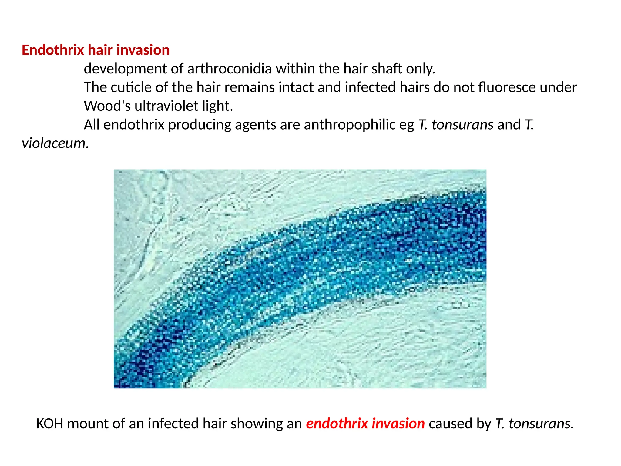
Endothrix hair invasion
developmentof arthroconidia within the hair shaft only.
The cuticle of the hair remains intact and infected hairs do not fluoresce under
Wood's ultraviolet light.
All endothrix producing agents are anthropophilic eg T. tonsurans and T.
violaceum.
KOH mount of an infected hair showing an endothrix invasion caused by T. tonsurans.
11.
Favus
• produces favus-likecrusts or scutula and corresponding hair loss.
• usually caused by T. schoenleinii, M. gypseum
• The fungus is entirely confined within the hair shaft but does NOT fragment into
arthroconidia (thus the infected hair commonly grows to normal lengths). The
relatively few hyphae run intact through the hair, forming tunnels within its
structure. When first immersed in KOH,
air is initially trapped around the hyphae forming the characteristic, long air spaces.
These rapidly fill in with KOH, when the hyphae themselves become visible.
CULTURE
• Aerobic
• Slowgrowth on SDA, DTM with some species
requiring additional growth factor.
• Tolerate cycloheximide in media
• Colonies are often pigmented
• Macroconidia formed in cultures.
14.
Obverse: white to
buffwith bright
orange periphery.
Reverse: yelowish
orange or yelowish
brown.
Dysgonic type M. canis
Obverse: yellow brown, heaped
and folded.
Reverse: colorless pale yellow
Macroconidia: usually absent
Obverse: flat,
spreading suede-like
todowny in testure
and white to buff in
color
Reverse: deep-yellow
submerged fringe
and reverse, later
becomes dark red in
the centre
Abundant microconidia, sessile and pyriform,
stallked or spherical
Clavate, smooth, thin-walled, variable
size, rarely produced
17.
Abundant microconidia, and
macroconidiais cigar shaped
with 7 septa.
Obverse: flat, white
to cream color,
powdery to granular
surface, red-brown
fringed periphery
Reverse: dark red
reverse pigment