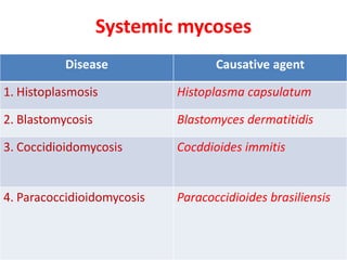
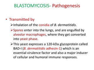
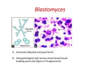
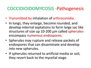
Systemic mycoses
- DR. ANKUR KUMAR
Systemic mycoses
Disease Causative agent
1. Histoplasmosis Histoplasma capsulatum
2. Blastomycosis Blastomyces dermatitidis
3. Coccidioidomycosis Cocddioides immitis
4. Paracoccidioidomycosis Paracoccidioides brasiliensis
HISTOPLASMOSIS
• Systemic granulomatous disease caused by a
dimorphic fungus, Histoplasma capsulatum.
• Species name is misnomer as it is not
capsulated.
• Also known as Darling's disease (because they
was first reported by Samuel Darling in1905 )
HISTOPLASMOSIS- Pathogenesis
• Transmitted by- inhalation of spores (i.e.
microconidia) which usually circulate in the air
after the contaminated soil is disturbed.
• Spores enter into the lungs engulfed
inside the alveolar macrophages
transform into yeast forms .
• Yeasts survive within the phagolysosome of
the macrophage by producing alkaline
substances (bicarbonate and ammonia).
HISTOPLASMOSIS- Clinical Manifestations
• Pulmonary hlstoplasmosis:
 Most common form.
 Acute form presents with mild flu like illness, pulmonary infiltrates
in chest X-ray with hilar or mediastinal lymphadenopadiy.
 Chronic cavitary histoplasmosis may be seen in smokers with
underlying structural lung disease.
• Mucocutaneous histoplasmosis
 Skin and oral mucosal lesions may develop secondary to
pulmonary infection.
 Oral lesions are particularly seen in Indian patients.
• Disseminated histoplasmosis:
 Develops if CMI is very low (e.g. untreated HIV infected people or
following organ transplantation).
 Common sites affected- bone marrow, spleen, liver, eyes and
adrenal glands
HISTOPLASMOSIS- Laboratory Diagnosis
• Specimens:
Sputum, aspirate from bone marrow & lymph
node, blood and biopsies from skin & mucosa.
• Direct microscopy:
Histopathological staining (PAS, Giemsa or GMS
stain) – Shows tiny oval yeast cells (2-4 µm size)
with narrow based budding within macrophages
with an underlying granulomatous response.
HISTOPLASMOSIS- Laboratory Diagnosis
• Culture:
Gold standard method of diagnosis.
Media- SDA, blood agar and BHI agar
Incubated simultaneously at 25°C and 37°C.
At 25°C:
 Mycelial phase
 Produces white to buff brown colonies, consist of 2 types of
conidia or spores:
1) Tuberculate macroconidia- typical thick walls and finger-like
projections which is a characteristic feature
2) Microconidia are smaller, thin, and smooth-walled.
At 37°C:
 Gets converted into yeast form (creamy white colonies), which is
best developed in special media like Kelley's media.
HISTOPLASMOSIS- Laboratory Diagnosis
• Serology:
Antibodies in serum - detected by CFT and
imununodiffusion test.
Antibodies appear after 1 month of infection; hence
are more useful in chronic stage (often negative in
early course and in disseminated stage).
 False positive result may occur due to past infection
or cross infection with blastomyces.
• Skin test:
 May be done to demonstrate delayed type hypersensitivity
response to histoplasmin antigen, which indicates prior
exposure
Histoplasma capsulatum (schematic diagram)
A. Yeast form;
B. Mycelial form;
C. 2-4 µm yeast cells with narrow based budding (Giemsa stain)
D. Mold form, septate thin hyphae with tuberculate macroconidia (arrows showing)
HISTOPLASMOSIS- TREATMENT
• Liposomal amphotericin B
 Is the antifungal agent of choice in acute pulmonary and
disseminated histoplasmosis.
• ltraconazole
Recommended for chronic cavitary pulmonary
histoplasmosis.
BLASTOMYCOSIS
• Also known as North American blastomycosis
or Gilchrist's disease or Chicago disease
• Is a fungal infection of humans and other
animals also- dogs and cats, caused,
Blastomyces dermatitidis.
BLASTOMYCOSIS- Pathogenesis
• Transmitted by
Inhalation of the conidia of B. dermatitidis.
Spores enter into the lungs, and are engulfed by
alveolar macrophages, where they get converted
into yeast phase.
This yeast expresses a 120-kDa glycoprotein called
BAD-I (B. dermatitidis adhesin-1) which is an
essential virulence factor and also a major inducer
of cellular and humoral immune responses.
BLASTOMYCOSIS- Clinlcal Manifestations
• Acute pulmonary blastomycosis - Most common
form.
• Extrapulmonary manifestations
Skin involvement - Most common extrapulmonary
form; characterized by either verrucous (more
common) or ulcerative type of skin lesions.
Osteomyelitis may develop along with contiguous soft
tissue abscesses and draining sinuses.
Prostate and epididymis involvement in men.
 Central nervous system (CNS) involvement has been
reported in - 40% of AIDS patients.
Brain abscess is the usual presentation, followed by
cranial or spinal epidural abscess and meningitis.
BLASTOMYCOSIS- Laboratory Diagnosis
• Antibody detection: lmmunodiffusion test
specific for B. dermatitidis has been developed
against yeast phase antigens such as antigen-A,
BAD- I and ASWS antigen (alkali soluble water
soluble).
• Antigen detection assay- detect Blastomyces
antigen in urine (more sensitive) and in serum is
commercially available.
• Molecular methods- DNA probe hybridization and
real time PCR
BLASTOMYCOSIS- Laboratory Diagnosis
• Histopathological staining of the tissue biopsy
specimens reveals thick-walled round yeast cells
of 8- 15 µm size with single broad-based budding
(figure of 8 appearance)
• Culture media - SDA, blood agar and BH I.
At 250C- Mycelia form containing hyphae with small
pear-shaped conidia are produced;
At 370C- mold to yeast conversion takes place.
• Skin test: done to demonstrate delayed type
hypersensitivity to blastomycin antigen.
Blastomyces
A. Schematic (Mycelial and yeast form);
B. Histopathological stain (arrow shows) broad based
budding yeast cells (figure o f 8 appearance)
BLASTOMYCOSIS- TREATMENT
• Liposomal amphotericin B
 is the antifungal agent of choice in most of the cases.
• ltraconazole
in immunocompetent patients with mild
pulmonary or non CNS extrapulmonary
blastomycosis
COCCIDIOIDOMYCOSIS
• Also called desert rheumatism or Valley fever
or California fever
• Is a systemic fungal disease caused by a
dimorphic soil dwelling fungus- Coccidioides
(C. immitis and C. posadasii).
COCCIDIOIDOMYCOSIS -Pathogenesis
• Transmitted by inhalation of arthroconidia.
• In lungs, they enlarge, become rounded, and
develop internal septations to form large sac like
structures of size up 10-200 µm called spherules-
encompass numerous endospores.
• Spherules may rupture and release packets of
endospores that can disseminate and develop
into new spherules.
• If spherules returned to artificial media or soil,
they revert back to the mycelial stage.
COCCIDIOIDOMYCOSIS - Clinlcal Manifestations
• Asymptomatic- in most of patients (60%).
• Pulmonary coccidioidomycosis - presents as
pneumonia, cavities, pleural effusion or nodule
formation.
• Skin lesions such as rashes or erythema nodosum and
arthritis with joint pain may appear secondary to
pulmonary infection particularly in women.
• Disseminated form:
Males and persons with low CMI (HIV infected patients
with CD4+ T cell count <250/ µI) are at higher risk.
Common sites for dissemination include skin, bone, joints,
soft tissues, and meninges.
COCCIDIOIDOMYCOSIS - Laboratory Diagnosis
• Histopathological staining
H and E stain, PAS or CMS of sputum or tissue biopsy
specimens demonstrates spherules which are large
sac like structures (20-80 µm size), have thick, double
refractile wall, and are filled with endospores
• Cultures on SDA –
 produces mycelial growth, described as fragmented hyphae
consisting of barrel-shaped arthrospores with alternate cells
distorted (empty cells)
 Coccidioides differs from other dimorphic fungi as it grows as
mold at both 25°C and 37°C in usual culture media (forms
spherules at 37°C in certain special culture media only).
 Cultures are highly infectious: lead to accidental inhalation of
spores in laboratories, require biosafety level-3 precautions.
COCCIDIOIDOMYCOSIS - Laboratory Diagnosis
• Serology: Antibodies are detected by
immunodiffusion test and CFT.
• Skin test: It is done by fungal extaracts
(coccidioidin or spherulin);
 produces at least a 5 mm induration within 48
hours after injection (delayed hypersensitivity
reaction) indicates past infection.
Cocddioides
A. Spherules (schematic);
B. Hyphae with arthroconidia (schematic);
C. Spherules (PAS staining);
D. Hyphae with arthroconidia (LPCB mount)
TREATMENT- Coccidioidomycosis
• Itraconazole -drug of choice
except for diffuse pneumonia with pulmonary
sequelae where amphotericin B is recommended.
PARACOCCIDIOIDOMYCOSIS
• Also known as South American blastomycosis,
Lutz-Splendore-de Almeida disease
• Is a systemic disease caused by the dimorphic
fungus Paracoccidioides brasiliensis.
Pathogenesis and clinical Manifestations
• Transmission- by inhalation of spores,
• Transform into the yeast phase in lungs.
• Occurs as two major forms.
1. Acute form (or juvenile type):
affects young adults under 30 years age.
less common variety, but more severe form, manifests as
disseminated infection involving multiple viscera and is
refractory to t/t.
2. Chronic form (or adult form):
accounts for 90% of cases
predominantly affects older men.
results from reactivation of quiescenct lung lesions.
less severe form, manifested as progressive pulmonary
disease affecting lower lobes, with fibrosis.
Skin, oral mucosal lesions and cervical lymphadenopathy
PARACOCCIDIOIDOMYCOSIS-Laboratory Diagnosis
• Histopathological staining - of pus, tissue biopsies or
sputum
 reveals round thick-walled yeasts, which multiple narrow-
necked buds attached circumferencially giving rise to Mickeiy
mouse or pilot wheel appearance.
• Culture on SDA
 yields mycelial form at 250C
 converts in to yeast phase at 370C when grown in BHI agar
supplemented with blood and glutamine.
• Serology:
 Antibodies are detected by immunodiffusion, and by ELISA,
using gp43 antigen of P. brasiliensis.
• Skin test:
 demonstrates delayed hypersensitivity response against
paracoccidioidin antigen.
Paracoccidioidomycosis
A. Schematic representation of mycelial and yeast forms;
B. Methenamine silver staining shows yeast from (pilot
wheel appearance)
TREATMENT- Paracoccidioidomycosis
• ltraconazole - is the treatment of choice
except for the seriously ill patients where
Amphotericin B is recommended.
Sulfonamides are effective, but the response is
slow with frequent relapses.
THANK
YOU

Systemic mycoses

  • 1.
  • 2.
    Systemic mycoses Disease Causativeagent 1. Histoplasmosis Histoplasma capsulatum 2. Blastomycosis Blastomyces dermatitidis 3. Coccidioidomycosis Cocddioides immitis 4. Paracoccidioidomycosis Paracoccidioides brasiliensis
  • 3.
    HISTOPLASMOSIS • Systemic granulomatousdisease caused by a dimorphic fungus, Histoplasma capsulatum. • Species name is misnomer as it is not capsulated. • Also known as Darling's disease (because they was first reported by Samuel Darling in1905 )
  • 4.
    HISTOPLASMOSIS- Pathogenesis • Transmittedby- inhalation of spores (i.e. microconidia) which usually circulate in the air after the contaminated soil is disturbed. • Spores enter into the lungs engulfed inside the alveolar macrophages transform into yeast forms . • Yeasts survive within the phagolysosome of the macrophage by producing alkaline substances (bicarbonate and ammonia).
  • 5.
    HISTOPLASMOSIS- Clinical Manifestations •Pulmonary hlstoplasmosis:  Most common form.  Acute form presents with mild flu like illness, pulmonary infiltrates in chest X-ray with hilar or mediastinal lymphadenopadiy.  Chronic cavitary histoplasmosis may be seen in smokers with underlying structural lung disease. • Mucocutaneous histoplasmosis  Skin and oral mucosal lesions may develop secondary to pulmonary infection.  Oral lesions are particularly seen in Indian patients. • Disseminated histoplasmosis:  Develops if CMI is very low (e.g. untreated HIV infected people or following organ transplantation).  Common sites affected- bone marrow, spleen, liver, eyes and adrenal glands
  • 6.
    HISTOPLASMOSIS- Laboratory Diagnosis •Specimens: Sputum, aspirate from bone marrow & lymph node, blood and biopsies from skin & mucosa. • Direct microscopy: Histopathological staining (PAS, Giemsa or GMS stain) – Shows tiny oval yeast cells (2-4 µm size) with narrow based budding within macrophages with an underlying granulomatous response.
  • 7.
    HISTOPLASMOSIS- Laboratory Diagnosis •Culture: Gold standard method of diagnosis. Media- SDA, blood agar and BHI agar Incubated simultaneously at 25°C and 37°C. At 25°C:  Mycelial phase  Produces white to buff brown colonies, consist of 2 types of conidia or spores: 1) Tuberculate macroconidia- typical thick walls and finger-like projections which is a characteristic feature 2) Microconidia are smaller, thin, and smooth-walled. At 37°C:  Gets converted into yeast form (creamy white colonies), which is best developed in special media like Kelley's media.
  • 8.
    HISTOPLASMOSIS- Laboratory Diagnosis •Serology: Antibodies in serum - detected by CFT and imununodiffusion test. Antibodies appear after 1 month of infection; hence are more useful in chronic stage (often negative in early course and in disseminated stage).  False positive result may occur due to past infection or cross infection with blastomyces. • Skin test:  May be done to demonstrate delayed type hypersensitivity response to histoplasmin antigen, which indicates prior exposure
  • 9.
    Histoplasma capsulatum (schematicdiagram) A. Yeast form; B. Mycelial form; C. 2-4 µm yeast cells with narrow based budding (Giemsa stain) D. Mold form, septate thin hyphae with tuberculate macroconidia (arrows showing)
  • 10.
    HISTOPLASMOSIS- TREATMENT • Liposomalamphotericin B  Is the antifungal agent of choice in acute pulmonary and disseminated histoplasmosis. • ltraconazole Recommended for chronic cavitary pulmonary histoplasmosis.
  • 11.
    BLASTOMYCOSIS • Also knownas North American blastomycosis or Gilchrist's disease or Chicago disease • Is a fungal infection of humans and other animals also- dogs and cats, caused, Blastomyces dermatitidis.
  • 12.
    BLASTOMYCOSIS- Pathogenesis • Transmittedby Inhalation of the conidia of B. dermatitidis. Spores enter into the lungs, and are engulfed by alveolar macrophages, where they get converted into yeast phase. This yeast expresses a 120-kDa glycoprotein called BAD-I (B. dermatitidis adhesin-1) which is an essential virulence factor and also a major inducer of cellular and humoral immune responses.
  • 13.
    BLASTOMYCOSIS- Clinlcal Manifestations •Acute pulmonary blastomycosis - Most common form. • Extrapulmonary manifestations Skin involvement - Most common extrapulmonary form; characterized by either verrucous (more common) or ulcerative type of skin lesions. Osteomyelitis may develop along with contiguous soft tissue abscesses and draining sinuses. Prostate and epididymis involvement in men.  Central nervous system (CNS) involvement has been reported in - 40% of AIDS patients. Brain abscess is the usual presentation, followed by cranial or spinal epidural abscess and meningitis.
  • 14.
    BLASTOMYCOSIS- Laboratory Diagnosis •Antibody detection: lmmunodiffusion test specific for B. dermatitidis has been developed against yeast phase antigens such as antigen-A, BAD- I and ASWS antigen (alkali soluble water soluble). • Antigen detection assay- detect Blastomyces antigen in urine (more sensitive) and in serum is commercially available. • Molecular methods- DNA probe hybridization and real time PCR
  • 15.
    BLASTOMYCOSIS- Laboratory Diagnosis •Histopathological staining of the tissue biopsy specimens reveals thick-walled round yeast cells of 8- 15 µm size with single broad-based budding (figure of 8 appearance) • Culture media - SDA, blood agar and BH I. At 250C- Mycelia form containing hyphae with small pear-shaped conidia are produced; At 370C- mold to yeast conversion takes place. • Skin test: done to demonstrate delayed type hypersensitivity to blastomycin antigen.
  • 16.
    Blastomyces A. Schematic (Mycelialand yeast form); B. Histopathological stain (arrow shows) broad based budding yeast cells (figure o f 8 appearance)
  • 17.
    BLASTOMYCOSIS- TREATMENT • Liposomalamphotericin B  is the antifungal agent of choice in most of the cases. • ltraconazole in immunocompetent patients with mild pulmonary or non CNS extrapulmonary blastomycosis
  • 18.
    COCCIDIOIDOMYCOSIS • Also calleddesert rheumatism or Valley fever or California fever • Is a systemic fungal disease caused by a dimorphic soil dwelling fungus- Coccidioides (C. immitis and C. posadasii).
  • 19.
    COCCIDIOIDOMYCOSIS -Pathogenesis • Transmittedby inhalation of arthroconidia. • In lungs, they enlarge, become rounded, and develop internal septations to form large sac like structures of size up 10-200 µm called spherules- encompass numerous endospores. • Spherules may rupture and release packets of endospores that can disseminate and develop into new spherules. • If spherules returned to artificial media or soil, they revert back to the mycelial stage.
  • 20.
    COCCIDIOIDOMYCOSIS - ClinlcalManifestations • Asymptomatic- in most of patients (60%). • Pulmonary coccidioidomycosis - presents as pneumonia, cavities, pleural effusion or nodule formation. • Skin lesions such as rashes or erythema nodosum and arthritis with joint pain may appear secondary to pulmonary infection particularly in women. • Disseminated form: Males and persons with low CMI (HIV infected patients with CD4+ T cell count <250/ µI) are at higher risk. Common sites for dissemination include skin, bone, joints, soft tissues, and meninges.
  • 21.
    COCCIDIOIDOMYCOSIS - LaboratoryDiagnosis • Histopathological staining H and E stain, PAS or CMS of sputum or tissue biopsy specimens demonstrates spherules which are large sac like structures (20-80 µm size), have thick, double refractile wall, and are filled with endospores • Cultures on SDA –  produces mycelial growth, described as fragmented hyphae consisting of barrel-shaped arthrospores with alternate cells distorted (empty cells)  Coccidioides differs from other dimorphic fungi as it grows as mold at both 25°C and 37°C in usual culture media (forms spherules at 37°C in certain special culture media only).  Cultures are highly infectious: lead to accidental inhalation of spores in laboratories, require biosafety level-3 precautions.
  • 22.
    COCCIDIOIDOMYCOSIS - LaboratoryDiagnosis • Serology: Antibodies are detected by immunodiffusion test and CFT. • Skin test: It is done by fungal extaracts (coccidioidin or spherulin);  produces at least a 5 mm induration within 48 hours after injection (delayed hypersensitivity reaction) indicates past infection.
  • 23.
    Cocddioides A. Spherules (schematic); B.Hyphae with arthroconidia (schematic); C. Spherules (PAS staining); D. Hyphae with arthroconidia (LPCB mount)
  • 24.
    TREATMENT- Coccidioidomycosis • Itraconazole-drug of choice except for diffuse pneumonia with pulmonary sequelae where amphotericin B is recommended.
  • 25.
    PARACOCCIDIOIDOMYCOSIS • Also knownas South American blastomycosis, Lutz-Splendore-de Almeida disease • Is a systemic disease caused by the dimorphic fungus Paracoccidioides brasiliensis.
  • 26.
    Pathogenesis and clinicalManifestations • Transmission- by inhalation of spores, • Transform into the yeast phase in lungs. • Occurs as two major forms. 1. Acute form (or juvenile type): affects young adults under 30 years age. less common variety, but more severe form, manifests as disseminated infection involving multiple viscera and is refractory to t/t. 2. Chronic form (or adult form): accounts for 90% of cases predominantly affects older men. results from reactivation of quiescenct lung lesions. less severe form, manifested as progressive pulmonary disease affecting lower lobes, with fibrosis. Skin, oral mucosal lesions and cervical lymphadenopathy
  • 27.
    PARACOCCIDIOIDOMYCOSIS-Laboratory Diagnosis • Histopathologicalstaining - of pus, tissue biopsies or sputum  reveals round thick-walled yeasts, which multiple narrow- necked buds attached circumferencially giving rise to Mickeiy mouse or pilot wheel appearance. • Culture on SDA  yields mycelial form at 250C  converts in to yeast phase at 370C when grown in BHI agar supplemented with blood and glutamine. • Serology:  Antibodies are detected by immunodiffusion, and by ELISA, using gp43 antigen of P. brasiliensis. • Skin test:  demonstrates delayed hypersensitivity response against paracoccidioidin antigen.
  • 28.
    Paracoccidioidomycosis A. Schematic representationof mycelial and yeast forms; B. Methenamine silver staining shows yeast from (pilot wheel appearance)
  • 29.
    TREATMENT- Paracoccidioidomycosis • ltraconazole- is the treatment of choice except for the seriously ill patients where Amphotericin B is recommended. Sulfonamides are effective, but the response is slow with frequent relapses.
  • 30.