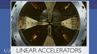
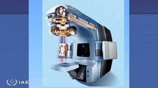
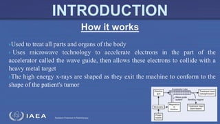
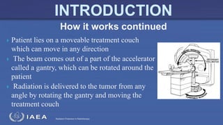
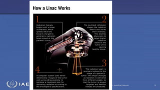
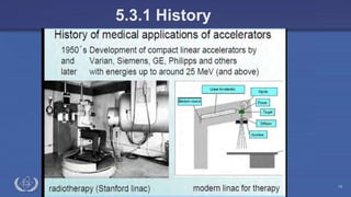
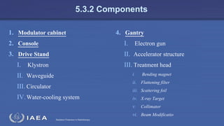
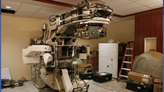
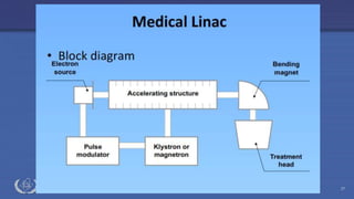
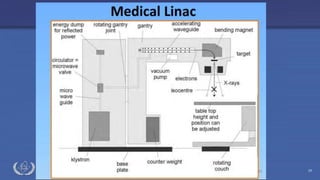
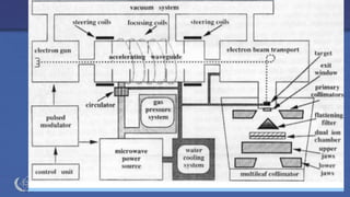
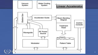
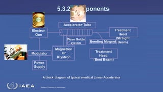
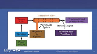
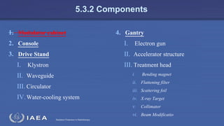
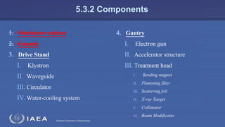
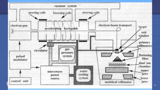
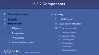
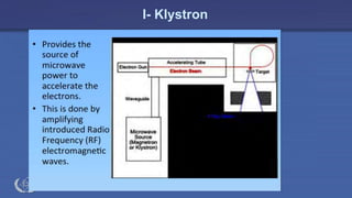
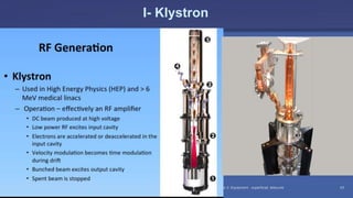
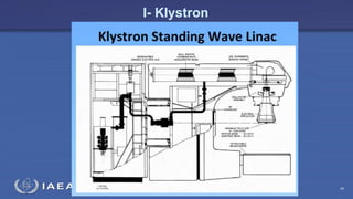
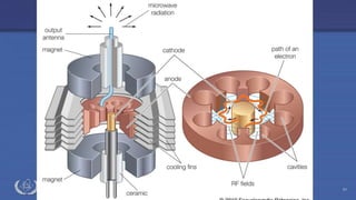
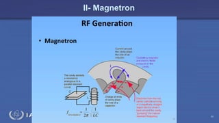
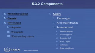
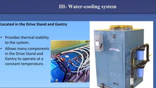
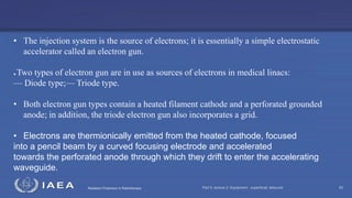
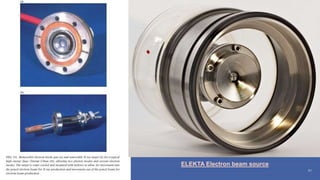
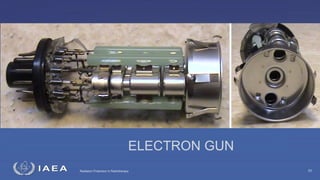
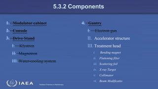
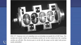
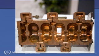
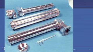
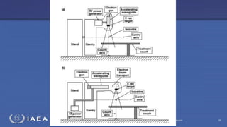
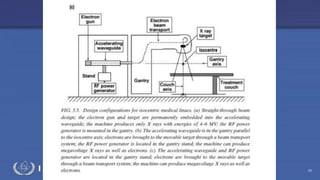
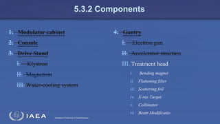
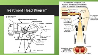
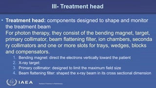
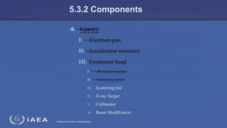
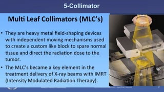
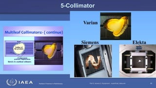
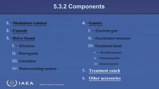
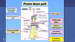
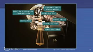
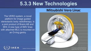
The document discusses the components and operation of a linear accelerator used for radiation therapy. It describes the key parts of a linear accelerator including the electron gun, accelerator structure, treatment head and how they work together to generate photon or electron beams for radiation treatment. The linear accelerator uses microwave technology to accelerate electrons which are then converted into x-ray or electron beams that can be aimed at a tumor from multiple angles using the rotating gantry.
5. Radiation Protection in Radiotherapy
INTRODUCTION
Part 5, lecture 2: Equipment - superficial, telecurie 5
6. Radiation Protection in Radiotherapy Part 5, lecture 2: Equipment - superficial, telecurie 6
INTRODUCTION
7. Radiation Protection in Radiotherapy
•Photon Beam (X-Ray):
•4 MV To 22 MV.
•Single Beam.
•Duel Beams
•Electron Beam:
•Multi-Beams with energy
grope between: 4- 22 MeV.
INTRODUCTION
8. Radiation Protection in Radiotherapy
WHAT IS LINAC
A linear accelerator is a device that uses high
Radio‐Frequency (RF)electromagnet waves to
accelerate charged particles (i.e. electrons) to
high energies in a linear path, inside a tube like
structure called the accelerator waveguide.
The resonance cavity frequency of the medical
linacs is about 3 billion Hertz (cycles/sec)
Part 5, lecture 2: Equipment - superficial, telecurie 8
INTRODUCTION
9. Radiation Protection in Radiotherapy
Used to treat all parts and organs of the body
Uses microwave technology to accelerate electrons in the part of the
accelerator called the wave guide, then allows these electrons to collide with a
heavy metal target
The high energy x-rays are shaped as they exit the machine to conform to the
shape of the patient's tumor
How it works
INTRODUCTION
10. Radiation Protection in Radiotherapy
X-ray treatments are designed in a way that destroy the cancer cells
while sparing the surrounding normal tissue
High energy photons enter the patient's body and aim to break the
DNA in all the cells within the treatment area
The good cells are able to mend themselves
The cancerous cells are unable to do this and therefore die
How It Works Continued
11. Radiation Protection in Radiotherapy
Patient lies on a moveable treatment couch
which can move in any direction
The beam comes out of a part of the accelerator
called a gantry, which can be rotated around the
patient
Radiation is delivered to the tumor from any
angle by rotating the gantry and moving the
treatment couch
How it works continued
12. Radiation Protection in Radiotherapy Part 5, lecture 2: Equipment - superficial, telecurie 12
13. Radiation Protection in Radiotherapy
Advantages: particles are able to reach very high energies
without the need for extremely high voltages
Linear accelerators attack the affected area with higher
doses of radiation than other machines
Advantages
14. Radiation Protection in Radiotherapy
Disadvantages: A linear accelerator can cost anywhere between one million
and three million dollars. Operating the machine costs about $900,000
annually.
The particles travel in a straight line, each accelerating segment is used only
once. The segments run in short pulses, limiting the average current output
and forcing the experimental detectors to handle data coming in short bursts,
thus increasing the maintenance expense
Disadvantages
15. Radiation Protection in Radiotherapy
5.3.1 History
Part 5, lecture 2: Equipment - superficial, telecurie 15
16. Radiation Protection in Radiotherapy Part 5, lecture 2: Equipment - superficial, telecurie 16
5.3.1 History
17. Radiation Protection in Radiotherapy Part 5, lecture 2: Equipment - superficial, telecurie 17
5.3.1 History
18. Radiation Protection in Radiotherapy Part 5, lecture 2: Equipment - superficial, telecurie 18
5.3.1 History
19. Radiation Protection in Radiotherapy Part 5, lecture 2: Equipment - superficial, telecurie 19
5.3.1 History
20. Radiation Protection in Radiotherapy Part 5, lecture 2: Equipment - superficial, telecurie 20
5.3.1 History
Early Accelerators
(1953-1961):
Extremely large and bulky
Limited gantry motion
21. Radiation Protection in Radiotherapy Part 5, lecture 2: Equipment - superficial, telecurie 21
5.3.1 History
Second Generations
(1962-1982):
360 degree rotational
Allow treatment to a patent from
any gantry angle
Improvement in accuracy and
dose delivery
22. Radiation Protection in Radiotherapy Part 5, lecture 2: Equipment - superficial, telecurie 22
5.3.1 History
• Third generation accelerators:
• Improved accelerator guide
• Magnet systems
• Beam-modifying systems to
provide wide ranges of beam
energy, dose rate, field size
• Operating modes with improved
beam characteristics
• Highly reliable
• Compact design
• May include: dual photon energies,
multileaf collimation, several
electron energies & electronic
portal verification systems
23. Radiation Protection in Radiotherapy Part 5, lecture 2: Equipment - superficial, telecurie 23
5.3.1 History
24. Radiation Protection in Radiotherapy Part 5, lecture 2: Equipment - superficial, telecurie 24
5.3.2 Components
25. Radiation Protection in Radiotherapy
5.3.2 Components
1. Modulator cabinet
2. Console
3. Drive Stand
I. Klystron
II. Waveguide
III. Circulator
IV.Water-cooling system
4. Gantry
I. Electron gun
II. Accelerator structure
III. Treatment head
i. Bending magnet
ii. Flattening filter
iii. Scattering foil
iv. X-ray Target
v. Collimator
vi. Beam Modificatio
26. Radiation Protection in Radiotherapy Part 5, lecture 2: Equipment - superficial, telecurie 26
27. Radiation Protection in Radiotherapy Part 5, lecture 2: Equipment - superficial, telecurie 27
28. Radiation Protection in Radiotherapy Part 5, lecture 2: Equipment - superficial, telecurie 28
29. Radiation Protection in Radiotherapy Part 5, lecture 2: Equipment - superficial, telecurie 29
30. Radiation Protection in Radiotherapy Part 5, lecture 2: Equipment - superficial, telecurie 30
31. Radiation Protection in Radiotherapy
Treatment
Head
(Straight
Beam)
Power
Supply
Modulator
Electron
Gun
Magnetron
Or
Klystron
Wave Guide
system
Treatment
Head
(Bent Beam)
Bending Magnet
Accelerator Tube
A block diagram of typical medical Linear Accelerator
5.3.2 Components
32. Radiation Protection in Radiotherapy Part 5, lecture 2: Equipment - superficial, telecurie 32
33. Radiation Protection in Radiotherapy
A power supply Provides DC power to the modulator
Modulator
the magnetron or klystron
Deliver the pulses to the electron gun
Pulsedmicrowaves
the accelerator tube
or structure via a waveguide systems.
electrons
Assignment: Briefly describe the process within a linear accelerator starting
from the power supply to the production of a 3 mm pencil beam.
34. Radiation Protection in Radiotherapy
5.3.2.1 Modulator Cabinet
• Modulator cabinet: contains components that distribute and monitor
primary electrical power and high-voltage pulses to the magnetron or
klystron
• Located in the treatment room
• Three major components:
• The fan control: automatically turns the fans off and on as the need arises for
cooling the power distribution
• Auxiliary power-distribution system: contains the emergency off button that
shuts off the power to the treatment unit.
• Primary power-distribution system
35. Radiation Protection in Radiotherapy
5.3.2 Components
1. Modulator cabinet
2. Console
3. Drive Stand
I. Klystron
II. Waveguide
III. Circulator
IV.Water-cooling system
4. Gantry
I. Electron gun
II. Accelerator structure
III. Treatment head
i. Bending magnet
ii. Flattening filter
iii. Scattering foil
iv. X-ray Target
v. Collimator
vi. Beam Modificatio
36. Radiation Protection in Radiotherapy
5.3.2.2 Console
• Console electronic cabinet: provides a central location for
monitoring and controlling the linac
• Take the form of a digital display, push button panel or video display
terminal (VDT)
• All interlocks must be satisfied for the machine to allow the beam to be
started
• Provides a digital display for prescribe dose (monitor units),
mechanical beam parameters such as collimator setting or gantry angle
37. Radiation Protection in Radiotherapy
5.3.2 Components
1. Modulator cabinet
2. Console
3. Drive Stand
I. Klystron
II. Waveguide
III. Circulator
IV.Water-cooling system
4. Gantry
I. Electron gun
II. Accelerator structure
III. Treatment head
i. Bending magnet
ii. Flattening filter
iii. Scattering foil
iv. X-ray Target
v. Collimator
vi. Beam Modificatio
38. Radiation Protection in Radiotherapy
5.3.2.3 Drive Stand
• Drive Stand: a stand containing the apparatus that drives the linear accelerator
• Open on both sides with swinging doors for easy access to gauges, valves, tanks, and
buttons
• Klystron/Magnetron: power source used to generate electromagnetic waves for the
accelerator guides
• Waveguide: hollow tube-like structure that guide the electromagnetic waves from the
magnetron to the accelerating guide where electrons are accelerated
• Circulator: directs the RF energy into the waveguide and prevents any reflected
microwaves from returning to the klystron
• Water-cooling system: allows many components in the gantry and drive stand to operate
at a constant temperature
39. Radiation Protection in Radiotherapy Part 5, lecture 2: Equipment - superficial, telecurie 39
40. Radiation Protection in Radiotherapy
5.3.2 Components
1. Modulator cabinet
2. Console
3. Drive Stand
I. Klystron
II. Magnetron
III. Waveguide
IV.Water-cooling system
4. Gantry
I. Electron gun
II. Accelerator structure
III. Treatment head
i. Bending magnet
ii. Flattening filter
iii. Scattering foil
iv. X-ray Target
v. Collimator
vi. Beam Modificatio
42. Radiation Protection in Radiotherapy Part 5, lecture 2: Equipment - superficial, telecurie 42
43. Radiation Protection in Radiotherapy Part 5, lecture 2: Equipment - superficial, telecurie 43
I- Klystron
44. Radiation Protection in Radiotherapy Part 5, lecture 2: Equipment - superficial, telecurie 44
45. Radiation Protection in Radiotherapy Part 5, lecture 2: Equipment - superficial, telecurie 45
46. Radiation Protection in Radiotherapy Part 5, lecture 2: Equipment - superficial, telecurie 46
I- Klystron
47. Radiation Protection in Radiotherapy
5.3.2 Components
1. Modulator cabinet
2. Console
3. Drive Stand
I. Klystron
II. Magnetron
III. Water-cooling system
4. Gantry
I. Electron gun
II. Accelerator structure
III. Treatment head
i. Bending magnet
ii. Flattening filter
iii. Scattering foil
iv. X-ray Target
v. Collimator
vi. Beam Modificatio
48. Radiation Protection in Radiotherapy
II- Magnetron
• Magnetron: device that provides high-frequency microwave
power that is used to accelerate the electrons in the
accelerating waveguide.
• Electrons are emitted from the cathode and spiral in the
perpendicular magnetic field. The interaction of the spiraling
electrons with the cavities in the anode creates the high-
frequency EM waves.
• oscillator and amplifier used in low-energy
49. Radiation Protection in Radiotherapy Part 5, lecture 2: Equipment - superficial, telecurie 49
II- Magnetron
50. Radiation Protection in Radiotherapy Part 5, lecture 2: Equipment - superficial, telecurie 50
II- Magnetron
51. Radiation Protection in Radiotherapy Part 5, lecture 2: Equipment - superficial, telecurie 51
52. Radiation Protection in Radiotherapy Part 5, lecture 2: Equipment - superficial, telecurie 52
II- Magnetron
53. Radiation Protection in Radiotherapy Part 5, lecture 2: Equipment - superficial, telecurie 53
• Waveguides are evacuated or gas filled metallic structures of rectangular
or circular cross-section used in the transmission of microwaves.
• Two types of waveguide are used in linacs: RF power transmission
waveguides and accelerating waveguides.
• The power transmission waveguides transmit the RF power from the
power source to the accelerating waveguide in which the electrons are
accelerated.
• The electrons are accelerated in the accelerating waveguide by means
of an energy transfer from the high power RF fields, which are set up
in the accelerating waveguide and are produced by the RF power
generators.
II- RF power transmission Waveguide
54. Radiation Protection in Radiotherapy
5.3.2 Components
1. Modulator cabinet
2. Console
3. Drive Stand
I. Klystron
II. Waveguide
III. Water-cooling system
4. Gantry
I. Electron gun
II. Accelerator structure
III. Treatment head
i. Bending magnet
ii. Flattening filter
iii. Scattering foil
iv. X-ray Target
v. Collimator
vi. Beam Modificatio
55. Radiation Protection in Radiotherapy Part 5, lecture 2: Equipment - superficial, telecurie 55
III- Water-cooling system
56. Radiation Protection in Radiotherapy
5.3.2 Components
1. Modulator cabinet
2. Console
3. Drive Stand
I. Klystron
II. Waveguide
III. Water-cooling system
4. Gantry
I. Electron gun
II. Accelerator structure
III. Treatment head
i. Bending magnet
ii. Flattening filter
iii. Scattering foil
iv. X-ray Target
v. Collimator
vi. Beam Modificatio
57. Radiation Protection in Radiotherapy
5.3.2.4 Gantry
• Gantry: responsible for directing the photon (x-ray) energy or electron
beam at a patients tumor.
it rotates 360 degrees around a line/point, called the Isocenter.
• Electron gun: produce electrons and injects them into the accelerator structure
• Accelerator structure: a special type of wave guide in which electrons are
accelerated.
• Treatment head: components designed to shape and monitor the treatment
beam
58. Radiation Protection in Radiotherapy
5.3.2 Components
1. Modulator cabinet
2. Console
3. Drive Stand
I. Klystron
II. Magnetron
III. Water-cooling system
4. Gantry
I. Electron gun
II. Accelerator structure
III. Treatment head
i. Bending magnet
ii. Flattening filter
iii. Scattering foil
iv. X-ray Target
v. Collimator
vi. Beam Modificatio
59. Radiation Protection in Radiotherapy
I – Electron Gun
Part 5, lecture 2: Equipment - superficial, telecurie 59
Electron gun: produce
electrons and injects them into
the accelerator structure
60. Radiation Protection in Radiotherapy Part 5, lecture 2: Equipment - superficial, telecurie 60
• The injection system is the source of electrons; it is essentially a simple electrostatic
accelerator called an electron gun.
● Two types of electron gun are in use as sources of electrons in medical linacs:
— Diode type;— Triode type.
• Both electron gun types contain a heated filament cathode and a perforated grounded
anode; in addition, the triode electron gun also incorporates a grid.
• Electrons are thermionically emitted from the heated cathode, focused
into a pencil beam by a curved focusing electrode and accelerated
towards the perforated anode through which they drift to enter the accelerating
waveguide.
63. Radiation Protection in Radiotherapy
5.3.2 Components
1. Modulator cabinet
2. Console
3. Drive Stand
I. Klystron
II. Magnetron
III. Water-cooling system
4. Gantry
I. Electron gun
II. Accelerator structure
III. Treatment head
i. Bending magnet
ii. Flattening filter
iii. Scattering foil
iv. X-ray Target
v. Collimator
vi. Beam Modificatio
64. Radiation Protection in Radiotherapy
II- Accelerator Structure/ waveguide
• Microwave power (produced in the klyston) is transported
to the accelerator structure in which corrugations are used
to slow up the waves synchronous with the flowing
electrons. After the flowing electrons leave the accelerator
structure, they are directed toward the target (for photon
production) or scattering foil (for electron production)
located in the treatment head.
65. Radiation Protection in Radiotherapy Part 5, lecture 2: Equipment - superficial, telecurie 65
66. Radiation Protection in Radiotherapy Part 5, lecture 2: Equipment - superficial, telecurie 66
67. Radiation Protection in Radiotherapy Part 5, lecture 2: Equipment - superficial, telecurie 67
68. Radiation Protection in Radiotherapy Part 5, lecture 2: Equipment - superficial, telecurie 68
69. Radiation Protection in Radiotherapy Part 5, lecture 2: Equipment - superficial, telecurie 69
70. Radiation Protection in Radiotherapy
5.3.2 Components
1. Modulator cabinet
2. Console
3. Drive Stand
I. Klystron
II. Magnetron
III. Water-cooling system
4. Gantry
I. Electron gun
II. Accelerator structure
III. Treatment head
i. Bending magnet
ii. Flattening filter
iii. Scattering foil
iv. X-ray Target
v. Collimator
vi. Beam Modificatio
72. Radiation Protection in Radiotherapy
III- Treatment head
• Treatment head: components designed to shape and monitor
the treatment beam
For photon therapy, they consist of the bending magnet, target,
primary collimator, beam flattening filter, ion chambers, seconda
ry collimators and one or more slots for trays, wedges, blocks
and compensators.
1. Bending magnet: direct the electrons vertically toward the patient
2. X-ray target:
3. Primary collimator: designed to limit the maximum field size
4. Beam flattening filter: shaped the x-ray beam in its cross sectional dimension
73. Radiation Protection in Radiotherapy
III- Treatment head
5. Ion chamber: monitors the beam for its symmetry in the right-left
and inferior-superior direction
6.Secondary collimators: upper and lower collimator jaws
7. Field light: outlines the dimensions of the radiation field as it
appears on the patient, allows accurate positioning of the radiation
field in relationship to skim marks or other reference points
74. Radiation Protection in Radiotherapy
5.3.2 Components
1. Modulator cabinet
2. Console
3. Drive Stand
I. Klystron
II. Magnetron
III. Water-cooling system
4. Gantry
I. Electron gun
II. Accelerator structure
III. Treatment head
i. Bending magnet
ii. Flattening filter
iii. Scattering foil
iv. X-ray Target
v. Collimator
vi. Beam Modificatio
75. Radiation Protection in Radiotherapy Part 5, lecture 2: Equipment - superficial, telecurie 75
1- Bending Magnet
76. Radiation Protection in Radiotherapy
5.3.2 Components
4. Gantry
I. Electron gun
II. Accelerator structure
III. Treatment head
i. Bending magnet
ii. Flattening filter
iii. Scattering foil
iv. X-ray Target
v. Collimator
vi. Beam Modification
77. Radiation Protection in Radiotherapy
2- Flattening Filter
• Flattening filter: (lead, steel, copper etc.)
• Modifies the narrow, non-uniform photon beam at the isocenter into a
clinically useful beam through a combination of attenuation of the
center of the beam and scatter into the periphery of the beam
• Measured in percent at a particular depth in a phantom (10 cm)
• Must be carefully positioned in the beam or the beam hitting the
patient will be non-uniform, resulting in hot and cold spots
78. Flattening filter :
• The flattening filter is a cone shaped
• change the beam profile at depth
• Absorbs photons on the central axis
• Producing a more uniform beam
profile at the treatment distance.
79. Radiation Protection in Radiotherapy
2- Flattening Filter
• Flatness: a wide beam that is nearly uniform in intensity from one
side to the other (+/- 6%)
• Symmetry: the measure of intensity difference between its
opposite sides (+/- 4%)
• Causes include the use of a wedge, misalignment of the flattening
filter, and misdirection of the electron beam before hitting the target.
80. Radiation Protection in Radiotherapy
5.3.2 Components
4. Gantry
I. Electron gun
II. Accelerator structure
III. Treatment head
i. Bending magnet
ii. Flattening filter
iii. Scattering foil
iv. X-ray Target
v. Collimator
vi. Beam Modification
81. Radiation Protection in Radiotherapy
3- Scattering foil
• Scattering foil: thin metal sheets provide electrons with
which they can scatter, expanding the useful size of the
beam
82. Scattering Foils:
• Typically consist of dual lead foils.
• To ensure minimize the bremsstrahlung x-rays
• narrow beam is usually spread by two scattering foils
• This converts the beam from a pencil beam to a usable wide beam
83. Ionization chambers:
• Ionization chambers embedded in linac clinical x-rays and
electrons for dose monitoring for safety of the patients
• Two separately ion chambers
• It position between the flattening filters or scattering foils and
secondary collimators
84. Radiation Protection in Radiotherapy
5.3.2 Components
4. Gantry
I. Electron gun
II. Accelerator structure
III. Treatment head
i. Bending magnet
ii. Flattening filter
iii. Scattering foil
iv. X-ray Target
v. Collimator
vi. Beam Modification
85. Radiation Protection in Radiotherapy Part 5, lecture 2: Equipment - superficial, telecurie 85
4- X-ray Target
86. Radiation Protection in Radiotherapy Part 5, lecture 2: Equipment - superficial, telecurie 86
5-Collimator
87. Radiation Protection in Radiotherapy Part 5, lecture 2: Equipment - superficial, telecurie 87
5-Collimator
88. Radiation Protection in Radiotherapy Part 5, lecture 2: Equipment - superficial, telecurie 88
5-Collimator
89. Radiation Protection in Radiotherapy
5.3.2 Components
4. Gantry
I. Electron gun
II. Accelerator structure
III. Treatment head
i. Bending magnet
ii. Flattening filter
iii. Scattering foil
iv. X-ray Target
v. Collimator
vi. Beam Modification
90. Radiation Protection in Radiotherapy Part 5, lecture 2: Equipment - superficial, telecurie 90
6- Beam Modification
91. Radiation Protection in Radiotherapy Part 5, lecture 2: Equipment - superficial, telecurie 91
6- Beam Modification
93. Radiation Protection in Radiotherapy
• SHEILDING BLOCKS
• Aims of Shielding:
• Protect critical organs
• Avoid unnecessary radiation to surrounding normal tissue
• Matching adjacent fields
• An ideal shielding material should have the following characteristics:
• High atomic number.
• High-density.
• Easily available.
• Inexpensive.
The most commonly used shielding material for photons is Lead (Pb).
94. Radiation Protection in Radiotherapy Part 5, lecture 2: Equipment - superficial, telecurie 94
6- Beam Modification
95. Radiation Protection in Radiotherapy Part 5, lecture 2: Equipment - superficial, telecurie 95
6- Beam Modification
96. Radiation Protection in RadiotherapySeminar on Beam Modification Devices. Moderator : Dr. S.C. Sharma. Department of Radiotherapy.
Compensators
• A beam modifying device which evens
out the skin surface contours, while
retaining the skin-sparing advantage.
• It allows normal depth dose data to be
used for such irregular surfaces.
• Compensators can also be used for
• To compensate for tissue
heterogeneity.
• To compensate for dose irregularities
arising due to reduced scatter near
the field edges (example mantle
fields), and horns in the beam profile.
Notice the reduction in
the hot spot
97. Radiation Protection in RadiotherapySeminar on Beam Modification Devices. Moderator : Dr. S.C. Sharma. Department of Radiotherapy.
Compensators
h'
h
d
d
h’/h
1
• Can be constructed using thin
sheets of lead, lucite or
aluminum.
• This results in production of a
laminated filter
98. Radiation Protection in RadiotherapySeminar on Beam Modification Devices. Moderator : Dr. S.C. Sharma. Department of Radiotherapy.
Compensators
106. Radiation Protection in Radiotherapy Part 5, lecture 2: Equipment - superficial, telecurie 106
6- Beam Modification
107. Radiation Protection in Radiotherapy Part 5, lecture 2: Equipment - superficial, telecurie 107
6- Beam Modification
108. Radiation Protection in RadiotherapySeminar on Beam Modification Devices. Moderator : Dr. S.C. Sharma. Department of Radiotherapy.
Bolus
• A tissue equivalent material used
to reduce the depth of the
maximum dose (Dmax).
• Better called a “build-up bolus”.
• A bolus can be used in place of a
compensator for kilovoltage
radiation to even out the skin
surface contours.
• In megavoltage radiation bolus is
primarily used to bring up the
buildup zone near the skin in
treating superficial lesions.
110. Radiation Protection in Radiotherapy
Bolus
• Commonly used materials are:
• Cotton soaked with water.
• Paraffin wax.
• Other materials that have been used:
• Mix- D (wax, polyethylene, mag oxide)
• Temex rubber (rubber)
• Lincolnshire bolus (sugar and mag carbonate in form of spheres)
• Spiers Bolus (rice flour and soda bicarb)
• Commercial materials:
• Superflab: Thick and doesn't undergo elastic deformation. Made of synthetic oil gel.
• Superstuff: Add water to powder to get a pliable gelatin like material.
• Bolx Sheets: Gel enclosed in plastic sheet.
111. Radiation Protection in Radiotherapy
5.3.2 Components
1. Modulator cabinet
2. Console
3. Drive Stand
I. Klystron
II. Waveguide
III. Circulator
IV.Water-cooling system
4. Gantry
I. Electron gun
II. Accelerator structure
III. Treatment head
i. Bending magnet
ii. Flattening filter
iii. Scattering foil
5. Treatment couch
6. Other accessories
112. Radiation Protection in Radiotherapy
5.3.2.5 Treatment Couch
• Treatment couch: mounted on a rotational axis around the isocenter
• Also called patient support assembly (PSA)
• Move mechanically in a horizontal and lengthwise direction- must be smooth
and accurate allowing for precise and exact positioning of the isocenter during
treatment positioning
• Support up to 450 lbs
• Range in width from 45-50 cm
• Racket-like frame should be periodically tightened to provide more patient
support and reduce the amount of sag during treatment positioning.
113. Radiation Protection in Radiotherapy Part 5, lecture 2: Equipment - superficial, telecurie 113
114. Radiation Protection in Radiotherapy Part 5, lecture 2: Equipment - superficial, telecurie 114
115. Radiation Protection in Radiotherapy
5.3.3 The Linac X-Ray Beam
• Production of x-rays
• Electrons are incident on a target of a high-Z material (e.g. tungsten)
• Target – need water cooled & thick enough to absorb most of the incident
electrons
• Bremsstrahlung interactions
• Electrons energy is converted into a spectrum of x-rays energies
• Max energy of x-rays = energy of incident energy of electrons
• Average photon energy = 1/3 of max energy of x-rays
116. Radiation Protection in Radiotherapy
5.3.3 The Linac X-Ray Beam
• Designation of energy of electron beam and x-rays
• Electron beam - MeV (million electron volts,
monoenergetic)
• X-ray beam – MV (megavolts, voltage across an x-ray
tube, heterogeneous in energy)
117. Radiation Protection in Radiotherapy
x-rays produced from
high energy electrons
impinging on a target
tend to be scattered in
the forward direction
x-rays produced by
lower energy electrons
tend to be scattered at
right angle to the
direction of the
electron beam
X-Ray Emission
5.3.3 The Linac X-Ray Beam
118. Radiation Protection in Radiotherapy
The electron beam
• Electron beam exits the window of accelerator tube is
narrow pencil beam
• In electron mode, instead of striking the target, is made
strike an electron scattering foil in order to spread the beam
as well as get a uniform electron fluence across the
treatment field
5.3.3 The Linac X-Ray Beam
119. Radiation Protection in Radiotherapy
Uniform electron
fluence across the
treatment field
e.g. lead
Narrow pencil about 3
mm in diameter
Electron scatter
readily in air
Beam collimator
must be achieved
close to the skin
surface
120. Radiation Protection in Radiotherapy Part 5, lecture 2: Equipment - superficial, telecurie 120
Q: With an aid of a diagram, explain
the production of X-ray beam and
electron beam in a linear accelerator
machine.
121. Radiation Protection in Radiotherapy Part 5, lecture 2: Equipment - superficial, telecurie 121
Answer:
1.POWER DISTRIBUTION
Modulator cabinet: contains components that distribute and monitor primary electrical
power and high-voltage pulses to the magnetron or klystron and electron gun
2.PRODUCTION OF ELECTRONS
Electrons are produced in an electron gun. A hot cathode emits electrons, which are
accelerated towards an anode, passing through an aperture to reach the accelerating
waveguide.
3. ACCELERATION OF ELECTRONS
Upon entering the accelerating waveguide, the electrons are accelerated by a
magnetic field generated by microwaves. These microwaves are produced in either a
klystron or a magnetron and are supplied to the waveguide.
122. Radiation Protection in Radiotherapy Part 5, lecture 2: Equipment - superficial, telecurie 122
4. BEAM TRANSPORT
The length of waveguides capable of generating high megavoltage photon beams (> 6 MV) makes a
straight beam treatment head unfeasible. High energy linacs use a beam transport system to deliver
the electrons to the treatment head. This is accomplished using strong electromagnets which bend the
beam through 270o. The three turns cause the electrons to initially diverge and then converge upon
the scattering foil as a pencil beam.
5. TREATMENT HEAD
PRODUCTION OF X-RAYS BEAM
• Electrons are incident on a target of a high-Z material.
• A typical spectrum of a clinical X-ray beam consists of line spectra that are characteristic of the
target material and that are superimposed on to the continuous bremsstrahlung spectrum. The
bremsstrahlung spectrum originates in the X ray target, while the characteristic line spectra
originate in the target and in any attenuators placed into the beam
PRODUCTION OF ELECTRON BEAM
• Electron beam exits the window of accelerator tube is narrow pencil beam
• In electron mode, instead of striking the target, is made strike an electron scattering foil in order to
spread the beam as well as get a uniform electron fluence across the treatment field
• Electrons are not collimated by the secondary or tertiary collimators. This is due to the lateral scatter
of electrons would cause a significant geometric penumbra at the target surface.
123. Radiation Protection in Radiotherapy
A power supply Provides DC power to the modulator
Modulator
the magnetron or klystron
Deliver the pulses to the electron gun
Pulsedmicrowaves
the accelerator tube
or structure via a waveguide systems.
electrons
Assignment: Briefly describe the process within a linear accelerator starting
from the power supply to the production of a 3 mm pencil beam.
124. Radiation Protection in Radiotherapy Part 5, lecture 2: Equipment - superficial, telecurie 124
125. Radiation Protection in Radiotherapy Part 5, lecture 2: Equipment - superficial, telecurie 125
5.3.3 New Technologies
126. Radiation Protection in Radiotherapy
1. Three-dimensional conformal therapy (3D-CRT):
the field shape and beam angle change as the gantry moves
around the patient
2. Intensity Modulated radiation therapy (IMRT):
beneficial in escalating the dose to the tumor volume and
reducing the dose to normal tissue
5.3.3 New Technologies
127. Radiation Protection in Radiotherapy
• Independent collimators (dual asymmetrical jaws):
provide increased flexibility in treatment planning
• MLCs allow an increased number of treatment fields
without the use of heavy Cerrobend blocking
• Dynamic wedge: computerized shaping of the
treatment field
• Electronic portal imaging: provides feedback on
single-event setup accuracy or observation of
treatment in near real-time
5.3.3 New Technologies
128. Radiation Protection in Radiotherapy
• Verification and record devices:
• Allow incorrect setup parameters to be corrected before the machine is turned
on
• Provide data in computer assisted setup
• Recording of patient data
• Allowing for data transfer from the simulator or treatment planning computer
• Assisting with quality control
• Stereotactic radiation therapy: involves the aiming and delivery of a
well defined narrow beam to extremely hard to reach places
5.3.3 New Technologies
129. Radiation Protection in Radiotherapy Part 5, lecture 2: Equipment - superficial, telecurie 129
5.3.3 New Technologies
Flattening Filter Free (FFF)
FFF X-rays is to provide much higher
dose rates available for treatments.
For example, FFF X-rays from Varian
TrueBEAM can deliver 1400
MU/minute for 6 MV X-rays and 2400
MU/minutes for 10 MV X-rays.
130. Radiation Protection in Radiotherapy Part 5, lecture 2: Equipment - superficial, telecurie 130
5.3.3 New Technologies
The VERO system, a novel
platform for image guided
stereotactic body radiotherapy, is
a joint product of BrainLAB and
MHI. A new type of 6MV linac
with attached MLC is mounted on
an O-ring gantry