Downloaded 140 times

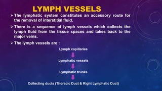

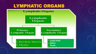


The lymphatic system is a crucial component of the circulatory and immune systems, functioning as a drainage system that collects tissue fluids and returns them to the bloodstream. It includes lymph, lymphatic organs like the thymus and spleen, and a network of lymph vessels that facilitate fluid circulation, immune response, and absorption of nutrients. Key roles of the lymphatic system involve maintaining fluid balance, filtering pathogens, and producing immune cells.


![Lymphatic system ppt[1].pptx .](https://cdn.slidesharecdn.com/ss_thumbnails/lymphaticsystemppt1-250112055350-023a9ad6-thumbnail.jpg?width=640&height=640&fit=bounds)


