THE SENSORY ORGANS
Prof.Nagamani.T
CONTENTS
Applied Anatomy ( 4 hrs)
 Structure of skin, eye, ear, nose and tongue
Application and implications in Nursing
Applied Physiology ( 4 hrs)
 Functions of skin
 Vision, hearing, taste and smell
 Errors of refraction, aging changes
 Application and implications in Nursing
INTRODUCTION
 Humans can perceive various types of sensations, and
with this information, our motor movement is determined.
We become aware of the world by way of sensation.
Sensations can also be protective to the body, by
registering eg environmental cold or warm, and painful
needle prick.
 Sense organs are specialized organs that help to perceive
the world around us. They are an integral part of our lives
and it is the only way that enables us to perceive the
environment.
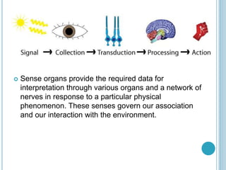
 Sense organs provide the required data for
interpretation through various organs and a network of
nerves in response to a particular physical
phenomenon. These senses govern our association
and our interaction with the environment.
We have five sense organs, namely:
 Eyes
 Ears
 Nose
 Tongue
 Skin
 These five sense organs contain receptors that relay
information through the sensory neurons to the
appropriate places within the nervous system.
 The receptors could be classified into two parts viz. the
general and special receptors. The former is present
throughout the body while the latter includes chemo
receptors, photoreceptors and mechanoreceptors.
 Broadly, these sensations can classify into two
categories.
 General sensations which include touch, pain,
temperature, proprioception, and pressure.
 Special Senses: Vision, hearing, taste, and smell
which convey sensations to the brain through cranial
nerves.
 The peripheral nervous system (PNS) consists of
sensory receptors that extend from the central nervous
system (CNS) to communicate with other parts of the
body. These receptors respond to changes and stimuli
in the environment. Sense organs (made up of sensory
receptors and other cells ) operate the senses of vision,
hearing, equilibrium, smell, and taste
SKIN
INTRODUCTION TO SKIN
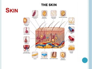
 The skin is the body’s largest organ, made of water, protein,
fats and minerals.
 It is an impressive and vital organ. It is a fleshy surface with
hair, nerves, glands and nails. It consists of hair follicles which
anchor hair strands into the skin.
 It acts as a barrier between outside and inside environment.
 The skin has different thicknesses and textures. E.g. the skin
under the eyes is as thin as paper but is thick at the soles of
the feet and palm.
 The skin acquires an area of 20 square feet on our body
surface.
 It protects us from external elements, regulates the body
temperature by releasing water in the form of sweat, and
allows sensations such as touch, heat, and cold. It also guards
the bones, muscles and other vital organs of our body.
 Structure Of Skin
 It provides a protective covering throughout our body
and acts as the body’s initial barrier against external
harmful substances or foreign particles. The hair is
made up of a protein called keratin, and the same
protein is found in hooves, horns, claws and nails of
other animals too.
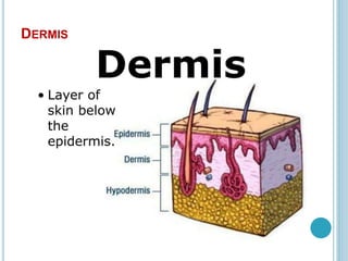
The structure of the skin is made up of three layers
of, namely:
 Epidermis
 Dermis
 Hypodermis
Epidermis
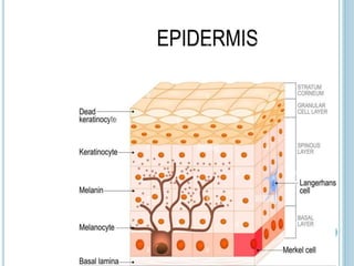
 It is the outermost layer of the skin. The cells in this
layer are called keratinocytes. The keratinocytes are
composed of a protein called keratin. Keratin
strengthens the skin and makes it waterproof.
 Melanocytes that produce melanin are also present in
this layer. In addition, Merkel cells essential for light-
touch sensation and Langerhans cells, part of the
immune system are also present on the skin.
 The epidermis is subdivided into the following layers:
 Stratum corneum
 Granular cell layer
 Spinous cell layer
 Basal cell layer
 Basal layer ( Stratum Basale)
 It is the deepest of all other layers of the epidermis, where
new cells are produced continuously.
 It is made up of a single layer of columnar cells that rest
on a basal lamina.
 These cells undergo mitotic divisions and give off cells
called keratinocytes, which form the more superficial
layers of the epidermis.
 They grow actively because of the blood supply below this
layer due to the presence of blood vessels.
 Spinous Layer ( Stratum Spinosum)
 It consists of several layers of polygonal keratinocytes.
 These cells appear to have a number of ‘ spines’
 That is why this layer is called stratum spinosum.
 Granular layer ( Startum Granulosum)
 It consists of 1-5 layers of flattened cells, containing deeply
staining granules in their cytoplasm.
 The granules consist of a protein called Keratohyalin.
 Stratum Lucidum ( Lucid= Clear)
 This layer appears clear, homogenous, with indistinct cell
boundaries.
 Stratum Corneum
 The outermost layer of the epidermis, which consists of dead
and uniform scale-like cells which are overlapped.
 These cells contain abundant keratin and provide rigidity to the
skin.
 These cells are regularly peeled off the skin’s surface which is
balanced by the cells of the basal layer.
 They are held together by a layer of lipid, which makes the
later water proof.
Types of Epidermal Cells
There are three main cell types in the epidermis:
 Melanocytes – These cells are located in the basal cell
layer and produce a pigment called melanin which is
absorbed by the new cells to protect against harmful
sun rays. The two factors that govern the melanin
quantity in one’s body is – genes and amount of
exposure to sunlight.
 Keratinocytes – They are the main cells which are
produced in the basal layer and approach towards the
outermost layer.
 Langerhans – These cells are specialized to protect
the body against foreign particles and hence is part of
the immune system.
 The epidermis and the dermis layer are connected
through a dermo-epidermal junction and contain the
basement membrane.
DERMIS
Dermis
 Beneath the epidermis is the dermis layer.
 It consists of papillae which form finger-like projections.
 This layer constitutes of fat, fibres and collagen which
makes the skin flexible and strong. Dermis synthesizes
Vitamin D to absorb calcium on exposure to sunlight.
 It consists of blood vessels which supply blood for the
formation of new cells.
 They are also vital in regulating body temperature.
Nerves in the dermis are sensitive to environmental
factors such as pressure and temperature. Also, it
contains the hair follicles and sebaceous oil gland which
produces sebum.
 It acts as a lubricant and protects the skin by acting
against the microbes.
 Sweat glands are produced all over the skin and
release sweat through specialized ducts. They help the
body to eliminate salts and minerals such as urea.
 An arrector pili muscle is attached to each hair follicle.
This helps in the erection of hair when we experience
cold or strong emotions.
Hypodermis
 This subcutaneous layer is made up of fat and forms
the innermost layer.
 Its thickness is dependant on the region where they
appear and vary. For example, the area around the eye
is comparatively thinner for the easy movement of the
eye.
 Fat stored provides energy and is crucial in reacting to
ambient temperature. It insulates the body from heat
and cold. It cushions the internal organs, muscles and
bones, and protects them from any injuries
BLOOD SUPPLY TO THE SKIN
 Blood vessels and lymphatic vessels are found in the dermal layer of
the skin.
 Blood supply to the skin is an arrangement of two plexuses, the first
lies between the papillary and reticular layers of the dermis and the
second lie between the dermis and subcutaneous tissues.
 Supply to the epidermis is by way of the superficial arteriovenous
plexus (subepidermal/papillary plexus). These vessels are important
for temperature regulation.
 The mechanism by which the body regulates temperature through the
skin is very effective and works by increased blood flow to the skin,
transferring heat from the body to the environment.
 The changes in blood flow are controlled by the autonomic nervous
system, sympathetic stimulation resulting in vasoconstriction (heat
retention) and while vasodilation results in heat loss.
 Vasodilation of the blood vessels is the response to increased body
temperature and is the result of inhibition of the sympathetic centers in
the posterior hypothalamus whereas decreased body temperature will
cause vasoconstriction of skin blood vessels.
NERVES
 Nerves of the skin include both somatic and
autonomic nerves. The somatic sensory system is
responsible for pain (nociceptors), temperature, light
touch, discriminative touch, vibration, pressure etc.
 The autonomic innervation is responsible for the
control of the tone of the vasculature, pilomotor
stimulation at the hair root, and sweating. The free
nerve endings extend into the epidermis and sense
pain, heat, and cold. They are most numerous in the
stratum granulosum layer and surround most hair
follicles.
FUNCTION OF SKIN
Protection from the Environment
 This is foremost and the most important function of the skin.
It keeps the pathogens away so that they do not enter into
the skin and cause any harm.
Prevents Water Loss
Humans possess thick skin that loses less water. In deserts,
the human skin gets thicker to prevent water loss to dry air.
 Organisms with thin skin have the possibility of losing water
all the time and need to stay near water to prevent it from
drying.
Sensation
 Skin is the main sense organ that can sense touch, heat,
pressure, cold, pain, and pleasure. A network of nerves
transmits these signals to the brain. Thus we can respond
appropriately to a particular stimulus.
Regulation of Temperature
 Our skin loses water through perspiration and cools itself,
thereby, removing heat from the body. It also allows the hot
blood to move to the surface of the skin, where its heat is
radiated out of the skin. The phenomenon of “goosebumps” is
also a temperature regulation response.
Storage
 The skin can store fats and water in its tissues. These provide
extra insulation to our body.
 The animals found in colder regions develop thick layers of fat
to prevent themselves from the outside cold.
Excreting Scent Signals
 The sweat secreted by our skin can also act as a signal to
other organisms. Many animals mark their territories by
secreting some scent from the glands in their skin which
contains information about its age, health, gender, and
availability to the mate.
Summary of Functions
 Protection of the body against mechanical, chemical, thermal, and
bacterial agents, this occurs through the heavily keratinized cells of
the stratum corneum.
 Protection of the body against water loss or gain (waterproof-barrier
function), this occurs through the lipid-rich extracellular material in
stratum granulosum and corneum.
 Screening against sun ultraviolet radiation through melanin pigments.
 Perception of different stimuli from the environment through the
presence of various receptors (sensory nerve endings) in the skin.
 Regulation of the body temperature:
– In hot weather, cooling of the body is enhanced by increase sweat
secretion and evaporation with vasodilation of the dermal capillaries
and closure of the arteriovenous (A-V) shunts, these mechanisms
allow maximum cutaneous blood flow and heat loss.
– In cold weather, constriction of dermal blood vessels and opening of
the A-V shunts to reduce cutaneous blood flow and reduce heat loss.
 Excretion of nitrogenous products and sodium chloride in sweat.
 Formation of vitamin D in the epidermis (mainly in stratum basale and
spinosum) when the skin is exposed to sunlight.
SKIN APPENDAGES
 Skin appendages are epidermal & dermal derived
components of the skin that include hair, nails, sweat
glands, and sebaceous glands.
 The skin, along with hair and nails, is the protective
covering of the body, the skin prevents germs from
entering the body and damaging the internal organs,
the skin manufactures vitamin D when exposed to the
sun’s ultraviolet rays, and vitamin D is an essential
vitamin for healthy skin.
 Skin appendages are skin-associated structures, they
serve a particular function including sensation,
contractility, lubrication, and heat loss.
 Skin appendages (or adnexa) are derived from
the skin, and they are adjacent to it, The skin supports
the life of all other body parts and plays a role in
maintaining the immune system.
 Types of appendages include hair, glands, and
nails. hairs are used for sensation, heat loss, filter for
breathing & protection, arrector pilli (smooth muscles
that pull hairs straight), sebaceous glands secrete
sebum onto hair follicle, which oils the hair, sweat
glands can secrete sweat with a strong odour
(apocrine) or with a faint odour (merocrine or eccrine),
and nails (protection).
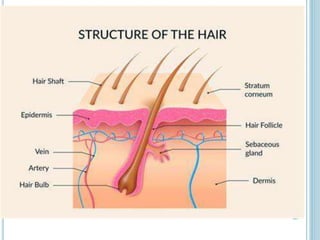
Hair
 The hair follicle is a skin appendage located deep in
the dermis of the skin.
 Hairs are horny threads varying in length, thickness,
color, and distribution according to the race, sex, and site
of skin.
Histological features:
 The hair shaft develops from the hair follicle which is a
cylindrical down growth of the epidermis into the dermis,
hair follicle ends by the hair bulb.
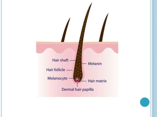
 The hair bulb is invaginated from below by avascular
connective tissue dermal papilla, which if destroyed, the
hair follicle dies and no hair will grow again.
 Its function is to produce hair and enclose the hair shaft.
 A hair follicle consists of two main layers, an inner
(epithelial) root sheath and an outer (fibrous) root sheath.
At the base of the hair follicle is the hair bulb, which
houses the dermal papillae and hair matrix cells.
 The hair bulb and inner epithelial layer generates hair.
 More specifically, the hair matrix keratinocytes proliferate
to form the growing hair shaft while the dermal papilla
regulates the hair’s growth and the inner root sheath
provides a tube shaped channel for the hair.
 The outer fibrous layer surrounds the hair follicle. It is
formed from dermal connective tissue, supplies blood to
the epithelial sheath and holds undifferentiated hair stem
cells.
 The cells of the hair follicle actively change during the
growth cycle of a hair.
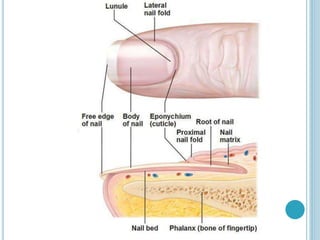
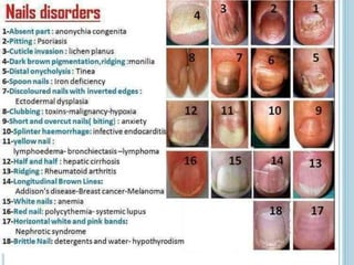
Nail
 Nails are horny plates present on the dorsal surfaces of the
terminal phalanges of fingers and toes, they consist of closely-
packed hard keratin scales.
 Nails are homologous to the stratum corneum of the epidermis and
contain a variety of minerals, such as calcium. They consist of
compacted and layered keratin-filled squames (scales). The
arrangement and cohesion of the squames are responsible for the
hardness of nails. A nail consists of: the nail plate, nail folds, nail
matrix, nail bed and hyponychium.
 Nail plate
 The nail plate is a rectangular and convex structure embedded
within the nail folds. It originates from the nail matrices, found at
the base of the nails. The nail plate is completely free distally to
the onychodermal band (distal margin of the nail bed).
 Nail folds
 The nail folds are the borders of the nail plate, located laterally and
proximally, which are continuous around the nail plate.
The cuticle (eponychium) is an extension of the proximal nail fold
located on the dorsal aspect of the nail plate, overlying the root of
the nail.
 Nail matrix
 The nail matrix is the structure out of which the nail plate
grows. Cornified cells from the matrix are gradually extruded
distally to form the nail plate.
 Nail bed
 The nail bed extends between the lunula (crescent shaped,
white area of the nail bed) and the hyponychium (area under
the free edge of the nail plate).
 The distal margin of the nail bed is called the onychodermal
band.
 There is a perfect match between the nail bed and plate,
forming a seal, which prevents microbial invasion and debris
collection.
 The nail bed consists of two layers: the epidermis and dermis.
 The dermis is directly attached to the periosteum of the distal
phalanx and it is richly vascularized.
 It also contains numerous sensory nerve endings, such
as Merkel endings and Meissner’s corpuscles.
Sebaceous glands
 These are simple branched alveolar, holocrine glands that
develop from the upper third of the hair follicle; therefore, they
are usually present where the hair is present. They are most
numerous over the head region and the anogenital area.
Histological features:
1- The secretory portion: It is a pale flask-shaped structure
formed of one or more alveoli with a common duct.
 The alveolus is lined with a layer of flattened germinal cells
lying on a basement membrane, these cells proliferate by
mitosis, enlarge by accumulating fatty material, become
polyhedral, and are pushed to the center, where they
degenerate and discharge sebum (holocrine mode), thus;
sebum is a mixture of fatty substances, cell debris, and
keratin.
2- The excretory duct is short and wide, it opens obliquely into
the upper third of the hair follicle or directly into the epidermal
surface. The duct is lined by stratified squamous epithelium.
The function of the sebaceous gland
 Sebaceous glands secrete sebum which lubricates the
skin surface and hair, also has a bactericidal effect.
 Sebaceous glands are relatively inactive until puberty
when they are stimulated by the rising levels of sex
hormones.
 A disturbance in the normal flow of sebum is one of the
reasons for the development of acne during
adolescence, Acne is a chronic inflammation of
obstructed sebaceous glands.
 Sweat glands
 They are simple coiled tubular glands, they are found deep in
the dermis or the hypodermis, there are two types of sweat
glands; the eccrine and the apocrine glands.
 The eccrine sweat glands
 They are distributed all over the body, they secrete a watery
secretion rich in sodium chloride.
 Histological features:
 1- The secretory portion appears in histological sections the
form of small rounded acini with a narrow lumen and lined by
stratified cuboidal epithelium composed of three cell types:
 The dark pyramidal cells secreting glycoprotein mucoid
substance.
 The clear cuboidal cells secreting a watery secretion.
 The myoepithelial cells to move the secretion into the ducts.
 2- The long excretory duct is lined by stratified cuboidal cells,
It ascends in a helical course to the epidermis where it opens
on the skin surface.
 The apocrine sweat glands
 They are present only in the skin of the axilla, areola of
the breast, and perianal region, they begin their
secretory function at puberty, their secretion is viscous
and has a characteristic odor.
 Histological features:
 The acini of the secretory portion are larger in size and
of wider lumen than the eccrine glands, the secretory
portion is composed of only two cell types; large
cuboidal cells with apical secretory granules and
myoepithelial cells.
 The excretory duct is lined by stratified cuboidal cells
and opens into the hair follicle.
 The secretory cells undergo merocrine, not apocrine,
secretion, thus; the glands are misnamed.
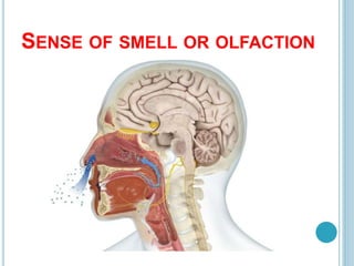
SENSE OF SMELL OR OLFACTION
 Nose is the fleshy protrusion located in the middle of the
face that works as a principal part of the respiratory
system, in addition to being the primary organ of smell
 Like taste, the sense of smell, or olfaction, is also
responsive to chemical stimuli. The olfactory receptor
neurons are located in a small region within the superior
nasal cavity- in the olfactory epithelium — a patch of
tissue about the size of a postage stamp located high in
the nasal cavity.
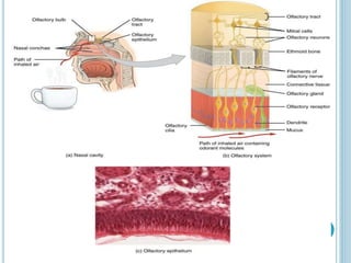
 The olfactory epithelium is made up of three kinds of
cells: sensory neurons each with a primary cilium
supporting cells between them basal cells that divide
regularly producing a fresh crop of sensory neurons to
replace those that die.
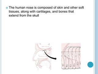
 The human nose is composed of skin and other soft
tissues, along with cartilages, and bones that
extend from the skull
 Smell depends on sensory receptors that respond to
airborne chemicals. In humans,
these chemoreceptors are located in the olfactory
epithelium — a patch of tissue about the size of a
postage stamp located high in the nasal cavity.
 The olfactory epithelium is made up of three kinds of
cells:
 sensory neurons each with a primary cilium
 supporting cells between them
 basal cells that divide regularly producing a fresh crop
of sensory neurons to replace those that die (and
providing an exception to the usual rule that neurons
seldom are replaced)
ANATOMY OF THE NOSE
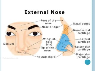
The External Nose
 The external part of the nose has a triangular or
pyramidal shape, with the highest point of it referred
to as the apex or the tip of the nose.
 The thin superior part that blends with the forehead
is called the root of the nose, while the region
between the apex and the root is called
the dorsum. The small curve just beneath the root
is known as the nose bridge.
 Beneath the apex are the nostrils or nares,
surrounded by the nasal septum and the
cartilaginous ala nasi or wings of nose
Nasal Cavity
 The nasal cavity is the space behind the nostrils and the
beginning of the respiratory tract. It is separated into the
right and left nasal cavities by the nasal septum. There are
three sections of the cavity:
 Nasal Vestibule: The area lying just behind the nasal
cavities, the nasal vestibule is lined with skin tissue, unlike
the rest of the nasal cavity. It is surrounded by the wings of
the nose and the nasal septum, while its floor is formed of
the hard palate.
 Respiratory Region: The wide space within the nose
covered with cilia and mucus-secreting goblet cells.
 Olfactory Region: The highest point of the nasal cavity, the
olfactory region contains the olfactory receptor neurons.
Humans have around 5 to 6 million of these receptors (as
opposed to dogs who have 220 million). There are two such
patches of olfactory receptors in each nasal cavity, located
beneath the two olfactory bulbs, the region that connects the
nose with olfactory (smelling region) of the brain
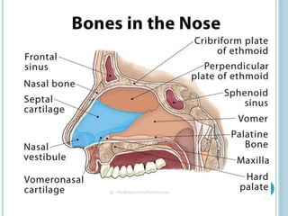
Nasal Skeleton:
 The nasal walls are formed of three paired and two
unpaired bones; the paired ones are
the nasal, palatine and maxillary bones, while
the vomer and ethmoid bones are unpaired. Also,
there are three pairs of nasal conchae (nasal turbinates)
that protrudes spirally from the sides of the nasal cavity
on both sides.
 The primary cartilages in the human nose include
the lateral, septal, and the greater and lesser alar
cartilages
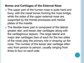
Bones and Cartilages of the External Nose
 The upper part of the human nose is quite hard and
bony, with the nasal bones forming the nose bridge,
while the sides of the upper external nose are
supported by the frontal processes and medial
plates of the maxilla
 The flexible lower part is composed of the lateral,
greater alar, and lesser alar cartilages along with
the cartilaginous septum. The large lateral and
major alar cartilages (two of each for the two sides
of the nose) play the primary role in shaping up the
nose. The number of the lesser alar cartilage often
vary from person to person, usually ranging from
three to four on each side.
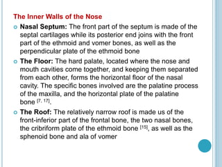
The Inner Walls of the Nose
 Nasal Septum: The front part of the septum is made of the
septal cartilages while its posterior end joins with the front
part of the ethmoid and vomer bones, as well as the
perpendicular plate of the ethmoid bone
 The Floor: The hard palate, located where the nose and
mouth cavities come together, and keeping them separated
from each other, forms the horizontal floor of the nasal
cavity. The specific bones involved are the palatine process
of the maxilla, and the horizontal plate of the palatine
bone [7, 17].
 The Roof: The relatively narrow roof is made us of the
front-inferior part of the frontal bone, the two nasal bones,
the cribriform plate of the ethmoid bone [15], as well as the
sphenoid bone and ala of vomer
Nasal Conchae
 There are three pairs of nasal
conchae: inferior, middle and superior nasal
conchae. The inferior conchae are the largest among
the three (almost as long as an index finger), while
the superior conchae are the smallest. The middle
conchae are about the size of a little finger
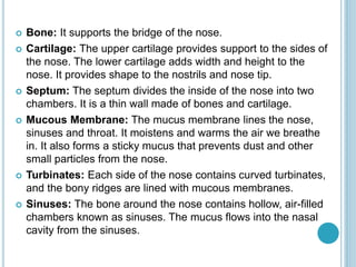
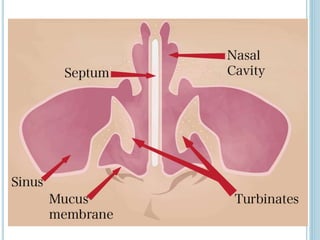
 Bone: It supports the bridge of the nose.
 Cartilage: The upper cartilage provides support to the sides of
the nose. The lower cartilage adds width and height to the
nose. It provides shape to the nostrils and nose tip.
 Septum: The septum divides the inside of the nose into two
chambers. It is a thin wall made of bones and cartilage.
 Mucous Membrane: The mucus membrane lines the nose,
sinuses and throat. It moistens and warms the air we breathe
in. It also forms a sticky mucus that prevents dust and other
small particles from the nose.
 Turbinates: Each side of the nose contains curved turbinates,
and the bony ridges are lined with mucous membranes.
 Sinuses: The bone around the nose contains hollow, air-filled
chambers known as sinuses. The mucus flows into the nasal
cavity from the sinuses.
Blood Supply to the Nose
 Arterial Supply
 The nose has a rich blood supply provided by arterial
branches from both the internal and external carotid [8].
The former includes the posterior and anterior
ethmoidal arteries [8], while the latter includes the
sphenopalatine artery, greater palatine artery, superior
labial artery, and angular artery
 The superior labial artery, a branch of the facial artery,
supplies the external nose region [10]. The vascular
supply of the dorsal and sellar (the area in the sphenoid
bone housing the pituitary gland) regions come from
the internal maxillary and ophthalmic arteries
 Internal Vasculature
 The sphenopalatine artery, along with the anterior and
posterior ethmoid arteries supplies the lateral wall of the
nose. These arteries also bring up blood supply to the
nasal septum and sidewalls, with additional sources of
vascular supply including the greater palatine artery
(posteriorly) and the superior labial artery (anteriorly).
 Venous Supply
 The venous system of the nose follows the arteries,
draining into the ophthalmic and facial veins as well as
the pharyngeal and pterygoid plexuses
 Nasal Innervation
 Nerve supply to the human nose can be classified into two
sections according to their functions:
 Special Sensory Innervation: This refers to the sense of
smell controlled by the olfactory nerves (CN1). The olfactory
sensory fibers run through the cribriform plate of the ethmoid
bone to connect to the olfactory bulb located superiorly to
the nasal cavity . The neural receptors in this vein come in
contact with any particles and chemicals entering the nose,
and send them to the olfactory region of the brain to ‘smell’.
 General Sensory Innervation: This refers to the general
senses of the external nose and nasal cavity. The lateral
walls of the nasal cavity and the septum are innervated by
the maxillary nerve branch known as the nasopalatine nerve,
and the ophthalmic nerve branch called the nasociliary nerve.
The trigeminal nerve innervates the outer skin of the human
nose [
FUNCTIONS OF NOSE
Helps in Inhalation
 The process of respiration starts in the nose. The oxygen
enters the nose through the nostrils and exits the same
way during exhalation. The nasal cavities open into a
space called choana, which further opens into the
nasopharynx. The air then enters the oropharynx and
finally reaches the lungs via the larynx, trachea and
bronchi.
Purification of Inhaled Air
 The walls of the nasal cavity are covered with hair or
cilia that trap the dust and harmful particles and purify
the inhaled air. The back and forth movement of cilia
helps in moving the dust particles to the throat where
they are swallowed or excreted through the nasal
cavity.
 The nose hair also moisturizes and warms the air, so
that it resembles the air temperature and moisture
within the lungs. During exhalation, the heat and
moisture present in the carbon dioxide is absorbed by
the nasal hair and then released into the atmosphere.
 The nasal conchae, which are spiral in structure, keep
whirling the air for a longer time within the nasal cavity
so that it is humidified and purified properly.
Organ of Smell
 The inhaled air comes in contact with the olfactory
epithelium and the nerve fibres extending from the
olfactory receptors accumulate the molecules
containing the odour to send the signals to the olfactory
bulbs through the receptors. These signals are carried
to the olfactory region of the brain and are decoded so
that the smell is identified.
Sense of Taste
 While chewing, the food releases certain chemicals that
travel up to the nose and activate the olfactory
receptors inside the nose. They work in coordination
with the taste buds to identify the actual flavour of the
food.
THE EYE
INTRODUCTION
 An eye is the sense organ of vision. It perceives the light
energy and converts it into electrical energy, thereby helping
us to ‘’see‘’.
 Vision is by far the most used of the five senses and is one
of the primary means that we use to gather information from
our surroundings.
 More than 75% of the information we receive about the world
a
 The eye is often compared to a camera. Each gathers light
and then transforms that light into a "picture." Both also have
lenses to focus the incoming light.
 Just as a camera focuses light onto the film to create a
picture, the eye focuses light onto a specialized layer of
cells, called the retina around us consists of visual
information, to produce an image.
THE STRUCTURE OF THE EYE
 The structure of the eye is an important topic to
understand as it one of the important sensory organs in
the human body.
 It is mainly responsible for vision, differentiation of colour
(the human eye can differentiate approximately 10 – 12
million colours) and maintaining the biological clock of the
human body.
 The human eye can be compared to a camera as both
works by gathering, focusing and transmitting the light
through the lens for creating an image of an object.
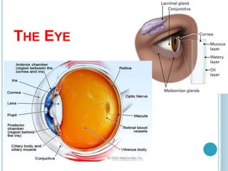
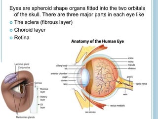
Eyes are spheroid shape organs fitted into the two orbitals
of the skull. There are three major parts in each eye like
 The sclera (fibrous layer)
 Choroid layer
 Retina
The sclera
 It makes up the outermost part of eye anatomy. It is
made of a dense, strong fibrous wall consisting of the
sclera that is 5/6th and the cornea that is anterior 1/6th of
the eyeball.
 The sclera is the outermost layer and it gives a definite
shape to the eye. The gap between the sclera and the
orbitals of the skull is filled with adipose tissue.
 This sclera on the anterior side extends as a
transparent epithelium called the cornea. The junction
of the cornea and sclera is called the limbus.
The choroid layer
 This is the second layer forming the eyeball. It consists
of a densely capillary-rich layer supplying blood to the
eyeball.
 The choroid layer also acts as a black screen which
prevents extra reflections inside the eyeball so that we
can get a perfect image.
 Parts are the iris, ciliary body and ligaments.
Iris
 This is the extension of the choroid layer towards the
anterior side of the eye. This iris is pigmented, due to
which there appears color in the eyes.
 The pigment is genetically determined and can be black,
brown, green, blue, etc.
 The iris has a small orifice at the center called the pupil.
 Both parasympathetic and sympathetic nerves supply
this iris.
 It dilates due to sympathetic stimulation for distant
vision.
 It constricts by parasympathetic stimulation for short
vision.
 This iris separates the aqueous chamber into anterior
and posterior chambers.
Lens
 The lens is a biconvex, transparent and elastic structure
that can alter its shape. It helps to focus the objects
based on their distance and concentrate the refracted
light onto the retina.
 When it constricts, it becomes thicker and when it
dilates, it becomes thin.
Retina
 This is the innermost layer of the eye made of nerve cells.
This neural coat of the eye is an essential part of the eye.
 This retina is the only part of the eye that perceives the
light and converts it into electrical energy.
 It consists of photosensitive cells, namely the rods and cones.
 The rods are light-sensitive and recognize the dark and bright
light.
 The cones are color sensitive and recognize the colors.
 The light received from the lens is converted into a nerve
impulse and carried backward as the optic nerve into the
brain for further processing.
 The point at which the nerves from the retina converge is
called the optic spot.
 The retinal chamber is filled with vitreous humor.
ACCESSORY PARTS OF THE EYE
These are the parts that help in the safety and
smooth functioning of the eye. They are
 The eyebrows
 The eyelids and eyelashes
 The lachrymal bodies
Eyebrows:
 These are arch-shaped ridges projecting from the
frontal bone. They have hair that is bent to one side.
These help to prevent sweat, dust, and other waste
from falling over the eye and the head.
Eyelids and lashes:
 Eyelids are delicate layers of tissue that cover the eye.
They protect the eyes from dust, insects, etc., by their
reflection action.
 The eyelashes are hair-like outgrowth projecting from
the eyelids. They protect the eyes from insects and dust
particles.
Lachrymal bodies:
 These are the glands that secrete tears. These tears
are rich in salts, lysozyme, and immunoglobulins. They
help
 a) Protection from irritants and dust.
 b) Prevent microbial growth
 c) Keep the eye moist, and the oil in it slows the
evaporation.
 Different parameters of the eyeball are given below:
 Anteroposterior diameter – 24mm
 Horizontal diameter – 23.5mm
 Vertical diameter – 23mm
 Circumference – 75mm
 Volume- 6.5ml
 Weight-7g
Segments and Chambers of Eyeball: Eyeball is divided into two
segments, the anterior one and the posterior one.
 Anterior segment: The segmentation inside the eye is based on the
position of the lens. An anterior segment consists of a crystalline lens
hanging from the ciliary body by zonules and all structures in front of
it, viz. iris, cornea, and two aqueous humor-filled spaces, i.e.,
anterior and posterior chambers.
 Anterior Chamber: The boundaries of the anterior chamber are
anteriorly the back of the cornea and posteriorly the anterior surface
of the iris and part of the ciliary body. It is 2.5mm deep in adults but
varies in hypermetropes and myopes. It communities with the
posterior chamber, located just behind it via the pupil, i.e., opening
through the iris.
 Posterior Chamber: It is a small triangular structure located just
behind the anterior chamber guarded by the posterior surface of the
iris and parts of the ciliary body anteriorly and by the crystalline lens
and its zonules posteriorly.
 Posterior Segment: It consists of all structures that are posterior to
the lens, i.e., vitreous humor, retina, and choroid optic disc.
Extraocular Muscles: Extraocular muscles are
responsible for the continuous movement of the eyeball
to increase the field of vision. They can be divided into
two groups of muscles:
 The Recti: There are four recti muscles :
 Superior Rectus
 Inferior Rectus
 Medial Rectus
 Lateral Rectus
 The Oblique: There are 2 oblique muscles :
 Superior Oblique
 Inferior Oblique.
PHYSIOLOGY OF VISION
Vision is the special sense of sight that is based on
the transduction of light stimuli received through the
eyes.
The physiology of Vision consists of three main
mechanisms, they are:
 Initiation of vision (phototransduction), which is a
function of photoreceptors, i.e., Rods and cones
 Processing and transmission of visual sensation is
a function of image-processing cells of the retina
and visual pathway.
 Visual perception is a function of the visual cortex
and related areas of the cerebral cortex.
 Layers of the retina: The retina is the primary light
perceiving the area of the eyeball covering the whole
inner surface of the eyeball. They are:
 The inner limiting membrane (ILM);
 the nerve fiber layer (NFL);
 the ganglion cell layer (GCL);
 the inner plexiform layer (IPL);
 the inner nuclear layer (INL);
 the outer plexiform layer (OPL);
 the outer nuclear layer (ONL);
 the outer limiting membrane (OLM);
 the photoreceptor layer (PL),
 The retinal pigmented epithelium (RPE) monolayer.
All these layers have individual functions in the perception
of light.
 Phototransduction: The rods and cones are the cells
that are designed to cause chemical changes when light
falls on them. They serve as sensory nerve ending for
visual sensation. When the light falls on them, a cascade
of visual reactions starts resulting in the generation of
electrical changes. Rods contain a pigment
called Rhodopsin or Visual Purple, while cones contain
Iodopsin. Rods are responsible for dim light vision or
monochromatic vision, while cones are responsible for
color vision.
 Photochemical changes: The Photochemical changes
include
 Rhodopsin bleaching
 Rhodopsin regeneration
 and Transmission of visual Impulse
 A receptor potential is developed in the photoreceptors, which
is transmitted by electronic conduction, i.e., direct flow of
current, not as an action potential, to other cells such as
amacrine cells, horizontal cells, and ganglion cells. Now,
these ganglion cells transmit electric impulses to an action
potential to neurons of lateral geniculate bodies and later to
the primary cortex.
Visual perception
 It consists of some thresholds, for example:
Light sense: It refers to minimal brightness required to evoke a
sensation of light, called light minimum. It should be
measured after at least a dark adaptation for 20 to 30mins.
The two types of visual adaptations are
 Light adaptation
 Dark adaptation
 Rods are more sensitive to low light vision, i.e.,
scotopic vision. Therefore rods are used more in dim
light vision, whereas cones are used more in bright
light vision, i.e., photopic vision.
 The dark adaptation depends on the presence of
vitamin A. So, a deficiency of Vit A causes night
blindness.
 The eye is the only organ of photoreception. It lets us
see the world. Many diseases are caused to the eye,
starting from minor ailments like hypermetropia,
myopia, and cataracts to diseases of significant
concern like glaucoma, retinopathy, etc.
THE EAR
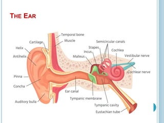
 The ear is a complex part of sensory system.
 It is situated bilaterally on the human skull, at the same
level as the nose.
 The main functions of the ear are, of course, hearing, as
well as constantly maintaining balance.
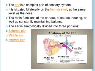
 The ear is anatomically divided into three portions:
 External ear
 Middle ear
 Internal ear
External ear
 The external ear is the part of the ear we can see with
the naked eye. It is attached to the lateral aspect of the
head and is characteristically divided into two parts:
 the auricle or pinna and
 the external acoustic meatus (more commonly
referred to as the ear canal)
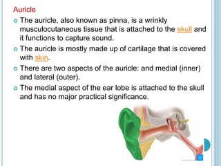
Auricle
 The auricle, also known as pinna, is a wrinkly
musculocutaneous tissue that is attached to the skull and
it functions to capture sound.
 The auricle is mostly made up of cartilage that is covered
with skin.
 There are two aspects of the auricle: and medial (inner)
and lateral (outer).
 The medial aspect of the ear lobe is attached to the skull
and has no major practical significance.
 The lateral aspect is concave and presents numerous
grooves and ridges.
 The outer rim of the auricle is called the helix, which
then inferiorly ends as soft tissue known as the lobule
of auricle (or ear lobe).
 The outer curve of the auricle is called the helix, which
connects with the lobule of the ear. Parallel and anterior
to the helix is a smaller curve, aptly called the antihelix.
 In the centre of the auricle, there is a depression called
the concha of the auricle, which gives rise to
the external acoustic meatus.
 Anterior to the opening of the external acoustic meatus is
an elevation of cartilage called the tragus.
 The tragus forms a protective barrier to the external
acoustic meatus, partially covering it to prevent the
passage of large foreign objects down the external
acoustic meatus. Opposite the tragus is the antitragus.
Innervation of the auricle
 The auricle has several sources of sensory
innervation:
 The superficial surface is supplied by the great
auricular nerve and lesser occipital nerve, both of which
are branches of the cervical plexus (C2 & C3), and the
auriculotemporal branch of the mandibular nerve, which
is a branch of the trigeminal nerve (cranial nerve V)
 The deep part of the auricle is supplied by the vagus
nerve (cranial nerve X) and facial nerve (cranial nerve
VII)
Vascular supply and lymphatic drainage
 The auricle is supplied by three main arteries:
 The external carotid artery gives rise to the posterior
auricular artery, which supplies the posterior aspect of
the auricle
 The superficial temporal artery gives rise to the anterior
auricular arteries, which supplies the anterior aspect of
the auricle
 Occipital artery
 Lymphatic drainage from the auricle passes into
the parotid and mastoid lymph nodes and eventually
into the upper deep cervical nodes.
Function of the auricle
 The shape of the auricle helps to capture sound
waves and direct them into the external acoustic meatus
and assist in determining where the sound is coming
from.
THE EXTERNAL ACOUSTIC MEATUS
 The external acoustic meatus is a tube that travels from
the deepest aspect of the concha to the tympanic
membrane, which marks the divide of the external and
middle ear.
 The composition of the walls of the external acoustic
meatus changes the deeper it travels:
 The lateral one-third of the external acoustic meatus is
cartilaginous, which extends slightly deeper on the
inferior portion external acoustic meatus
 The medial two-thirds of the external acoustic meatus
is bony, derived from the temporal bone of the skull
 There is a slightly raised constriction located at the
junction of the cartilaginous and bony portion
 The entire length of the external acoustic meatus is
covered with skin (keratinised squamous epithelium).
The outer third (cartilaginous portion) contains hair and
cerumen producing glands.
Blood supply
 The external acoustic meatus has the same blood
supply as the auricle: the superficial temporal
artery and the posterior auricular artery
FUNCTION OF THE EXTERNAL ACOUSTIC MEATUS
 The external acoustic meatus transmits sound to the
middle ear.
 The slight bend or constriction in the external acoustic
meatus acts as a protective mechanism to prevent
the passage of foreign objects. In addition, the cerumen
produced by the glands acts as a disinfectant to kill any
pathogenic organisms.
TYMPANIC MEMBRANE
 The tympanic membrane, or eardrum, marks
the border between the external and middle ear.
 It is formed of a middle layer of connective
tissue with a layer of skin on its lateral surface
(facing the external acoustic meatus) and mucous
membrane on its medial surface (facing the middle
ear).
 The tympanic membrane lies at an oblique angle
that helps maximise the efficiency in sound
localisation. The external surface of the tympanic
membrane is concave and the internal surface
convex.
 Surrounding the tympanic membrane is
a fibrocartilaginous ring that attaches to the temporal
bone, this is called the annulus.
 At the centre of the tympanic membrane is the umbo,
which is a concave depression that attaches to the
handle of the malleus, one of the auditory ossicles in the
middle ear. Located anterior-inferior to the umbo is the
cone of light, which is created due to the conical shape
of the tympanic membrane.
 The tympanic membrane can be divided into two
regions:
 The pars flaccida: where the membrane is thin and
slack (small upper portion)
 The pars tensa: where the membrane is thick and taut
(larger lower portion)
 Innervation of the tympanic membrane
 The medial surface of the tympanic membrane is
supplied by the vagus (cranial nerve
X) and glossopharyngeal nerve (IX).
 The lateral surface of the tympanic nerve is supplied by
the auriculotemporal nerve (a branch of the
mandibular division of trigeminal nerve) and facial
nerve.
 Function of the tympanic membrane
 The tympanic membrane transmits sound waves from
the external ear to the ossicles of the middle ear. The
tympanic membrane and the middle ear amplify
sound reaching the inner ear.
MIDDLE EAR
 The middle ear, also referred to as the tympanic
cavity, is an air-filled section in the temporal
bone and is lined with a mucous membrane.
 It can be split into two main sections:
 The tympanic cavity proper: this is the space
directly internal to the tympanic membrane
 The epitympanic recess: this is the space behind
pars flaccida
 Anterior to the tympanic cavity is the auditory tube
(Eustachian tube) which extends to the pharynx, while
posterior to the cavity are the mastoid cells of
the mastoid process of the temporal bone. Mastoid cells
are covered with mucosa that has extended from
the nasopharynx through the auditory tube and then
through the tympanic cavity.
 The anatomical communication between the
pharynx, middle ear, and mastoid cells is a possible
path of spreading infections.
 Tympanic cavity
 This is a bony-membranous cavity. It is shaped like a
biconcave lens, but from an anatomical aspect it is
usually described as a six-sided prism. Along with these
six walls, the auditory ossicles can also be described
within the anatomy of the tympanic cavity.
 Tegmental wall (roof)Tegmen tympani - formed by both
squamous and petrous parts of the temporal bone, and it
separates the tympanic cavity from the cranial cavity.
 Jugular wall (floor)Separates the cavity from the jugular
fossa.
 Carotid wall (anterior)Corresponds with internal carotid artery
canal
Contains: petrotympanic fissure, orifice of the auditory tube,
orifice of the semicanal of the tensor tympani muscle
 Membranous wall (lateral)Medial aspect of the tympanic
membrane
 Labyrinthine wall (medial)Contains: oval window, round
window, promontory, prominence of the facial canal
 Mastoid wall (posterior)Corresponds with the mastoid
process and its pneumatic cavities
Contains: aditus to the mastoid antrum (entrance to the
mastoid antrum), the pyramidal eminence (contains the
stapedius muscle)
THE AUDITORY OSSICLES
 The auditory ossicles consist of three separate bones:
the malleus, incus and stapes. These connect to form a
mobile chain to join the tympanic membrane of the external
ear to the oval window of the inner ear.
 The handle of the malleus is attached to the tympanic
membrane, whilst its head links with the body of the incus in
the epitympanic recess.
 The incus consists of three parts: the body links with the
head of the malleus, the short limb attaches to the posterior
wall of the tympanic cavity and the long limb links with the
stapes.
 The stapes is the smallest bone in the human body and its
footplate attaches to the oval window of the inner ear.
 The function of the auditory ossicles is to transmit and
amplify the sound vibrations from the external ear to the
endolymph and perilymph of the inner ear