
Recommended
More Related Content
Similar to ANATOMY & PHYSIOLOGY OF THE LYMPHATIC SYSTEM
Similar to ANATOMY & PHYSIOLOGY OF THE LYMPHATIC SYSTEM (20)
Recently uploaded
Recently uploaded (20)
ANATOMY & PHYSIOLOGY OF THE LYMPHATIC SYSTEM
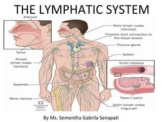
- 1. THE LYMPHATIC SYSTEM By Ms. Sementha Gabrila Senapati
- 2. INTRODUCTION • The body cells are bathed in interstitial ( tissue) fluid, which leaks constantly out of the blood stream through the permeable walls of blood capillaries. • Some tissue fluid diffuses through the more permeable walls of lymph capillaries, forming lymph. • Lymph passes through vessels of increasing size & a varying number of lymph nodes before returning to the blood.
- 3. The lymphatic system consist of:- • Lymph • Lymph vessels • Lymph nodes • Lymph organs(e.g.spleen & thymus) • Diffuse lymphoid tissue, e.g.tonsils • Bone marrow
- 5. FUNCTIONS OF LYMPHATIC SYSTEM:- • 1.TISSUE DRAINAGE:- • About 3-4 litres of fluid, is drained away by the lymphatic vessels. • 2.ABSORPTION IN THE SMALL INTESTINE:- • fat & fat soluble materials, e. g. the fat soluble vitamins, are absorbed into the central lacteals( lymphatic vessels) of the villi. • 3.IMMUNITY:- • The lymphatic organs helps in the production & maturation of lymphocytes, the WBC responsible for immunity. • Bone marrow is considered to be lymphatic tissue, since lymphocytes are produced there.
- 6. LYMPH & LYMPH VESSELS:- • Lymph is a clear watery fluid. • Lymph transports the plasma proteins that seep out of the capillary beds back to the blood stream. • It also carries away larger particles, eg. Bacteria & cell debris from damaged tissues, which can then be filtered out & destroyed by the lymph nodes. • Lymph contains lymphocytes(defence cells) • In the lacteals of the small intestine, fats absorbed into the lymphatic give the lymph(chyle), a milky appearance.
- 9. LYMPH CAPILLARIES • These originate as blind-end tubes in the interstitial spaces. • Nearly all tissues have a network of lymphatic vessels, except in the central nervous system, the cornea of the eye, the bones & the most superficial layers of the skin.
- 11. LARGER LYMPH VESSELS • Lymph vessels are found running alongside the arteries & veins serving the area. • Has a fibrous covering, a middle layer of smooth muscle & elastic tissue & an inner lining of endothelium. • Lymph vessels have numerous cup-shaped valves to ensure that lymph flows in a one way system towards the thorax. • The lymphatic pump:-the muscle layer in the walls of the large lymph vessels has an ability to contract rhythmically.
- 12. • Lymph vessels become larger as they join together, forming two large ducts:- • 1) thoracic duct • 2) right lymphatic duct, empty the lymph into the subclavian veins.
- 14. 1) THORACIC DUCT • Begins at the cisterna chyli, a dilated lymph channel situated in front of the first two lumbar vertebrae. • 40cm long, opens into the left subclavian vein in the root of the neck. • It drains lymph from both legs, the pelvic & abdominal cavities, the left half of the thorax, head & neck & the left arm.
- 16. RIGHT LYMPHATIC DUCT • Dilated lymph vessel about 1 cm long. • Lies in the root of the neck & opens into the right subclavian vein. • It drains lymph from the right half of the thorax, head & neck & the right arm.
- 18. LYMPHATIC ORGANS AND TISSUES • LYMPH NODES:- • oval or bean-shaped organs that lie often in groups. The lymph drains, usually 8-10 before returning to the venous circulation. • These nodes are as small as a pin head & some are the largest about the size of an almond.
- 20. STRUCTURE • Lymph nodes have an outer capsule of fibrous tissue that dips down into the node substance forming partition, or trabeculae. • The main substance of the node consists of reticular & lymphatic tissue containing many lymphocytes & macrophages. • Each node has a concave surface called the hilum where an artery enters & a vein and the efferent lymph vessel leave.
- 21. • Lymph from the head & neck passes through deep & superficial cervical nodes. • Lymph from the upper limbs passes through nodes situated in the elbow region then through the deep & superficial axillary nodes. • Lymph from organs & tissues in the thoracic cavity drains through groups of nodes situated close to the mediastinum, large airways, oesophagus & chest wall. • Most of the lymph from the breast passes through the axillary nodes.
- 22. • Lymph from the pelvic & abdominal cavity passes through many lymph nodes before entering the cisterna chyli. • The lymph from the lower limbs drains through the deep & superficial nodes including behind the knee & the groin.(inguinal nodes)
- 24. FUNCTIONS • 1) FILTERING & PHAGOCYTOSIS:- • Lymph is filtered by the reticular & lymphatic tissue. • Particulates include bacteria, dead and live phagocytes containing ingested microbes, cells from malignant tumors, worn out & damaged tissue cells & inhaled particles. • Organic material is destroyed in lymph nodes by macrophages & antibodies. • Some inorganic inhaled particles cannot be destroyed by phagocytosis. • These inorganic materials remain inside the macrophage, either causing no damage or killing the cell.
- 25. • Materials not filtered out & dealt with one lymph node passes onto successive nodes& by the time lymph enters the blood it is usually been cleared of foreign matter & cell debris. • In some cases where phagocytosis of bacteria is incomplete they may stimulate inflammation & enlargement of the node. (Lymphadenopathy) •
- 26. 2) PROLIFERATION OF LYMPHOCYTES • Activated T & B lymphocytes multiply in lymph nodes. • Antibodies produced by sensitized B lymphocytes enter lymph & blood draining the node.
- 28. SPLEEN • The spleen contains reticular & lymphatic tissue & is the largest lymph organ. • Position:-left hypochondriac region of the abdominal cavity between the fundus of the stomach & the diaphragm. • Purplish in colour, varies in size. • 12 cm long, 7cm wide & 2.5 cm thick. • It weighs about 200gm.
- 30. ORGANS ASSOCIATED WITH THE SPLEEN
- 31. STRUCTURE • Slightly oval in shape with the hilum on the lower medial border. • Anterior surface is covered with peritoneum. • Enclosed in a fibroelastic capsule that dips into the organ, forming trabeculae. • The cellular material consisting of lymphocytes & macrophages is called splenic pulp, & lies between the trabeculae. • Red pulp is the part suffused with blood & white pulp consist of areas of lymphatic tissue where there are sleeves of lymphocytes & macrophages around blood vessels.
- 33. • The structures entering & leaving the spleen at the hilum are:- • Splenic artery( branch of the coeliac artery) • Splenic vein( branch of the portal vein) • Lymph vessels ( efferent only) • Nerves. • Blood passing through the spleen flows in sinusoids( distinct pores between the endothelial cells,in close association with splenic pulp. • ThisThis is essential for the spleens function in removing ageing or damaged cells from the blood stream.
- 35. FUNCTIONS • 1) PHAHOCYTOSIS:-old & abnormal erythrocytes are mainly destroyed in the spleen, and the breakdown products, bilirubin and iron, are transported to the liver via the splenic & portal veins. Other cellular material eg. Leukocyte, platelets & bacteria are phagocysed in the spleen. • 2)STORAGE OF BLOOD:-the spleen contains up to 350 ml of blood, & in response to sympathetic stimulation can rapidly return most of this volume to the circulation. Eg. In hemorrhage.
- 36. • 3) IMMUNE RESPONSE:-The spleen contains T- & B- lymphocytes, which are activated by the presence of antigens. Eg. In infection. Lymphocytes proliferation during serious infection can cause enlargement of the spleen ( spleenomegaly) • 4) ERYTHROPOIESIS:-The spleen & liver are important sites of fetal cell production, & the spleen can also fulfil this function in adults in times of great need.
- 37. THYMUS GLAND • Position:- upper part of the mediastinum behind the sternum & extends upwards into the root of the neck. • Weight:-10-15 g at birth & grows until puberty, when it begins to atrophy. • It's maximum weight at puberty is between 30-40 g & at middle age it has returned to approx. It's weight at birth.
- 41. STRUCTURE • The thymus consist of two lobes joined by areolar tissue. The lobes are enclosed by a fibrous capsule which dips into their substance, dividing them into lobules that consist of an irregular branching framework of epithelial cells & lymphocytes.
- 42. FUNCTION • Lymphocytes originates from stem cells in red bone marrow. • Those that enter the thymus develop into activated T- lymphocytes. • Thymic processing produces mature T- lymphocytes that can distinguish self tissue from foreign tissue, and also provides each T- lymphocytes with the ability to react to only one specific antigen from the millions it encounter.
- 43. • T- lymphocytes then leave the thymus & enter the blood. • Some enter lymphoid tissues & other circulate in the blood stream. • The maturation of thethe thymus & other lymphoid tissue is stimulated by thymosin, a hormone secreted by the epithelial cells of the thymus gland.
- 44. MUCOSA ASSOCIATED LYMPHOID TISSUE (MALT) • MALT is found throughout the gastrointestinal tract, in the respiratory tract & in the genitourinary tract. • The main groups of MALT are the tonsils & aggregated lymphoid follicles( Peyers patch) • Tonsils:-these are located in the mouth & throat, & therefore destroy swallowed & inhaled antigens. • Aggregated lymphoid follicles(Peyers patches) these large collections of lymphoid tissue are found in the small intestine.
- 46. LYMPHATIC DISEASES • Lymphangitis • Lymphatic obstruction • Lymphadenitis • Lymphomas( Hodgkin's & hona hodgkins) • DISORDERS OF SPLEEN:- • spleenomegaly
- 47. ASSIGNMENT • 1.The lymphatic system consist of? • 2.List the function of lymphatic system. • 3. What is the shape of lymphnode? • 4.Lymph from head & neck drains in_________nodes. • 5.Write the function of lymphatic organs & tissues. • 6.Which is the largest lymph organ? • 7.What is the weight of the spleen.? • 8.What is the shape of a spleen? • 9.List the function of spleen. • 10.Write the position of thymus gland. • 11.The maturation of thymus is stimulated by which hormone? • 12.Write the fullform of MALT.
- 48. Thankyou