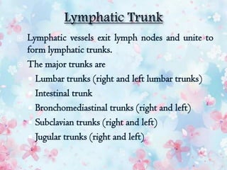
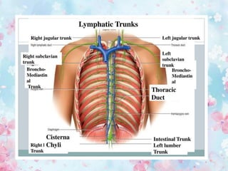
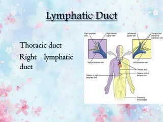
Download as PDF, PPTX
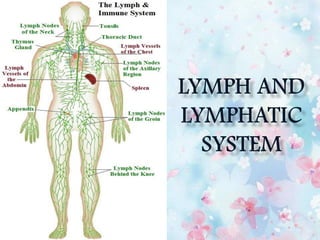
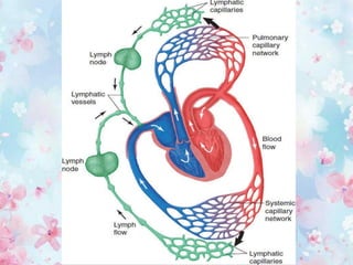


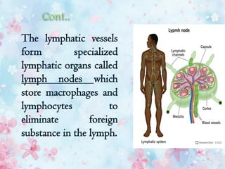


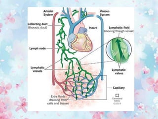

The lymphatic system removes excess fluid from tissues, absorbs fat and transports white blood cells and antigens. It comprises a network of lymphatic vessels that carry lymph fluid towards the heart. Lymph is filtered through lymph nodes which contain lymphocytes and phagocytes that help fight infection and disease. The major components are lymph, lymph vessels, lymphoid tissues and lymphocytes.


![Lymphatic system[1]](https://cdn.slidesharecdn.com/ss_thumbnails/v8tdil7slo1obvifzera-signature-460517c25b85fc4e63c8080c3e27df73c8dfae9e0c6544cc7ea6d9e8b5e79cc7-poli-180213064029-thumbnail.jpg?width=640&height=640&fit=bounds)


