The document provides an overview of the lymphatic system, including:
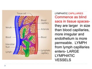
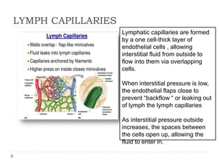
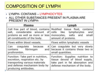
1. The formation and composition of lymph, which is a clear fluid that drains into the blood via the thoracic duct and transports lymphocytes.
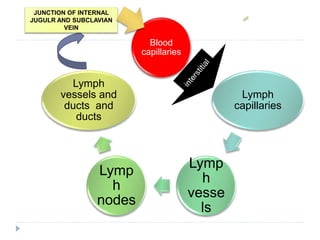
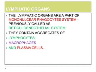
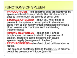
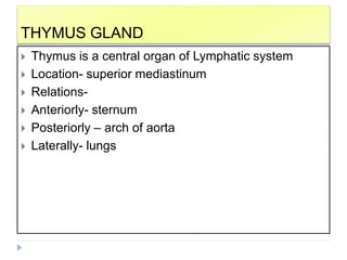
2. The components of the lymphatic system include lymph vessels, lymph organs such as the spleen and thymus gland, and lymph nodes which filter lymph.
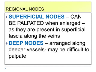
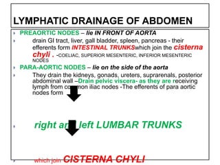
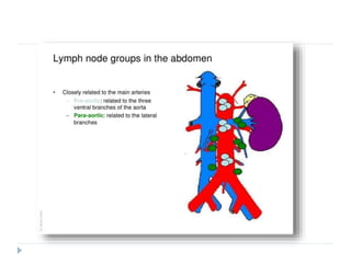
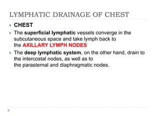
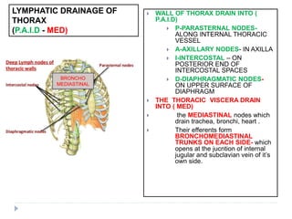
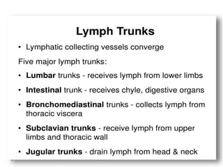
3. Lymphatic drainage patterns of different body regions including the head and neck, upper and lower limbs, chest, abdomen, and pelvis which drain via a series of lymph nodes and vessels to the thoracic duct.