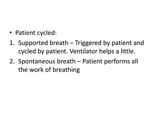
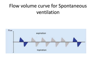
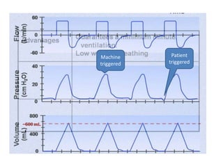
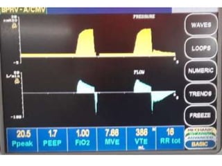
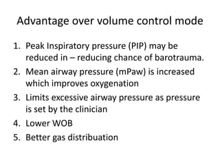
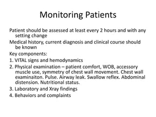
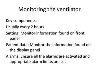
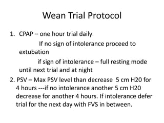
Basic Ventilator Management
Charipersion:
Assoc Prof Dr Md Mukhlesur Rahman
By
Dr Md Mashiul Alam
Phase B resident
Mechanical Ventilation: Short History
Negative pressure ventilation:
• The iron lung, also known as the Drinker and
Shaw tank, was developed in 1929
• Large scale use in 1940 during polio epidemic
Positive pressure ventilation:
by the military during World War II to supply
oxygen to fighter pilots in high altitude
In 1950 again during polio
epidemic
Types of mechanical Ventilation
• Negative pressure ventilation
• Positive pressure ventilation
- Noninvasive (NPPV)
- Invasive (IPPV)
IPPV vs NIPPV
• NPPV is the provision of any form of ventilatory
support applied without the use of an
endotracheal tube.
• With either a nasal or oral face mask.
• Patients can speak, eat and less chance of
pneumonia
Indication:
1. Acute exacerbation of COPD if the patient is alert
and no indication of ETT
2. Patient with hypoxemic respiratory failure but
hemodynamically stable and can protect his or
her airway.
3. After extubation to avoid reintubation
4. Patient with acute pulmonary edema in the
absence of MI to redistribute fluid from
alveolar space to vessel.
5. Obstructive sleep apnea
Exclusion criteria for NPPV:
1. Patient is not breathing spontaneously
2. Hemodynamically instable
3. Un co-operative patient
4. Facial burns or other trauma
5. Copious secretion
6. High risk of aspiration
Invasive PPV
Indication:
1. Primary indication- impending or existing
respiratory failure despite maximal treatment
2. General anesthesia
3. Cardiogenic shock to decrease work of
breathing
4. Increased ICP
General parameters to consider MV:
• Respiratory rate > 30/min or less than 8 bpm
• Vital capacity <1-1.5 L/min
• PaO2 <60 mmHg on FIO2 > 0.5
• PaCO2 high with repiratory acidosis (pH<7.2)
• Exhaustion
• Confusion
• Severe shock
• Severe LVF
• Raised ICP
Basic Ventilator Settings
• FIO2: Fraction of inspired oxygen
concentration
Initially it should be 0.7 to 1 to ensure adequate
tissue oxygenation.
After an initial ABGA obtained FiO2 may be
decreased to 0.5 or less while achieving PO2
more than 60 mmHg to prevent oxygen
toxicity
• VT: Tidal Volume
A starting point for VT is 8 to 10 ml/Kg ideal body weight.
An acceptable VT is one that results in a plateau
pressure of 30 mmHg
A VT of 12 ml/Kg or more is not usually needed
• EVT: Exhaled Tidal Volume
Regardless of mode of ventilation the most accurate
measure of the volume received by the patient is the
exhaled tidal volume.
The volume actually received by the patient must be
confirmed by monitoring EVT on the display panel of
the ventilator
• RR: Respiratory Rate
Near physiological as possible: 10 to 20 bpm. Typical
initial rate settings are between 8 and 12 breaths/min.
Slow rate may be useful in patients with obstructive
pulmonary disease because as the rate is decreased
more time is available for exhalation
Fast rates may be useful in patients with noncomplaint
lungs to prevent barotrauma with small VT
RR x VT = minute ventilation (mV)
When patient has adequate respiratory drive no RR set on
the ventilator
• Flow Rate:
The speed with which the VT is delivered.
Average: 40-60L /min
High flow rate – Decreased inspiratory time
Increased peak inspiratory pressure (PIP)
Low flow rate – Increased inspiratory time
Decreased PIP
Pressure modes
Volume mode Spontaneous
• I:E (Inspiratory to expiratory ratio)
The duration of inspiration in comparison with
expiration.
Inspiratory time of 1 sec is a good initial setting.
Generally I:E ratio is set at 1:2
1:3 or 1:4 or longer may be necessary to ventilate
the lungs in COPD
I:E ratio is a function of flow rate, inspiratory time
and RR
Inversed I:E ratio:
1:1 or 2:1 or 3:1 are called inverse I:E ratio
Employed when conventional strategies to
improve oxygenation fail
But it may increase mean airway pressure
(mPaw) and lead to hemondynamic
compromise
• PEEP (Positive End Expiratory Pressure)
A constant positive pressure in the airways at
the end of expiration to prevent alveolar
collapse.
Range 5 to 10 cmH2O
5 to 10 cmH2O in LVF, 10 to 20 cmH2O in ARDS
It prevents atelectasis, improve oxygenation,
redistribute lung water from alveoli to
perivascular space
Initial ventilator settings: Guidelines
Parameter Initial settings
FiO2 0.7 – 1.0
Tidal Volume 8-12 ml/Kg, 4-6 ml/Kg is ARDS
RR 10-20
Flow rate 40-60 L /min
Inspiratory time 1 sec (.8 to 2 sec)
I:E ratio 1:2 to 1:3
Sensitivity Pressure trigger : -0.5 to -1.5 below
baseline. Flow trigger: 1 – 3 L/min
PEEP 3 -5 cmH20
Correcting oxygenation and ventilator
problems
Problem ABG findings Adjustments
Excessive oxygenation PaO2 >100
SaO2 100%
Decrease FiO2
Keep I:E to 1:2
Decreased PEEP
Inadequate oxygenation PaO2 <60
SaO2 <90%
Increase FiO2
Increase PEEP
Prolong inspiratory time to
increase mPaw
Respiratory acidosis PaCO2 >45
pH≤ 7.35
Increase VT with PIP ≤ 40
mmHg
Increase RR
Respiratory alkalosis PaCO2<35
pH ≥ 7.45
Decrease VT
Decrease RR
Modes of mechanical ventilation
• Breath types
Machine cycled:
1. Mandatory – Ventilator perform all of the
work of breathing
2. Assisted- breath is triggered by the patient
and then rest are taken over by the machine
• Patient cycled:
1. Supported breath – Triggered by patient and
cycled by patient. Ventilator helps a little.
2. Spontaneous breath – Patient performs all
the work of breathing
Ventilatory support
• Full ventilatory support (FVS):
CMV or A/CMV
1. Apnea
2. Paralysed
3. Severe flail chest
4. Depressed neurological state
• Partial Ventilatory Support (PVS):
It allows the patient to respond to increase in
CO2 and promotes use of respiratory muscle
thereby preventing disuse atrophy
SIMV, PSV, CPAP
Control Modes
1. Volume Control
2. Pressure Control
3. Dual control
Flow volume curve for Spontaneous
ventilation
expiration
Inpiration
Volume control ventilation
A delivered breath is volume controlled when
volume is constant for every breath. The
pressure wave form varies because is pressure
is variable.
Types:
• CMV
• A/CMV
• SIMV
CMV (continuous mandatory vent.)
Patient receives a preset number of breaths per
minute of a preset tidal volume (VT) and
patient’s effort does not trigger a mechanical
breath
• Indication:
1. No respiratory effort
2. Anesthesia
3. Backup to assisted ventilation
V A/CMV
Asisst/ Control Mandatory vent.
The ventilator delivers a preset number of
breaths of a preset VT and patient may trigger
spontaneous breath
Machine
triggered
Patient
triggered
• Indication:
1. Normal respiratory drive but patient is too
weak or unable to perform work of breathing
(WOB)
2. To allow patient to set their own rate
V SIMV
• Synchronized Intermittent Mandatory Vent.
Patient receives a set number of breaths of a set
VT but patient may initiate spontaneous
breaths un assisted by the ventilator.
Patients effort is assisted if it falls in mandatory
breathing window
Mandatory breathing
window
• Indication:
1. Normal respiratory drive by unable to
perform all WOB
2. Allowing patients to set their own RR and
assist in maintaining a normal PaCO2
3. Weaning from mechanical ventilation
Pressure control ventilation
A delivered breath is pressure controlled when
pressure is constant for every breath.
Here the pressure wave form has a specific pattern
for every breath while volume waveform varies.
Types:
• PC or P A/C
• PSV / P SIMV
• CPAP
P- A/CMV
A mode of ventilation in which every breath is
augmented by a preset amount of inspiratory
pressure and there is no set VT. It may sense
patients spontaneous breathing effort and assist
with preset pressure.
Indication:
Providing full ventilatory support (FVS) in patients
with non compliant lungs who exhibit high Paw
and poor oxygenation in volume control mode.
Advantage over volume control mode
1. Peak Inspiratory pressure (PIP) may be
reduced in – reducing chance of barotrauma.
2. Mean airway pressure (mPaw) is increased
which improves oxygenation
3. Limits excessive airway pressure as pressure
is set by the clinician
4. Lower WOB
5. Better gas distribuation
P-SIMV/ PSV
The patients spontaneous respiratory activity is
augmented by the delivery of a preset amount
of inspiratory positive pressure.
It may used alone or in combination of SIMV. In
P SIMV only the spontaneous breaths are
pressure supported
P SIMV
• Indication
1. Weaning from mechanical ventilation. PSV is
used for spontaneous breathing trial
2. Non invasive ventilation
Trouble shooting patient-ventilator
system
Objective:
1. To evaluate the patient’s response to the
current level of ventilatory support
2. To determine accuracy and appropriateness
of the current ventilatory settings
3. To ensure the presence and proper
functioning of the necessary equipment
Monitoring Patients
Patient should be assessed at least every 2 hours and with any
setting change
Medical history, current diagnosis and clinical course should
be known
Key components:
1. VITAL signs and hemodynamics
2. Physical examination – patient comfort, WOB, accessory
muscle use, symmetry of chest wall movement. Chest wall
examinaiton. Pulse. Airway leak. Swallow reflex. Abdominal
distension. Nutritional status.
3. Laboratory and Xray findings
4. Behaviors and complaints
Monitoring the ventilator
Key components:
Usually every 2 hours
Setting: Monitor information found on front
panel
Patient data: Monitor the information found on
the display panel
Alarms: Ensure all the alarms are activated and
appropriate alarm limits are set
Weaning procedure
When patient is on ventilator for more than 3
days weaning procedure become for complex
If the condition requiring ventilation is fully
corrected and the patient is alert and
breathing spontaneously – extubation is
planned
Weaning trial screen should be done daily
1. Hemaodynamically stable ( no arrythmia, HR
≤ 120 bpm, absence of vasopressores except
low dose dopamine and dobutamine
2. FiO2 ≤ 0.5
3. PEEP ≤ 8
4. Patient is on MV less than 3 days
Wean Trial Protocol
1. CPAP – one hour trial daily
If no sign of intolerance proceed to
extubation
if sign of intolerance – full resting mode
until next trial and at night
2. PSV – Max PSV level than decrease 5 cm H20 for
4 hours ---if no intolerance another 5 cm H20
decrease for another 4 hours. If intolerance defer
trial for the next day with FVS in between.
• Sign of intolerance ( any one 3-5 mins
sustained)
1. RR ≥ 35 breaths/ min
2. SpO2 ≤ 90% or decrease by 4 %
3. HR ≥ 140 bpm
4. SBP ≥ 180 or ≤ 90
5. Excessive anxiety or agitation
6. Diaphoresis
Complications of mechanical
ventilation
• Problems related to positive pressure
Ventilator induced lung injury – Barotrauma,
Oxygen toxicity
Reduction in cardiac output and oxygen delivery
Alteration in renal function and positive fluid
balance
Impaired hepatic function
Increased ICP
V/Q mismatch
• Problems related to the artificial airway
Infection (Ventilator associated pneumonia)
• Gastric distress
Abdominal distention
Ulcers and gastritis
• Patient’s anxiety and stress
Take Home message
• Put any acutely distressed patients on V
A/CMV at first and Sedate the patient if
needed
• Send ABG not less 20 mins of a change
• Adjust the ventilator according to ABG report,
SpO2
• Start weaning trial when the precipitating
condition resolved ( Use SIMV, PSV modes)
“After all there is no match for natural
ventilators…..”
THANK YOU

Basic ventilator management

  • 1.
    Basic Ventilator Management Charipersion: AssocProf Dr Md Mukhlesur Rahman By Dr Md Mashiul Alam Phase B resident
  • 2.
    Mechanical Ventilation: ShortHistory Negative pressure ventilation: • The iron lung, also known as the Drinker and Shaw tank, was developed in 1929 • Large scale use in 1940 during polio epidemic
  • 5.
    Positive pressure ventilation: bythe military during World War II to supply oxygen to fighter pilots in high altitude In 1950 again during polio epidemic
  • 7.
    Types of mechanicalVentilation • Negative pressure ventilation • Positive pressure ventilation - Noninvasive (NPPV) - Invasive (IPPV)
  • 8.
    IPPV vs NIPPV •NPPV is the provision of any form of ventilatory support applied without the use of an endotracheal tube. • With either a nasal or oral face mask. • Patients can speak, eat and less chance of pneumonia Indication: 1. Acute exacerbation of COPD if the patient is alert and no indication of ETT
  • 9.
    2. Patient withhypoxemic respiratory failure but hemodynamically stable and can protect his or her airway. 3. After extubation to avoid reintubation 4. Patient with acute pulmonary edema in the absence of MI to redistribute fluid from alveolar space to vessel. 5. Obstructive sleep apnea
  • 10.
    Exclusion criteria forNPPV: 1. Patient is not breathing spontaneously 2. Hemodynamically instable 3. Un co-operative patient 4. Facial burns or other trauma 5. Copious secretion 6. High risk of aspiration
  • 12.
    Invasive PPV Indication: 1. Primaryindication- impending or existing respiratory failure despite maximal treatment 2. General anesthesia 3. Cardiogenic shock to decrease work of breathing 4. Increased ICP
  • 13.
    General parameters toconsider MV: • Respiratory rate > 30/min or less than 8 bpm • Vital capacity <1-1.5 L/min • PaO2 <60 mmHg on FIO2 > 0.5 • PaCO2 high with repiratory acidosis (pH<7.2) • Exhaustion • Confusion • Severe shock • Severe LVF • Raised ICP
  • 14.
  • 17.
    • FIO2: Fractionof inspired oxygen concentration Initially it should be 0.7 to 1 to ensure adequate tissue oxygenation. After an initial ABGA obtained FiO2 may be decreased to 0.5 or less while achieving PO2 more than 60 mmHg to prevent oxygen toxicity
  • 18.
    • VT: TidalVolume A starting point for VT is 8 to 10 ml/Kg ideal body weight. An acceptable VT is one that results in a plateau pressure of 30 mmHg A VT of 12 ml/Kg or more is not usually needed • EVT: Exhaled Tidal Volume Regardless of mode of ventilation the most accurate measure of the volume received by the patient is the exhaled tidal volume. The volume actually received by the patient must be confirmed by monitoring EVT on the display panel of the ventilator
  • 19.
    • RR: RespiratoryRate Near physiological as possible: 10 to 20 bpm. Typical initial rate settings are between 8 and 12 breaths/min. Slow rate may be useful in patients with obstructive pulmonary disease because as the rate is decreased more time is available for exhalation Fast rates may be useful in patients with noncomplaint lungs to prevent barotrauma with small VT RR x VT = minute ventilation (mV) When patient has adequate respiratory drive no RR set on the ventilator
  • 20.
    • Flow Rate: Thespeed with which the VT is delivered. Average: 40-60L /min High flow rate – Decreased inspiratory time Increased peak inspiratory pressure (PIP) Low flow rate – Increased inspiratory time Decreased PIP
  • 21.
  • 23.
    • I:E (Inspiratoryto expiratory ratio) The duration of inspiration in comparison with expiration. Inspiratory time of 1 sec is a good initial setting. Generally I:E ratio is set at 1:2 1:3 or 1:4 or longer may be necessary to ventilate the lungs in COPD I:E ratio is a function of flow rate, inspiratory time and RR
  • 24.
    Inversed I:E ratio: 1:1or 2:1 or 3:1 are called inverse I:E ratio Employed when conventional strategies to improve oxygenation fail But it may increase mean airway pressure (mPaw) and lead to hemondynamic compromise
  • 25.
    • PEEP (PositiveEnd Expiratory Pressure) A constant positive pressure in the airways at the end of expiration to prevent alveolar collapse. Range 5 to 10 cmH2O 5 to 10 cmH2O in LVF, 10 to 20 cmH2O in ARDS It prevents atelectasis, improve oxygenation, redistribute lung water from alveoli to perivascular space
  • 26.
    Initial ventilator settings:Guidelines Parameter Initial settings FiO2 0.7 – 1.0 Tidal Volume 8-12 ml/Kg, 4-6 ml/Kg is ARDS RR 10-20 Flow rate 40-60 L /min Inspiratory time 1 sec (.8 to 2 sec) I:E ratio 1:2 to 1:3 Sensitivity Pressure trigger : -0.5 to -1.5 below baseline. Flow trigger: 1 – 3 L/min PEEP 3 -5 cmH20
  • 27.
    Correcting oxygenation andventilator problems Problem ABG findings Adjustments Excessive oxygenation PaO2 >100 SaO2 100% Decrease FiO2 Keep I:E to 1:2 Decreased PEEP Inadequate oxygenation PaO2 <60 SaO2 <90% Increase FiO2 Increase PEEP Prolong inspiratory time to increase mPaw Respiratory acidosis PaCO2 >45 pH≤ 7.35 Increase VT with PIP ≤ 40 mmHg Increase RR Respiratory alkalosis PaCO2<35 pH ≥ 7.45 Decrease VT Decrease RR
  • 28.
    Modes of mechanicalventilation • Breath types Machine cycled: 1. Mandatory – Ventilator perform all of the work of breathing 2. Assisted- breath is triggered by the patient and then rest are taken over by the machine
  • 29.
    • Patient cycled: 1.Supported breath – Triggered by patient and cycled by patient. Ventilator helps a little. 2. Spontaneous breath – Patient performs all the work of breathing
  • 30.
    Ventilatory support • Fullventilatory support (FVS): CMV or A/CMV 1. Apnea 2. Paralysed 3. Severe flail chest 4. Depressed neurological state
  • 31.
    • Partial VentilatorySupport (PVS): It allows the patient to respond to increase in CO2 and promotes use of respiratory muscle thereby preventing disuse atrophy SIMV, PSV, CPAP
  • 32.
    Control Modes 1. VolumeControl 2. Pressure Control 3. Dual control
  • 33.
    Flow volume curvefor Spontaneous ventilation expiration Inpiration
  • 34.
    Volume control ventilation Adelivered breath is volume controlled when volume is constant for every breath. The pressure wave form varies because is pressure is variable. Types: • CMV • A/CMV • SIMV
  • 35.
    CMV (continuous mandatoryvent.) Patient receives a preset number of breaths per minute of a preset tidal volume (VT) and patient’s effort does not trigger a mechanical breath
  • 37.
    • Indication: 1. Norespiratory effort 2. Anesthesia 3. Backup to assisted ventilation
  • 38.
    V A/CMV Asisst/ ControlMandatory vent. The ventilator delivers a preset number of breaths of a preset VT and patient may trigger spontaneous breath
  • 39.
  • 40.
    • Indication: 1. Normalrespiratory drive but patient is too weak or unable to perform work of breathing (WOB) 2. To allow patient to set their own rate
  • 41.
    V SIMV • SynchronizedIntermittent Mandatory Vent. Patient receives a set number of breaths of a set VT but patient may initiate spontaneous breaths un assisted by the ventilator. Patients effort is assisted if it falls in mandatory breathing window
  • 42.
  • 43.
    • Indication: 1. Normalrespiratory drive by unable to perform all WOB 2. Allowing patients to set their own RR and assist in maintaining a normal PaCO2 3. Weaning from mechanical ventilation
  • 44.
    Pressure control ventilation Adelivered breath is pressure controlled when pressure is constant for every breath. Here the pressure wave form has a specific pattern for every breath while volume waveform varies. Types: • PC or P A/C • PSV / P SIMV • CPAP
  • 45.
    P- A/CMV A modeof ventilation in which every breath is augmented by a preset amount of inspiratory pressure and there is no set VT. It may sense patients spontaneous breathing effort and assist with preset pressure. Indication: Providing full ventilatory support (FVS) in patients with non compliant lungs who exhibit high Paw and poor oxygenation in volume control mode.
  • 47.
    Advantage over volumecontrol mode 1. Peak Inspiratory pressure (PIP) may be reduced in – reducing chance of barotrauma. 2. Mean airway pressure (mPaw) is increased which improves oxygenation 3. Limits excessive airway pressure as pressure is set by the clinician 4. Lower WOB 5. Better gas distribuation
  • 48.
    P-SIMV/ PSV The patientsspontaneous respiratory activity is augmented by the delivery of a preset amount of inspiratory positive pressure. It may used alone or in combination of SIMV. In P SIMV only the spontaneous breaths are pressure supported
  • 49.
  • 50.
    • Indication 1. Weaningfrom mechanical ventilation. PSV is used for spontaneous breathing trial 2. Non invasive ventilation
  • 51.
    Trouble shooting patient-ventilator system Objective: 1.To evaluate the patient’s response to the current level of ventilatory support 2. To determine accuracy and appropriateness of the current ventilatory settings 3. To ensure the presence and proper functioning of the necessary equipment
  • 52.
    Monitoring Patients Patient shouldbe assessed at least every 2 hours and with any setting change Medical history, current diagnosis and clinical course should be known Key components: 1. VITAL signs and hemodynamics 2. Physical examination – patient comfort, WOB, accessory muscle use, symmetry of chest wall movement. Chest wall examinaiton. Pulse. Airway leak. Swallow reflex. Abdominal distension. Nutritional status. 3. Laboratory and Xray findings 4. Behaviors and complaints
  • 53.
    Monitoring the ventilator Keycomponents: Usually every 2 hours Setting: Monitor information found on front panel Patient data: Monitor the information found on the display panel Alarms: Ensure all the alarms are activated and appropriate alarm limits are set
  • 55.
    Weaning procedure When patientis on ventilator for more than 3 days weaning procedure become for complex If the condition requiring ventilation is fully corrected and the patient is alert and breathing spontaneously – extubation is planned
  • 56.
    Weaning trial screenshould be done daily 1. Hemaodynamically stable ( no arrythmia, HR ≤ 120 bpm, absence of vasopressores except low dose dopamine and dobutamine 2. FiO2 ≤ 0.5 3. PEEP ≤ 8 4. Patient is on MV less than 3 days
  • 57.
    Wean Trial Protocol 1.CPAP – one hour trial daily If no sign of intolerance proceed to extubation if sign of intolerance – full resting mode until next trial and at night 2. PSV – Max PSV level than decrease 5 cm H20 for 4 hours ---if no intolerance another 5 cm H20 decrease for another 4 hours. If intolerance defer trial for the next day with FVS in between.
  • 58.
    • Sign ofintolerance ( any one 3-5 mins sustained) 1. RR ≥ 35 breaths/ min 2. SpO2 ≤ 90% or decrease by 4 % 3. HR ≥ 140 bpm 4. SBP ≥ 180 or ≤ 90 5. Excessive anxiety or agitation 6. Diaphoresis
  • 59.
    Complications of mechanical ventilation •Problems related to positive pressure Ventilator induced lung injury – Barotrauma, Oxygen toxicity Reduction in cardiac output and oxygen delivery Alteration in renal function and positive fluid balance Impaired hepatic function Increased ICP V/Q mismatch
  • 60.
    • Problems relatedto the artificial airway Infection (Ventilator associated pneumonia) • Gastric distress Abdominal distention Ulcers and gastritis • Patient’s anxiety and stress
  • 61.
    Take Home message •Put any acutely distressed patients on V A/CMV at first and Sedate the patient if needed • Send ABG not less 20 mins of a change • Adjust the ventilator according to ABG report, SpO2 • Start weaning trial when the precipitating condition resolved ( Use SIMV, PSV modes)
  • 62.
    “After all thereis no match for natural ventilators…..” THANK YOU