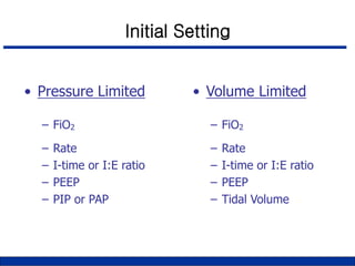
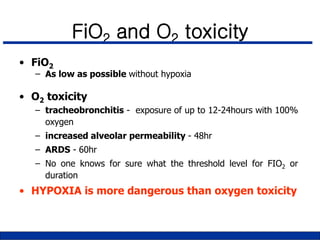
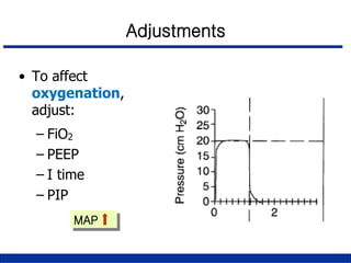
Basic Concepts & Application of
Mechanical Ventilation
2012. 06. 04
호흡기내과
이 상 민
• Mode of ventilation
• Setting & Monitoring
Contents
Modes
• Control Modes:
– every breath is fully supported by the ventilator
– in classic control modes, patients were unable to
breathe except at the controlled set rate
– in newer control modes, machines may act in
assist-control, with a minimum set rate and all
triggered breaths above that rate also fully
supported.
Controlled Mode
(Volume-Targeted Ventilation)
Preset VT
Volume Cycling
Dependent on
CL & Raw
Time (sec)
Flow
(L/m)
Pressure
(cm H2O)
Volume
(mL)
Preset Peak Flow
Assisted Mode
(Volume-Targeted Ventilation)
Time (sec)
Flow
(L/m)
Pressure
(cm H2O)
Volume
(mL)
Preset VT
Volume Cycling
Assist-Control Mode
(Volume-Targeted Ventilation)
• IMV Modes: intermittent mandatory
ventilation modes - breaths “above” set
rate not supported
• SIMV: vent synchronizes IMV “breath”
with patient’s effort
Modes
IMV
(Volume-Targeted Ventilation)
SIMV
(Volume-Targeted Ventilation)
Spontaneous Breaths
Flow
(L/m)
Pressure
(cm H2O)
Volume
(mL)
Whenever a breath is supported by the
ventilator, regardless of the mode, the
limit of the support is determined by a
preset pressure OR volume.
– Volume Limited: preset tidal volume
– Pressure Limited: preset PIP or PAP
Modes
Pressure vs. Volume
• Pressure Limited
– tidal volume by change
suddenly as patient’s
compliance changes
– this can lead to
hypoventilation or
overexpansion of the
lung
– if ETT is obstructed
acutely, delivered tidal
volume will decrease
• Volume Limited
– no limit per se on PIP
(usually vent will have
upper pressure limit)
– square wave(constant)
flow pattern results in
higher PIP for same
tidal volume as
compared to Pressure
modes
Pressure Support Ventilation
• Pressure augmented breathing
• Allows patient to determine the inflation
volume and respiratory cycle duration
• Uses: augment inflation during spontaneous
breathing or overcome resistance of breathing
through ventilator circuits (during weaning)
• Popular an a non-invasive mode of ventilation
via nasal or face masks
PSV
Time (sec)
Flow
(L/m)
Pressure
(cm H2O)
Volume
(mL)
Flow Cycling
Set PS
level
Continuous Positive Airway Pressure
• Spontaneous breathing
• Patient does not need to generate negative
pressure to receive inhaled gas
• CPAP replaced spontaneous PEEP
• Use: Non-intubated patients (OSA, COPD)
Positive End-Expiratory Pressure
• Alveolar pressure at end-expiration is above
atmospheric pressure : PEEP
• Extrinsic PEEP
• Auto PEEP
Positive End-Expiratory Pressure
• CLINICAL USES:
– Reduce toxic levels of FiO2 (ARDS)
– Low-volume ventilation
– Obstructive lung disease (Extrinsic=Occult PEEP)
• Mode of ventilation
• Setting & Monitoring
Contents
Setting Up the Ventilator
• Mode
• Triggering sensitivity
• Tidal volume
• Respiratory rate
• Flow rate and I:E ratio
• FIO2
Initial Setting
• Pressure Limited
– FiO2
– Rate
– I-time or I:E ratio
– PEEP
– PIP or PAP
• Volume Limited
– FiO2
– Rate
– I-time or I:E ratio
– PEEP
– Tidal Volume
• Minute ventilation (VE)
– Men VE = 4 X BSA
– Women VE = 3.5 X BSA
• Tidal volume (VT)
– Min 4mL/kg IBW ~ Max 12mL/kg IBW
– Keep alveolar pressure < 30 ~ 35cmH2O
• Respiratory frequency (f)
– f = VE / VT
Initial Setting (Volume Ventilation)
• Pressure Control Ventilation (PCV)
– Set pressure to achieve a target VT
– Set frequency to achieve same VE (f = VE / VT)
• Pressure Support Ventilation (PSV)
– Set pressure at 5~10cmH2O to overcome Rsys
– Set pressure to achieve a target VT for ventilatory support
Initial Setting (Pressure Ventilation)
Trigger
• How does the vent know when to give a breath?
- “Trigger”
– patient effort
– elapsed time
• The patient’s effort can be “sensed” as a
change in pressure or a change in flow (in the
circuit)
Triggering Sensitivity
• Pressure
– -1 to -2 cm H2O
• Flow
– 1 to 10L/min below the base flow
– less WOB
– faster response time than pressure triggering
Pattern of Flow Waveform
Change of Flow Waveform during VCV
Constant  Descending Same Volume, Increased TI
• Flow is greatest at beginning of inspiration
• TI will be longer
• Mean Paw is higher
• Peak pressure is lower
• Consequences: increase oxygenation, reduce dead space,
improve the distribution of gas in the lungs
• Occurs naturally in pressure ventilation
Advantage of Descending Flow
Flow Rate & I:E Ratio
• Conventional situation
– I:E ratio= 1:2
– Flow rate; 60 L/min
• Airway obstruction
– I:E ratio = 1:3
– Increase flow rate
• Hypoxemia
– Increase inspiratory time
Too short Te will not allow
to deliver max. possible
Vt at given P
 will induce PEEPi
 increases the risk for
hemodynamic instability
Too short Ti will
reduce delivered Vt
 unneccessary high
PIP will be applied
 unnecessary high
intrathoracic pressures
Change in Termination of Flow during PSV
Cycle off
Adjustment of Rising Time
Effect of Rise Time during PSV
Faster Rise Time Slow Rise Time
▪ Too fast
 pressure overshoot
 dyssynchrony
Respiratory Rate
• Btw 10 to 20 breath/min
• A/C mode
– 4 breaths lower than spontaneous
rate
• SIMV mode
– At least 80% of pt’s MV
FiO2 and O2 toxicity
• FiO2
– As low as possible without hypoxia
• O2 toxicity
– tracheobronchitis - exposure of up to 12-24hours with 100%
oxygen
– increased alveolar permeability - 48hr
– ARDS - 60hr
– No one knows for sure what the threshold level for FIO2 or
duration
• HYPOXIA is more dangerous than oxygen toxicity
Desired FiO2
Desired FiO2 =
PaO2 (known)
PaO2 (desired) X FiO2(known)
• Baseline ABGA is available
• Baseline ABGA is not available
– Start with high initial FiO2 setting (0.50 to 1.0)
– Reduce this as quickly as possible
100% Oxygen
• Continuous use of 100% O2 is not recommended
– Result in absorption atelectasis
– May cause oxygen toxicity
– 100% O2 should not be withheld if the patient is seriously ill
• Brief use of 100% O2
– Before and after suctioning
– During bronchoscopy
– During any procedure that might be risky for the patient
• To affect
oxygenation,
adjust:
– FiO2
– PEEP
– I time
– PIP
• To affect
ventilation,
adjust:
– Respiratory Rate
– Tidal Volume
MAP MV
Adjustments
• To affect
oxygenation,
adjust:
– FiO2
– PEEP
– I time
– PIP
MAP
Adjustments
Factors that affect on PaCO2 during MV
Inspiratory Time ↑
Factors that affect on PaCO2 during MV
Pressure-controlled ventilation
TI ↑  VT ↑till plateau
Airway Pressure during VCV
• End-inspiratory breath hold
: Pplateau
• End-expiratory breath hold
: Auto-PEEP
Waveforms
A : Normal
B : ↓ compliance
C : ↑ compliance
Volume-controlled ventilation
A: Normal
B: Compliance halved
C: Resistance doubled
A B C
Waveforms
Pressure-controlled ventilation
Pressure-Volume (P-V) loop
P-V loop according to Compliance
Pressure-controlled ventilation
P-V loop according to Compliance
Volume-controlled ventilation
Normal Flow-Volume (F-V) loop
Volume-controlled ventilation
Abnormal F-V loop
Airflow limitation
ex) COPD
Abnormal F-V loop
Air leaking
Proximal Airway Pressures
1. Peak Pressure (Pk)
Function of Inflation volume, recoil force of lungs and chest
wall, airway resistance
2. Plateau Pressure (Pl)
Occlude expiratory tubing at end-inspiration
Function of elastance alone
Monitoring Lung Mechanics
Proximal Airway Pressure
Pk increased Pl unchanged:
Tracheal tube obstruction
Airway obstruction from secretions
Acute bronchospasm
Rx: Suctioning and Bronchodilators
Use of Airway Pressure
Pk and Pl are both increased:
Pneumothorax
Lobar atelectasis
Acute pulmonary edema
Worsening pneumonia
ARDS
COPD with tachypnea and Auto-PEEP
Increased abdominal pressure
Asynchronous breathing
Use of Airway Pressure
Decreased Pk:
System air leak: Tubing disconnection, cuff leak
Rx: Manual inflation, listen for leak
Use of Airway Pressure
Volume
Pressure
D V
D P
C =
D V
D P
Compliance
C stat =
Pplat - PEEP
tidal volume
Static Compliance (Cstat):
Distensibility of Lungs and Chest wall
Cstat = Vt/Pl
Normal C stat: 50-80 ml/cm of water
Provides objective measure of severity of illness in a
pulmonary disorder
Dynamic Compliance:
Cdyn: Vt/Pk
*Subtract PEEP from Pl or Pk for compliance
measurement
Use Exhaled tidal volume for calculations
Compliance
1. Increased
Ppk-Pplat
2. Low and
prolonged
expiratory
flow
1. Elevated
Pplat-PEEP
2. Normal
expiratory
flow
Resistance ↑vs Compliance ↓
Take Home Message
1. Mode :
- pressure-limited vs volume-limited
- phase variables ; trigger, limit, cycle
2. Setting :
- to improve oxygenation ; mean airway pressure
- to improve ventilation ; minute ventilation
3. Monitoring :
- plateau pressure / static compliance / elastance
- peak pressure / dynamic compliance / + resistance
Thank you for your attention !

Mechanical ventilation

  • 1.
    Basic Concepts &Application of Mechanical Ventilation 2012. 06. 04 호흡기내과 이 상 민
  • 2.
    • Mode ofventilation • Setting & Monitoring Contents
  • 3.
    Modes • Control Modes: –every breath is fully supported by the ventilator – in classic control modes, patients were unable to breathe except at the controlled set rate – in newer control modes, machines may act in assist-control, with a minimum set rate and all triggered breaths above that rate also fully supported.
  • 4.
    Controlled Mode (Volume-Targeted Ventilation) PresetVT Volume Cycling Dependent on CL & Raw Time (sec) Flow (L/m) Pressure (cm H2O) Volume (mL) Preset Peak Flow
  • 5.
    Assisted Mode (Volume-Targeted Ventilation) Time(sec) Flow (L/m) Pressure (cm H2O) Volume (mL) Preset VT Volume Cycling
  • 6.
  • 7.
    • IMV Modes:intermittent mandatory ventilation modes - breaths “above” set rate not supported • SIMV: vent synchronizes IMV “breath” with patient’s effort Modes
  • 8.
  • 9.
  • 10.
    Whenever a breathis supported by the ventilator, regardless of the mode, the limit of the support is determined by a preset pressure OR volume. – Volume Limited: preset tidal volume – Pressure Limited: preset PIP or PAP Modes
  • 11.
    Pressure vs. Volume •Pressure Limited – tidal volume by change suddenly as patient’s compliance changes – this can lead to hypoventilation or overexpansion of the lung – if ETT is obstructed acutely, delivered tidal volume will decrease • Volume Limited – no limit per se on PIP (usually vent will have upper pressure limit) – square wave(constant) flow pattern results in higher PIP for same tidal volume as compared to Pressure modes
  • 12.
    Pressure Support Ventilation •Pressure augmented breathing • Allows patient to determine the inflation volume and respiratory cycle duration • Uses: augment inflation during spontaneous breathing or overcome resistance of breathing through ventilator circuits (during weaning) • Popular an a non-invasive mode of ventilation via nasal or face masks
  • 13.
  • 14.
    Continuous Positive AirwayPressure • Spontaneous breathing • Patient does not need to generate negative pressure to receive inhaled gas • CPAP replaced spontaneous PEEP • Use: Non-intubated patients (OSA, COPD)
  • 15.
    Positive End-Expiratory Pressure •Alveolar pressure at end-expiration is above atmospheric pressure : PEEP • Extrinsic PEEP • Auto PEEP
  • 16.
    Positive End-Expiratory Pressure •CLINICAL USES: – Reduce toxic levels of FiO2 (ARDS) – Low-volume ventilation – Obstructive lung disease (Extrinsic=Occult PEEP)
  • 17.
    • Mode ofventilation • Setting & Monitoring Contents
  • 18.
    Setting Up theVentilator • Mode • Triggering sensitivity • Tidal volume • Respiratory rate • Flow rate and I:E ratio • FIO2
  • 19.
    Initial Setting • PressureLimited – FiO2 – Rate – I-time or I:E ratio – PEEP – PIP or PAP • Volume Limited – FiO2 – Rate – I-time or I:E ratio – PEEP – Tidal Volume
  • 20.
    • Minute ventilation(VE) – Men VE = 4 X BSA – Women VE = 3.5 X BSA • Tidal volume (VT) – Min 4mL/kg IBW ~ Max 12mL/kg IBW – Keep alveolar pressure < 30 ~ 35cmH2O • Respiratory frequency (f) – f = VE / VT Initial Setting (Volume Ventilation)
  • 21.
    • Pressure ControlVentilation (PCV) – Set pressure to achieve a target VT – Set frequency to achieve same VE (f = VE / VT) • Pressure Support Ventilation (PSV) – Set pressure at 5~10cmH2O to overcome Rsys – Set pressure to achieve a target VT for ventilatory support Initial Setting (Pressure Ventilation)
  • 22.
    Trigger • How doesthe vent know when to give a breath? - “Trigger” – patient effort – elapsed time • The patient’s effort can be “sensed” as a change in pressure or a change in flow (in the circuit)
  • 23.
    Triggering Sensitivity • Pressure –-1 to -2 cm H2O • Flow – 1 to 10L/min below the base flow – less WOB – faster response time than pressure triggering
  • 24.
  • 25.
    Change of FlowWaveform during VCV Constant  Descending Same Volume, Increased TI
  • 26.
    • Flow isgreatest at beginning of inspiration • TI will be longer • Mean Paw is higher • Peak pressure is lower • Consequences: increase oxygenation, reduce dead space, improve the distribution of gas in the lungs • Occurs naturally in pressure ventilation Advantage of Descending Flow
  • 27.
    Flow Rate &I:E Ratio • Conventional situation – I:E ratio= 1:2 – Flow rate; 60 L/min • Airway obstruction – I:E ratio = 1:3 – Increase flow rate • Hypoxemia – Increase inspiratory time
  • 28.
    Too short Tewill not allow to deliver max. possible Vt at given P  will induce PEEPi  increases the risk for hemodynamic instability Too short Ti will reduce delivered Vt  unneccessary high PIP will be applied  unnecessary high intrathoracic pressures
  • 29.
    Change in Terminationof Flow during PSV Cycle off
  • 30.
  • 31.
    Effect of RiseTime during PSV Faster Rise Time Slow Rise Time ▪ Too fast  pressure overshoot  dyssynchrony
  • 32.
    Respiratory Rate • Btw10 to 20 breath/min • A/C mode – 4 breaths lower than spontaneous rate • SIMV mode – At least 80% of pt’s MV
  • 33.
    FiO2 and O2toxicity • FiO2 – As low as possible without hypoxia • O2 toxicity – tracheobronchitis - exposure of up to 12-24hours with 100% oxygen – increased alveolar permeability - 48hr – ARDS - 60hr – No one knows for sure what the threshold level for FIO2 or duration • HYPOXIA is more dangerous than oxygen toxicity
  • 34.
    Desired FiO2 Desired FiO2= PaO2 (known) PaO2 (desired) X FiO2(known) • Baseline ABGA is available • Baseline ABGA is not available – Start with high initial FiO2 setting (0.50 to 1.0) – Reduce this as quickly as possible
  • 35.
    100% Oxygen • Continuoususe of 100% O2 is not recommended – Result in absorption atelectasis – May cause oxygen toxicity – 100% O2 should not be withheld if the patient is seriously ill • Brief use of 100% O2 – Before and after suctioning – During bronchoscopy – During any procedure that might be risky for the patient
  • 36.
    • To affect oxygenation, adjust: –FiO2 – PEEP – I time – PIP • To affect ventilation, adjust: – Respiratory Rate – Tidal Volume MAP MV Adjustments
  • 37.
    • To affect oxygenation, adjust: –FiO2 – PEEP – I time – PIP MAP Adjustments
  • 38.
    Factors that affecton PaCO2 during MV
  • 39.
    Factors that affecton PaCO2 during MV Inspiratory Time ↑
  • 40.
    Factors that affecton PaCO2 during MV Pressure-controlled ventilation TI ↑  VT ↑till plateau
  • 41.
    Airway Pressure duringVCV • End-inspiratory breath hold : Pplateau • End-expiratory breath hold : Auto-PEEP
  • 42.
    Waveforms A : Normal B: ↓ compliance C : ↑ compliance Volume-controlled ventilation
  • 43.
    A: Normal B: Compliancehalved C: Resistance doubled A B C Waveforms Pressure-controlled ventilation
  • 44.
  • 45.
    P-V loop accordingto Compliance Pressure-controlled ventilation
  • 46.
    P-V loop accordingto Compliance Volume-controlled ventilation
  • 47.
    Normal Flow-Volume (F-V)loop Volume-controlled ventilation
  • 48.
    Abnormal F-V loop Airflowlimitation ex) COPD
  • 49.
  • 50.
    Proximal Airway Pressures 1.Peak Pressure (Pk) Function of Inflation volume, recoil force of lungs and chest wall, airway resistance 2. Plateau Pressure (Pl) Occlude expiratory tubing at end-inspiration Function of elastance alone Monitoring Lung Mechanics
  • 51.
  • 52.
    Pk increased Plunchanged: Tracheal tube obstruction Airway obstruction from secretions Acute bronchospasm Rx: Suctioning and Bronchodilators Use of Airway Pressure
  • 53.
    Pk and Plare both increased: Pneumothorax Lobar atelectasis Acute pulmonary edema Worsening pneumonia ARDS COPD with tachypnea and Auto-PEEP Increased abdominal pressure Asynchronous breathing Use of Airway Pressure
  • 54.
    Decreased Pk: System airleak: Tubing disconnection, cuff leak Rx: Manual inflation, listen for leak Use of Airway Pressure
  • 55.
    Volume Pressure D V D P C= D V D P Compliance C stat = Pplat - PEEP tidal volume
  • 56.
    Static Compliance (Cstat): Distensibilityof Lungs and Chest wall Cstat = Vt/Pl Normal C stat: 50-80 ml/cm of water Provides objective measure of severity of illness in a pulmonary disorder Dynamic Compliance: Cdyn: Vt/Pk *Subtract PEEP from Pl or Pk for compliance measurement Use Exhaled tidal volume for calculations Compliance
  • 57.
    1. Increased Ppk-Pplat 2. Lowand prolonged expiratory flow 1. Elevated Pplat-PEEP 2. Normal expiratory flow Resistance ↑vs Compliance ↓
  • 58.
    Take Home Message 1.Mode : - pressure-limited vs volume-limited - phase variables ; trigger, limit, cycle 2. Setting : - to improve oxygenation ; mean airway pressure - to improve ventilation ; minute ventilation 3. Monitoring : - plateau pressure / static compliance / elastance - peak pressure / dynamic compliance / + resistance
  • 59.
    Thank you foryour attention !