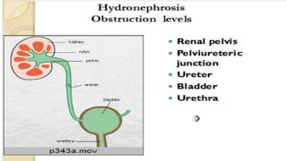
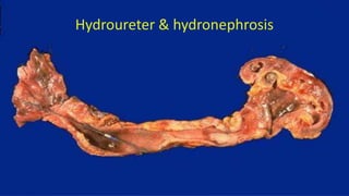
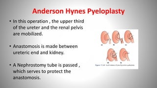
Hydronephrosis
Submitted by ; Vinayak Ambekar
Definition
• Hydronephrosis is defined as aseptic dilatation of the
whole or a part of the pelvi-calyceal system of the
kidney due to partial or intermittent interruption to
the outflow of urine.
Hydronephrosis of the kidney, with marked dilation of the pelvis and calyces and
thinning of the renal parenchyma.
Causes of hydronephrosis
• Hydronephrosis refers to dilatation of renal
pelvis and calyces with accompanying atrophy
of parenchyma.
• Cause by obstruction to outflow of urine,
Causes of hydronephrosis
Congenital
• Atresia of urethra,
• Aberrant renal artery compressing the
ureter,
• Renal ptosis with kinking of ureter.
Causes of hydronephrosis
Acquired
• Foreign Body: Stones
• Tumors: cancer prostate & bladder tumors
• Inflammation: prostatitis, ureteritis, urethritis
• Neurogenic: spinal cord damage with paralysis of
bladder.
Causes of hydronephrosis
• The kidney is massively enlarged with greatly
distended pelvi-calyceal system.
• The renal parenchyma is compressed and atrophied
with obliteration of the papilla and fattening of the
pyramids.
• Depending on obstruction one or both ureter may also
dilated (hydroureter)
Morphology :
Hydroureter & hydronephrosis
Severe hydronephrosis .
The kidney is markedly enlarged.
Hydronephrosis affecting mainly the lower pole of kidney due to stone at this site .The upper pole of the
kidney is normal
• Bilateral complete obstruction produce anuria
which need soon medial attention
• Incomplete bilateral obstruction produce polyuria
rather than oliguria as a result of defect in tubular
concentrating mechanism
Clinical course
• Unilateral hydronephrosis may be silent for long
period unless other kidney is affected.
• Bilateral hydronephrosis usually lead to uremia.
• Early removal of obstruction can return the kidney
function.
• However with the time the changes become
irreversible.
Clinical course
Treatment of Hydronephrosis secondary to a cause
• Stones  Pyelolithotomy, Ureterolithotomy
• Stricture  Stricturoplasty or excision and end to end anastomosis
• Aberrant Vessel  Transection of the ureter and anastomosis in
front of the vessel
• Benign Prostatic Hyperplasia  Transurthral resection of Prostate
(TURP)
• Carcinoma of Prostate  TURP+ Hormonal Therapy
• Urethral Stricture  Urethroplasty
• Meatal Stenosis  Meatoplasty
• Phimosis  Circumcision
A Case of Hydronephrosis
• Middle aged female presented with symptoms of loin pain, ??
Ultrasonography of abdomen revealed dilatation of pelvic-calyceal
system and renal cortical thickness as 5mm.
• Indications for Surgery in case of Hydronephrosis are
 Pain
 Atrophy of kidney (Cortical thinning)
 Infection
 Nephrosis (Increasing Hydronephrosis)
**Indications can be remembered by mnemonic ‘PAIN’
Management of Hydronephrosis in this case
Cortical Thickness is
adequate i.e. more than
or equal to 5mm
Preliminary
Nephrostomy should be
done to decompress the
system
Reassessment of renal
function to be done
after few days
If Renal function
improves , depending
on the cause definitive
surgery for
hydronephrosis can be
done
If the renal function
doesn’t improve and
the opposite kidney is
normal , nephrectomy
is done**If the Kidney is non-functioning with thinned out cortex i.e. less than
5mm ,nephrectomy should be performed.
Management of Renal Stones
• Considering that after nephrostomy, renal function has
improved and the cause of hydronephrosis in this case was
renal calculus. Further management can be done as follows :
Management of
Renal Stones
Modern Methods of
Stone Removal
Extra-corporeal shock
wave lithotripsy
Percutaneous
Nephrolithotomy
Open Surgery for
Stone Removal
Pyelolithotomy
Nephrolithotomy
Management of Congenital Hydronephrosis
• Patients with Congenital PUJ dysfunction ,present with congenital
Hydronephrosis.
• Congenital Hydronephrosis can be defined as more than 10mm
antero-posterior diameter of renal pelvis at 20 weeks of gestation.
• Management differs according to the grade of hydronephrosis,
which is based on renal pelvic diameter.
• Grading is as follows, Mild 11-20mm ,Moderate 21-35mm,
Severe >35mm.
• Mild hydronephrosis is managed conservatively with serial
monitoring of pelvic diameter by ultrasound and of renal
function
• Moderate hydronephrosis is also managed by serial
monitoring. Any deterioration of renal function is an indication
for surgical intervention.
• Severe Hydronephrosis should be treated early-Anderson
Hynes Pyeloplasty to prevent permanent damage to kidney.
Anderson Hynes Pyeloplasty
• In this operation , the upper third
of the ureter and the renal pelvis
are mobilized.
• Anastomosis is made between
ureteric end and kidney.
• A Nephrostomy tube is passed ,
which serves to protect the
anastomosis.
Hydronephrosis

Hydronephrosis

  • 1.
  • 2.
    Definition • Hydronephrosis isdefined as aseptic dilatation of the whole or a part of the pelvi-calyceal system of the kidney due to partial or intermittent interruption to the outflow of urine.
  • 3.
    Hydronephrosis of thekidney, with marked dilation of the pelvis and calyces and thinning of the renal parenchyma.
  • 4.
    Causes of hydronephrosis •Hydronephrosis refers to dilatation of renal pelvis and calyces with accompanying atrophy of parenchyma. • Cause by obstruction to outflow of urine,
  • 5.
    Causes of hydronephrosis Congenital •Atresia of urethra, • Aberrant renal artery compressing the ureter, • Renal ptosis with kinking of ureter.
  • 6.
    Causes of hydronephrosis Acquired •Foreign Body: Stones • Tumors: cancer prostate & bladder tumors • Inflammation: prostatitis, ureteritis, urethritis • Neurogenic: spinal cord damage with paralysis of bladder.
  • 7.
  • 10.
    • The kidneyis massively enlarged with greatly distended pelvi-calyceal system. • The renal parenchyma is compressed and atrophied with obliteration of the papilla and fattening of the pyramids. • Depending on obstruction one or both ureter may also dilated (hydroureter) Morphology :
  • 12.
  • 13.
    Severe hydronephrosis . Thekidney is markedly enlarged.
  • 14.
    Hydronephrosis affecting mainlythe lower pole of kidney due to stone at this site .The upper pole of the kidney is normal
  • 17.
    • Bilateral completeobstruction produce anuria which need soon medial attention • Incomplete bilateral obstruction produce polyuria rather than oliguria as a result of defect in tubular concentrating mechanism Clinical course
  • 18.
    • Unilateral hydronephrosismay be silent for long period unless other kidney is affected. • Bilateral hydronephrosis usually lead to uremia. • Early removal of obstruction can return the kidney function. • However with the time the changes become irreversible. Clinical course
  • 19.
    Treatment of Hydronephrosissecondary to a cause • Stones  Pyelolithotomy, Ureterolithotomy • Stricture  Stricturoplasty or excision and end to end anastomosis • Aberrant Vessel  Transection of the ureter and anastomosis in front of the vessel • Benign Prostatic Hyperplasia  Transurthral resection of Prostate (TURP) • Carcinoma of Prostate  TURP+ Hormonal Therapy • Urethral Stricture  Urethroplasty • Meatal Stenosis  Meatoplasty • Phimosis  Circumcision
  • 20.
    A Case ofHydronephrosis • Middle aged female presented with symptoms of loin pain, ?? Ultrasonography of abdomen revealed dilatation of pelvic-calyceal system and renal cortical thickness as 5mm. • Indications for Surgery in case of Hydronephrosis are  Pain  Atrophy of kidney (Cortical thinning)  Infection  Nephrosis (Increasing Hydronephrosis) **Indications can be remembered by mnemonic ‘PAIN’
  • 21.
    Management of Hydronephrosisin this case Cortical Thickness is adequate i.e. more than or equal to 5mm Preliminary Nephrostomy should be done to decompress the system Reassessment of renal function to be done after few days If Renal function improves , depending on the cause definitive surgery for hydronephrosis can be done If the renal function doesn’t improve and the opposite kidney is normal , nephrectomy is done**If the Kidney is non-functioning with thinned out cortex i.e. less than 5mm ,nephrectomy should be performed.
  • 22.
    Management of RenalStones • Considering that after nephrostomy, renal function has improved and the cause of hydronephrosis in this case was renal calculus. Further management can be done as follows : Management of Renal Stones Modern Methods of Stone Removal Extra-corporeal shock wave lithotripsy Percutaneous Nephrolithotomy Open Surgery for Stone Removal Pyelolithotomy Nephrolithotomy
  • 23.
    Management of CongenitalHydronephrosis • Patients with Congenital PUJ dysfunction ,present with congenital Hydronephrosis. • Congenital Hydronephrosis can be defined as more than 10mm antero-posterior diameter of renal pelvis at 20 weeks of gestation. • Management differs according to the grade of hydronephrosis, which is based on renal pelvic diameter.
  • 24.
    • Grading isas follows, Mild 11-20mm ,Moderate 21-35mm, Severe >35mm. • Mild hydronephrosis is managed conservatively with serial monitoring of pelvic diameter by ultrasound and of renal function • Moderate hydronephrosis is also managed by serial monitoring. Any deterioration of renal function is an indication for surgical intervention. • Severe Hydronephrosis should be treated early-Anderson Hynes Pyeloplasty to prevent permanent damage to kidney.
  • 25.
    Anderson Hynes Pyeloplasty •In this operation , the upper third of the ureter and the renal pelvis are mobilized. • Anastomosis is made between ureteric end and kidney. • A Nephrostomy tube is passed , which serves to protect the anastomosis.