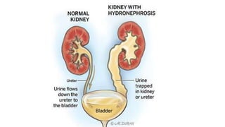
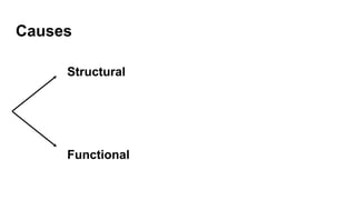
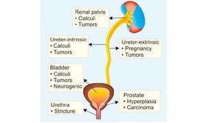
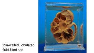
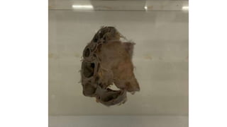
HYDRONEPHROSIS
Dr. Salman Ansari
Dept. of Pathology
Kanachur Institute of Medical Sciences
Contents
● Definition
● Causes
● Pathogenesis
● Morphology
● Clinical features
● Investigations, Treatment, Complications
Hydronephrosis
● Defined as aseptic dilation of collecting system
of kidney due to obstruction of urinary outflow
● Atrophy of kidney is seen
● Dilation of the ureter due to obstruction of
urine outflow is called hydroureter.
Causes
Structural
Functional
Structural causes:
● Urinary calculi
● Tumour of prostate, bladder, uterus, cervix
● BPH in men
● Congenital abnormalities like urethral stricture
● Inflammation: prostatitis, urethritis
● Pregnancy
Neurogenic causes:
Neurogenic bladder, due to spinal cord injury or diabetic
nephropathy
Pathogenesis
● Obstruction: complete or incomplete, which leads to
accumulation of urine proximal to obstruction
● Dilatation: accumulated urine causes dilatation of calyces
and pelvis due to back pressure, which is transmitted back
into renal tissue
● Reduced GFR: if obstruction continues, tubule loses
concentrating function and GFR decreases
● Inflammatory reaction in interstitium with fibrosis
Morphology
Types of obstruction:
- Sudden and complete obstruction: GFR is reduced
and mild dilation of renal pelvis and calyces occurs
- Intermittent or subtotal obstruction: GFR is not
affected and dilation is progressive
Level of obstruction:
- depending upon where the obstruction is, the first part
Gross features
- Obstruction can be unilateral or bilateral, with dilation
of ureter(called hydroureter)
- Kidney enlargement can be slight to massive
- Thin-walled kidney, with cysts
- Renal parenchyma shows destruction due to
severe pressure atrophy and thinning of cortex
thin-walled, lobulated,
fluid-filled sac
Microscopy
- Tubules: dilated and atrophied
- Glomeruli: partial or complete sclerosis
- Interstitium: fibrosis, chronic inflammatory cells
Clinical features
Features depend upon cause of obstruction
- Renal calculus presents with renal colic, whereas BPH
presents with bladder symptoms
- Unilateral hydronephrosis: can be asymptomatic
- Bilateral partial obstruction: polyuria, nocturia
- Bilateral complete obstruction: oliguria/anuria
Investigations: USG to diagnose obstructive uropathy
Treatment: depends upon cause and severity of
hydronephrosis
Complications:
- UTI
- pyelonephritis
References:
● Ramadas Nayak - Textbook of Pathology for Allied Health Sciences
Questions:
salman.s.ansari92@gmail.com
Scan for PPT →

Hydronephrosis - Pathology - Allied Health Sciences

  • 1.
    HYDRONEPHROSIS Dr. Salman Ansari Dept.of Pathology Kanachur Institute of Medical Sciences
  • 2.
    Contents ● Definition ● Causes ●Pathogenesis ● Morphology ● Clinical features ● Investigations, Treatment, Complications
  • 3.
    Hydronephrosis ● Defined asaseptic dilation of collecting system of kidney due to obstruction of urinary outflow ● Atrophy of kidney is seen ● Dilation of the ureter due to obstruction of urine outflow is called hydroureter.
  • 5.
  • 6.
    Structural causes: ● Urinarycalculi ● Tumour of prostate, bladder, uterus, cervix ● BPH in men ● Congenital abnormalities like urethral stricture ● Inflammation: prostatitis, urethritis ● Pregnancy
  • 7.
    Neurogenic causes: Neurogenic bladder,due to spinal cord injury or diabetic nephropathy
  • 9.
    Pathogenesis ● Obstruction: completeor incomplete, which leads to accumulation of urine proximal to obstruction ● Dilatation: accumulated urine causes dilatation of calyces and pelvis due to back pressure, which is transmitted back into renal tissue ● Reduced GFR: if obstruction continues, tubule loses concentrating function and GFR decreases ● Inflammatory reaction in interstitium with fibrosis
  • 10.
    Morphology Types of obstruction: -Sudden and complete obstruction: GFR is reduced and mild dilation of renal pelvis and calyces occurs - Intermittent or subtotal obstruction: GFR is not affected and dilation is progressive Level of obstruction: - depending upon where the obstruction is, the first part
  • 11.
    Gross features - Obstructioncan be unilateral or bilateral, with dilation of ureter(called hydroureter) - Kidney enlargement can be slight to massive - Thin-walled kidney, with cysts - Renal parenchyma shows destruction due to severe pressure atrophy and thinning of cortex
  • 12.
  • 14.
    Microscopy - Tubules: dilatedand atrophied - Glomeruli: partial or complete sclerosis - Interstitium: fibrosis, chronic inflammatory cells
  • 15.
    Clinical features Features dependupon cause of obstruction - Renal calculus presents with renal colic, whereas BPH presents with bladder symptoms - Unilateral hydronephrosis: can be asymptomatic - Bilateral partial obstruction: polyuria, nocturia - Bilateral complete obstruction: oliguria/anuria
  • 16.
    Investigations: USG todiagnose obstructive uropathy Treatment: depends upon cause and severity of hydronephrosis
  • 17.
  • 18.
    References: ● Ramadas Nayak- Textbook of Pathology for Allied Health Sciences Questions: salman.s.ansari92@gmail.com Scan for PPT →