This document provides an overview of urolithiasis (kidney stones). It discusses the epidemiology, classification, pathogenesis, clinical features, investigations, treatment modalities, complications, and prevention of kidney stones. Treatment depends on the location and size of the stone and includes extracorporeal shock wave lithotripsy, percutaneous nephrolithotomy, ureteroscopy, and open surgery. The goal is to remove stones while minimizing complications such as infection, obstruction, and loss of renal function. Prevention focuses on adequate fluid intake, dietary modifications, and medical management for certain stone types.
Urolithiasis is a common disease that is estimated to
produce medical costs of $2.1 billion per year in the United States alone.
Renal colic affects approximately 1.2 million people
each year in USA and accounts for approximately 1% of
all hospital admissions.
Most active emergency departments (EDs) manage
patients with acute renal colic every day.
Please find the power point on Renal and bladder stones. I tried present it on understandable way and all the contents are reviewed by experts and from very reliable references. Thank you
Hydronephrosis is the distension of the renal calyces and pelvis due to accumulation of the
urine as a result of the obstruction to the outflow of the urine mostly distal to renal pelvis.
This presentation comprises of congenital anomalies of kidney and urinary tract made concise and in depth for PG preparation. It contains all important topics of the regarding subject covered in detail.
continuation on the urinary tract disorders. congenital and acquired disorders well covered. pyelonephritis also forms part of the text. thanks for reading. remeber to like and follow
Nephrolithiasis, commonly known as kidney stones, refers to the formation of hard mineral and salt deposits within the kidneys or urinary tract. These stones can vary in size, ranging from tiny grains to larger, more substantial formations. Nephrolithiasis is a relatively common condition and can affect people of all ages, although it is more prevalent in adults.
HOT NEW PRODUCT! BIG SALES FAST SHIPPING NOW FROM CHINA!! EU KU DB BK substit...GL Anaacs
Contact us if you are interested:
Email / Skype : kefaya1771@gmail.com
Threema: PXHY5PDH
New BATCH Ku !!! MUCH IN DEMAND FAST SALE EVERY BATCH HAPPY GOOD EFFECT BIG BATCH !
Contact me on Threema or skype to start big business!!
Hot-sale products:
NEW HOT EUTYLONE WHITE CRYSTAL!!
5cl-adba precursor (semi finished )
5cl-adba raw materials
ADBB precursor (semi finished )
ADBB raw materials
APVP powder
5fadb/4f-adb
Jwh018 / Jwh210
Eutylone crystal
Protonitazene (hydrochloride) CAS: 119276-01-6
Flubrotizolam CAS: 57801-95-3
Metonitazene CAS: 14680-51-4
Payment terms: Western Union,MoneyGram,Bitcoin or USDT.
Deliver Time: Usually 7-15days
Shipping method: FedEx, TNT, DHL,UPS etc.Our deliveries are 100% safe, fast, reliable and discreet.
Samples will be sent for your evaluation!If you are interested in, please contact me, let's talk details.
We specializes in exporting high quality Research chemical, medical intermediate, Pharmaceutical chemicals and so on. Products are exported to USA, Canada, France, Korea, Japan,Russia, Southeast Asia and other countries.
New Directions in Targeted Therapeutic Approaches for Older Adults With Mantl...i3 Health
i3 Health is pleased to make the speaker slides from this activity available for use as a non-accredited self-study or teaching resource.
This slide deck presented by Dr. Kami Maddocks, Professor-Clinical in the Division of Hematology and
Associate Division Director for Ambulatory Operations
The Ohio State University Comprehensive Cancer Center, will provide insight into new directions in targeted therapeutic approaches for older adults with mantle cell lymphoma.
STATEMENT OF NEED
Mantle cell lymphoma (MCL) is a rare, aggressive B-cell non-Hodgkin lymphoma (NHL) accounting for 5% to 7% of all lymphomas. Its prognosis ranges from indolent disease that does not require treatment for years to very aggressive disease, which is associated with poor survival (Silkenstedt et al, 2021). Typically, MCL is diagnosed at advanced stage and in older patients who cannot tolerate intensive therapy (NCCN, 2022). Although recent advances have slightly increased remission rates, recurrence and relapse remain very common, leading to a median overall survival between 3 and 6 years (LLS, 2021). Though there are several effective options, progress is still needed towards establishing an accepted frontline approach for MCL (Castellino et al, 2022). Treatment selection and management of MCL are complicated by the heterogeneity of prognosis, advanced age and comorbidities of patients, and lack of an established standard approach for treatment, making it vital that clinicians be familiar with the latest research and advances in this area. In this activity chaired by Michael Wang, MD, Professor in the Department of Lymphoma & Myeloma at MD Anderson Cancer Center, expert faculty will discuss prognostic factors informing treatment, the promising results of recent trials in new therapeutic approaches, and the implications of treatment resistance in therapeutic selection for MCL.
Target Audience
Hematology/oncology fellows, attending faculty, and other health care professionals involved in the treatment of patients with mantle cell lymphoma (MCL).
Learning Objectives
1.) Identify clinical and biological prognostic factors that can guide treatment decision making for older adults with MCL
2.) Evaluate emerging data on targeted therapeutic approaches for treatment-naive and relapsed/refractory MCL and their applicability to older adults
3.) Assess mechanisms of resistance to targeted therapies for MCL and their implications for treatment selection
Anti ulcer drugs and their Advance pharmacology ||
Anti-ulcer drugs are medications used to prevent and treat ulcers in the stomach and upper part of the small intestine (duodenal ulcers). These ulcers are often caused by an imbalance between stomach acid and the mucosal lining, which protects the stomach lining.
||Scope: Overview of various classes of anti-ulcer drugs, their mechanisms of action, indications, side effects, and clinical considerations.
Title: Sense of Taste
Presenter: Dr. Faiza, Assistant Professor of Physiology
Qualifications:
MBBS (Best Graduate, AIMC Lahore)
FCPS Physiology
ICMT, CHPE, DHPE (STMU)
MPH (GC University, Faisalabad)
MBA (Virtual University of Pakistan)
Learning Objectives:
Describe the structure and function of taste buds.
Describe the relationship between the taste threshold and taste index of common substances.
Explain the chemical basis and signal transduction of taste perception for each type of primary taste sensation.
Recognize different abnormalities of taste perception and their causes.
Key Topics:
Significance of Taste Sensation:
Differentiation between pleasant and harmful food
Influence on behavior
Selection of food based on metabolic needs
Receptors of Taste:
Taste buds on the tongue
Influence of sense of smell, texture of food, and pain stimulation (e.g., by pepper)
Primary and Secondary Taste Sensations:
Primary taste sensations: Sweet, Sour, Salty, Bitter, Umami
Chemical basis and signal transduction mechanisms for each taste
Taste Threshold and Index:
Taste threshold values for Sweet (sucrose), Salty (NaCl), Sour (HCl), and Bitter (Quinine)
Taste index relationship: Inversely proportional to taste threshold
Taste Blindness:
Inability to taste certain substances, particularly thiourea compounds
Example: Phenylthiocarbamide
Structure and Function of Taste Buds:
Composition: Epithelial cells, Sustentacular/Supporting cells, Taste cells, Basal cells
Features: Taste pores, Taste hairs/microvilli, and Taste nerve fibers
Location of Taste Buds:
Found in papillae of the tongue (Fungiform, Circumvallate, Foliate)
Also present on the palate, tonsillar pillars, epiglottis, and proximal esophagus
Mechanism of Taste Stimulation:
Interaction of taste substances with receptors on microvilli
Signal transduction pathways for Umami, Sweet, Bitter, Sour, and Salty tastes
Taste Sensitivity and Adaptation:
Decrease in sensitivity with age
Rapid adaptation of taste sensation
Role of Saliva in Taste:
Dissolution of tastants to reach receptors
Washing away the stimulus
Taste Preferences and Aversions:
Mechanisms behind taste preference and aversion
Influence of receptors and neural pathways
Impact of Sensory Nerve Damage:
Degeneration of taste buds if the sensory nerve fiber is cut
Abnormalities of Taste Detection:
Conditions: Ageusia, Hypogeusia, Dysgeusia (parageusia)
Causes: Nerve damage, neurological disorders, infections, poor oral hygiene, adverse drug effects, deficiencies, aging, tobacco use, altered neurotransmitter levels
Neurotransmitters and Taste Threshold:
Effects of serotonin (5-HT) and norepinephrine (NE) on taste sensitivity
Supertasters:
25% of the population with heightened sensitivity to taste, especially bitterness
Increased number of fungiform papillae
Prix Galien International 2024 Forum ProgramLevi Shapiro
June 20, 2024, Prix Galien International and Jerusalem Ethics Forum in ROME. Detailed agenda including panels:
- ADVANCES IN CARDIOLOGY: A NEW PARADIGM IS COMING
- WOMEN’S HEALTH: FERTILITY PRESERVATION
- WHAT’S NEW IN THE TREATMENT OF INFECTIOUS,
ONCOLOGICAL AND INFLAMMATORY SKIN DISEASES?
- ARTIFICIAL INTELLIGENCE AND ETHICS
- GENE THERAPY
- BEYOND BORDERS: GLOBAL INITIATIVES FOR DEMOCRATIZING LIFE SCIENCE TECHNOLOGIES AND PROMOTING ACCESS TO HEALTHCARE
- ETHICAL CHALLENGES IN LIFE SCIENCES
- Prix Galien International Awards Ceremony
TEST BANK for Operations Management, 14th Edition by William J. Stevenson, Ve...kevinkariuki227
TEST BANK for Operations Management, 14th Edition by William J. Stevenson, Verified Chapters 1 - 19, Complete Newest Version.pdf
Lung Cancer: Artificial Intelligence, Synergetics, Complex System Analysis, S...Oleg Kshivets
RESULTS: Overall life span (LS) was 2252.1±1742.5 days and cumulative 5-year survival (5YS) reached 73.2%, 10 years – 64.8%, 20 years – 42.5%. 513 LCP lived more than 5 years (LS=3124.6±1525.6 days), 148 LCP – more than 10 years (LS=5054.4±1504.1 days).199 LCP died because of LC (LS=562.7±374.5 days). 5YS of LCP after bi/lobectomies was significantly superior in comparison with LCP after pneumonectomies (78.1% vs.63.7%, P=0.00001 by log-rank test). AT significantly improved 5YS (66.3% vs. 34.8%) (P=0.00000 by log-rank test) only for LCP with N1-2. Cox modeling displayed that 5YS of LCP significantly depended on: phase transition (PT) early-invasive LC in terms of synergetics, PT N0—N12, cell ratio factors (ratio between cancer cells- CC and blood cells subpopulations), G1-3, histology, glucose, AT, blood cell circuit, prothrombin index, heparin tolerance, recalcification time (P=0.000-0.038). Neural networks, genetic algorithm selection and bootstrap simulation revealed relationships between 5YS and PT early-invasive LC (rank=1), PT N0—N12 (rank=2), thrombocytes/CC (3), erythrocytes/CC (4), eosinophils/CC (5), healthy cells/CC (6), lymphocytes/CC (7), segmented neutrophils/CC (8), stick neutrophils/CC (9), monocytes/CC (10); leucocytes/CC (11). Correct prediction of 5YS was 100% by neural networks computing (area under ROC curve=1.0; error=0.0).
CONCLUSIONS: 5YS of LCP after radical procedures significantly depended on: 1) PT early-invasive cancer; 2) PT N0--N12; 3) cell ratio factors; 4) blood cell circuit; 5) biochemical factors; 6) hemostasis system; 7) AT; 8) LC characteristics; 9) LC cell dynamics; 10) surgery type: lobectomy/pneumonectomy; 11) anthropometric data. Optimal diagnosis and treatment strategies for LC are: 1) screening and early detection of LC; 2) availability of experienced thoracic surgeons because of complexity of radical procedures; 3) aggressive en block surgery and adequate lymph node dissection for completeness; 4) precise prediction; 5) adjuvant chemoimmunoradiotherapy for LCP with unfavorable prognosis.
Explore natural remedies for syphilis treatment in Singapore. Discover alternative therapies, herbal remedies, and lifestyle changes that may complement conventional treatments. Learn about holistic approaches to managing syphilis symptoms and supporting overall health.
These lecture slides, by Dr Sidra Arshad, offer a quick overview of physiological basis of a normal electrocardiogram.
Learning objectives:
1. Define an electrocardiogram (ECG) and electrocardiography
2. Describe how dipoles generated by the heart produce the waveforms of the ECG
3. Describe the components of a normal electrocardiogram of a typical bipolar leads (limb II)
4. Differentiate between intervals and segments
5. Enlist some common indications for obtaining an ECG
Study Resources:
1. Chapter 11, Guyton and Hall Textbook of Medical Physiology, 14th edition
2. Chapter 9, Human Physiology - From Cells to Systems, Lauralee Sherwood, 9th edition
3. Chapter 29, Ganong’s Review of Medical Physiology, 26th edition
4. Electrocardiogram, StatPearls - https://www.ncbi.nlm.nih.gov/books/NBK549803/
5. ECG in Medical Practice by ABM Abdullah, 4th edition
6. ECG Basics, http://www.nataliescasebook.com/tag/e-c-g-basics
- Video recording of this lecture in English language: https://youtu.be/lK81BzxMqdo
- Video recording of this lecture in Arabic language: https://youtu.be/Ve4P0COk9OI
- Link to download the book free: https://nephrotube.blogspot.com/p/nephrotube-nephrology-books.html
- Link to NephroTube website: www.NephroTube.com
- Link to NephroTube social media accounts: https://nephrotube.blogspot.com/p/join-nephrotube-on-social-media.html
3. INTRODUCTION
• Urolithiasis, kidney stones, renal stones, and renal calculi are used
interchangeably to refer to the accretion of hard, solid, nonmetallic
minerals in the urinary tract
• Passage of a urinary stone is the most common cause of acute ureteral
obstruction
• The pain may be some of the most severe pain that humans experience
• Complications of stone disease may result in severe infection; renal
failure; or, in rare cases, death.
• Urinary stones have afflicted humankind since antiquity
• The earliest recorded example being bladder and kidney stones detected
in Egyptian mummies dated to 4800 BC
• The specialty of urologic surgery was recognized even by Hippocrates, who
wrote, in his famous oath for the physician,
"I will not cut, even for the stone, but leave such procedures to the
practitioners of the craft“ (obviously, Hippocrates was not a urologist!!)
4. EPIDEMIOLOGY
• The prevalence of urinary tract stonedisease is estimated to be 2% to 3%.
• Rare in Blacks; Commoner in Whites and Asians
• The likelihood that a white man will develop stone disease by age 70 years
is about 1 in 8.
• The recurrence rate without treatment for calcium oxalate renal stones is
about
– 10% at 1 year
– 35% at 5 years, and
– 50% at 10 years
• Male : Female ratio is 3:1
• Peak at 20-40 years old
• Ingestion of excessive amounts of purines ,oxalates,calcium, phosphate,
and other elements often results in excessive excretion of these
components in urine
• A low fluid intake, with a subsequent low volume of urine production,
produces high concentrations of stone-forming solutes in the urine.
This is an important environmental factor in stone formation.
7. CLASSIFICATION…ctd
COMPARATIVE INCIDENCES OF FORMS OF URINARY LITHIASIS
Stone analysis in Percentage
Form of Lithiasis India USA Japan UK
Pure Calcium Oxalate 86.1 33 17.4 39.4
Mixed Calcium Oxalate and 4.9 34 50.8 20.2
Phosphate
Magnesium Ammonium 2.7 15 17.4 15.4
Phosphate (Struvite )
Uric Acid 1.2 8.0 4.4 8.0
Cystine 0.4 3.0 1.0 2.8
8. CLASSIFICATION…ctd
Oxalate (Calcium Oxalate)
• Also Called Mulberry Stone
• Covered With Sharp Projections
• Sharp Makes Kidney Bleed (Haematuria)
• Very Hard
• Radio – Opaque
• Under microscope looks like Hourglass or Dumbbell shape if
monohydrate and Like an Envelope if Dihydrate
9. CLASSIFICATION…ctd
Phosphate stones
• Usually Calcium Phosphate
• Sometimes Calcium Magnesium Ammonium Phosphate Or Triple
Phosphate
• Smooth Minimum Symptoms
• Dirty White
• Radio – Opaque
• Calcium Phosphate also called ‘Brushite’ appears like Needle shape under
microscope
• In Alkaline urineEnlarges rapidlyTake the shape of CalycesStaghorn
10. CLASSIFICATION…ctd
Uric Acid & Urate Stone
• Hard & Smooth
• Multiple
• Yellow or Red-brown
• Radio - Lucent (Use Ultrasound)
• Under microscope appear like irregular plates or rosettes
11. CLASSIFICATION…ctd
Cystine Stone
• Autosomal recesive disorder
• Usually in Young Girls
• Due To Cystinuria -
• Cystine Not Absorbed by Tubules
• Multiple
• Soft or Hard – can form stag-horns
• Pink or Yellow
• Radio-opaque
• Under microscope appears like hexagonal or benzene ring
12. PATHOGENESIS
• more than 1 of 3 general mechanisms is likely to
be active
– the possible presence or abundance of substances
that promote crystal and stone formation
– a possible relative lack of substances to inhibit crystal
formation;
– a possible excessive excretion or concentration of salts
in the urine, which leads to supersaturation of the
crystallizing salt.
The greater the degree of supersaturation, the greater
the rate of growth of the calculi
13. PATHOGENESIS…ctd
• Stasis or anatomic factors can also contribute to the
development of stone disease.
• ~ 85% of calcium stones are idiopathic, or primary.
– Idiopathic hypercalciuria occurs in more than one half of
patients with calcium oxalate stones.
– The remaining 15% of calcium stones are secondary to some
discernible etiology, most commonly, hyperparathyroidism
– Renal tubular acidosis (RTA) is an additional fairly common
secondary cause of calcium stones
– Immobilization of an individual causes rapid mobilization of the
calcium in bones, and this is an important mechanism in
patients with spinal cord injury
14. PATHOGENESIS…ctd
• Magnesium ammonium phosphate (struvite)
stones account for approximately 10-20% of
urinary stones.
– Sometimes they form complex with calcium
phosphate.
– Struvite stones are caused by urea-splitting bacteria
such as Proteus, Klebsiella, and Pseudomonas species.
– Combined obstruction and infection frequently cause
renal destruction and, potentially, renal failure if both
kidneys are affected
15. PATHOGENESIS…ctd
• Uric acid stones account for 5-10% of urinary
stones, Predisposing factors include
– acidic concentrated urine,
– excess urinary uric acid,
– small-bowel disease or resection,
– gout, and cell lysis
– Treatment and prevention for these stones is
alkalinization and dilution of the urine.
• Cystine stones account for only approximately 1%
of urinary stones.
– result from cystinuria (a rare autosomal recessive
metabolic disorder),
16. PATHOGENESIS…ctd
• Miscellaneous Stones
– Triamterene Stones
• potassium sparing diuretic
• 70% excreted in urine
• pure stone or nidus for CaOx/UA
– Indinavir Stones
• greatest incidence of protease inhibitors
• mean duration to stone 21.5 wks (6-50)
• 19% unchanged in urine
• fan shaped or starburst crystals
• not seen on IVU or CT
17. CLINICAL FEATURES
• Renal/Ureteral Colic (PAIN)
– Abrupt onset while asleep or at rest
– Crescendo of extreme pain
– Flank radiating laterally and downward to
groin/testicle or round ligament/labia majora
– Impossible to be still
• Mid ureter
– lateral flank and abdomen
• Lower ureter
– suprapubic and urethral
– urgency and frequency
18. CLINICAL FEATURES…ctd
• GI Symptoms
– Nausea and vomiting – autonomic n.s.
– Ileus or diarrhea
– DDX: gastroenteritis, appendicitis, colitis, diverticular
disease and salpingitis
• Hematuria
– gross or microscopic
– 15% no hematuria!
• Pyuria/Fever
– Pyuria even without infection
– Infection especially in females
19. CLINICAL FEATURES…ctd
• History
– Duration, characteristics, and location of pain
– History of urinary calculi
– Prior complications related to stone manipulation
– Urinary tract infections
– Loss of renal function
– Family history of calculi
20. INVESTIGATIONS
• Urinalysis- haematuria ~ 85% of pts
• FBP
– elevated WBC = renal/ systemic inf.
– low RBC= xnic d’se/ sev. haematuria
• serum eletrolytes, creatinine, calcium, uric acid,
phosphorus: to asses renal function and metabolic risk
factors for stone formation
• 24 hr urine collection for pH, Ca, oxalate, uric acid, Na,
phosphorus, citrate, magnesium, creatinine and total
volume
21. INVESTIGATION…ctd
• Plain abdominal radiograph
– KUB for assessing total stone burden ,the size, shape, and
location of urinary calculi in some patients.
– Calcium-containing stones (~85% of all upper urinary tract
calculi) are radiopaque,
– Pure uric acid, indinavir-induced, and cystine calculi are
relatively radiolucent on plain radiography
• Renal ultrasound
• IVU
– determine the size & location
– anatomical & functional assessment
• Helical CT-scan without contrast
25. MEDICAL RX
• The cornerstone of management of ureteral colic is analgesia
• Morphine sulfate is the narcotic analgesic drug of choice for parenteral
use.
• Antiemetic agents [metoclopramide ] may also be added as needed.
• The calcium channel blocker[ nifedipine] relaxes ureteral smooth muscle
and enhances stone passage
• The alpha blockers, [ terazosin], also relax musculature of the ureter and
lower urinary tract, markedly facilitating passage of ureteral stones
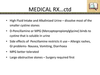
• Uric acid and cystine calculi can be dissolved with medical therapy
• stones are dissolved with alkalinization of the urine.
• Sodium bicarbonate can be used as the alkalinizing agent
26. MEDICAL RX…ctd
• High Fluid Intake and Alkalinized Urine – dissolve most of the
smaller cystine stones
• D-Pencillamine or MPG (Mercaptopropionylglycine) binds to
cystine that is soluble in urine
• Side effects of Pencillamine restricts it use – Allergic rashes,
GI problems- Nausea, Vomiting, Diarrhoea
• MPG better tolerated
• Large obstructive stones – Surgery required first
27. SURGERY
•Extracorporeal Shock Wave Lithotripsy (ESWL)
•Percutaneous Nephrolithotomy (PNL)
•Ureteroscopy
•Open surgery
Choice of approach depends on stone burden (size and
number), stone composition, and stone location.
28. ESWL
• Shock waves generated under water can travel through body
without any appreciable loss of energy.
• When they encounter stones, the changes in density causes energy
to be absorbed and reflected by the stone.
• This results in fragmentation of the stones.
• Before lithotripsy the stone is localized by either Ultrasound or
Flouroscopy.
• Complications:
– Haematuria – is quite common (hemorrhage and edema within
or around the kidney)
– Incomplete stone Fragmentation & Obstruction; “Stienstrasse” (
stone street ) usually due to a large “ Leading fragment” ( Stents
Recommended prior to ESWL for Calculi > 1.5 cm )
31. PNL• Percutaneous approach allows stone removal with less morbidity, shorter
convalescence, and reduced cost compared with open techniques
• PNL has replaced open surgical procedures for removal of large or complex
renal calculi at most institutions
• PNL can be performed with general, epidural, or local anesthesia
• The kidney should be approached from below the 12th rib to reduce the
risk of pleural complications
• The position of the retroperitoneal colon is usually anterior or anterolateral
to the lateral renal border. Therefore, risk of colon injury is minimal
• The liver and spleen may also be at risk of injury during percutaneous
access. However, in the absence of splenomegaly or hepatomegaly, injury to
these organs is extremely rare with a puncture below the 12th rib
• Once the point of puncture and the preferred calyx have been selected, a C-
arm fluouroscope is entered. The tract is dilated by special dilators
• The urologist can proceed with stone removal using endoscopic techniques
e.g with Randall's forceps, a grssper or stone baskets under fluoroscopic
guidance
32. PNL…. Ureteroscope
• There is a concurrence in the literature regarding the need for
postoperative drainage with a nephrostomy tube after percutaneous
procedures.
• The main function of a nephrostomy tube is the drainage of urine and
possibly the tamponade of bleeding originating from the structures
acutely expanded during dilatation.
URETEROSCOPY:
• A ureteroscope is passed through the ureteral orifices
• It is performed under general or regional anaesthesia
• Once the stone is visualized, fragmentation with of the stone can be done
with laser, or mechanically
• If significant ureteral edema or manipulation occurs, a stent should be
placed to prevent colic and obstruction
33. Open surgery
• Generally indicated for large stones that would require
multiple ESWL or PNL
• obese patients are poor candidates for ESWL and may be
difficult to manage with PNL; Open surgery might be the best
option
• Open surgery may be
– Pyelolithotomy
– Nephrolithotomy
– Ureterolithotomy
– Cystolithotomy
34. Summary
• Depending on the location of the stone, various
procedures are done for stone extraxtion
– In the kidney
• ESWL
• PNL
• Open methods
– Pyelolithotomy for a stone in the extrarenal pelvis
– Nephrolithotomy for a stone deep into the renal parenchyma
– Partial nephrectomy if there is a stone impacted into the lower most
calyx
– In the ureter
• Upper ureter: ESWL is ideal
• Mid ureter: ESWL, ureteroscopy or ureterolithotomy
• Lower Ureter: Ureteroscope or ureterolithotomy
35. Summary
– In the Bladder
• Litholapaxy:
through a cystoscopy, the stone is grasped firmly and broken.
Small fragments are evacuated by evacuator
• Suprapubic cystolithotomy
if the stone is too big or too hard
36. Complications
• Ureteral scarring and stenosis
• Nidus for infectionserious infection of the
kidney that diminishes renal function
• Urinary fistula formation
• Ureteral perforation
• Extravasation
• Urinary outflow obstruction
hydronephrosisCRF
37. Prevention
• High Fluid Intake
• Restrict Salt
• Avoid high intake of purine food
• Increased citrus fruits may help
• If hypercalciuria restrict Ca intake