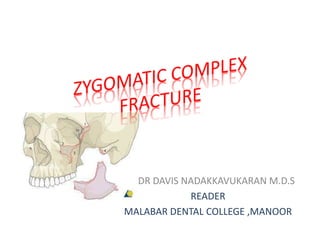
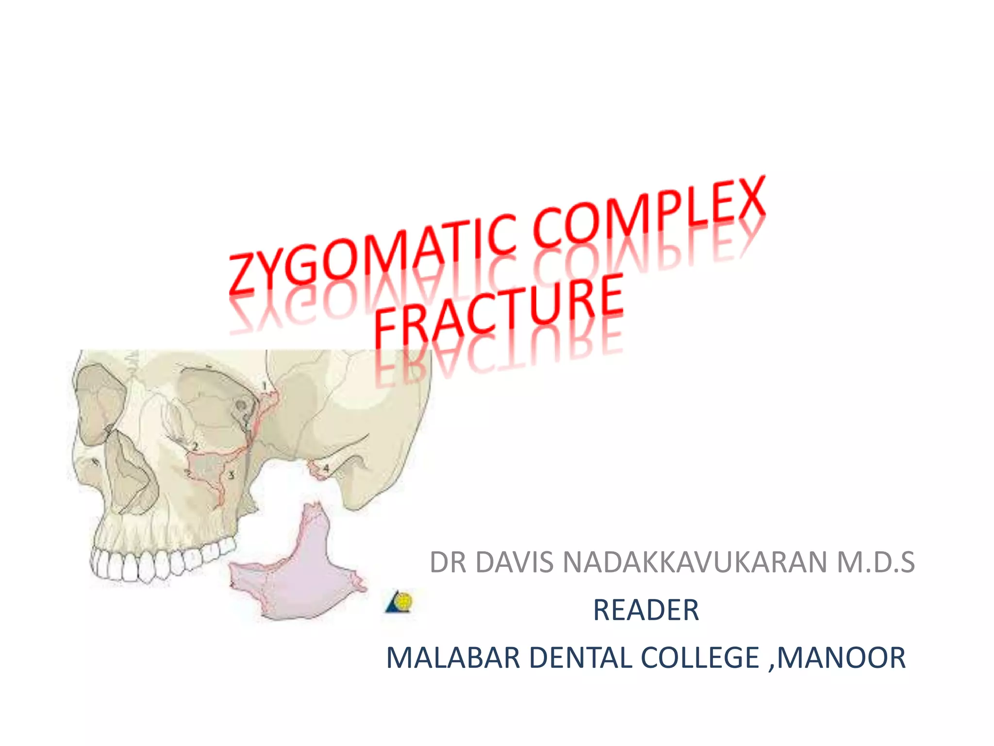
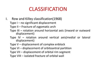
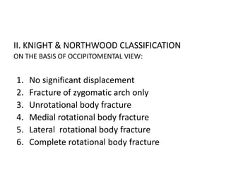
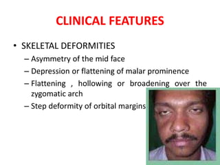
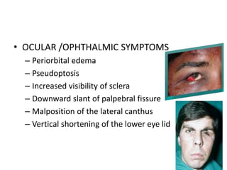
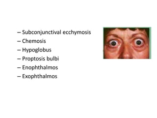
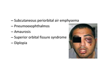
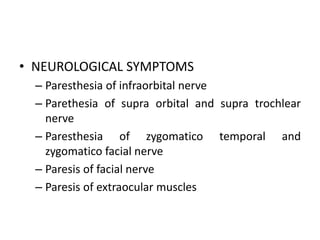
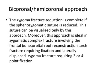
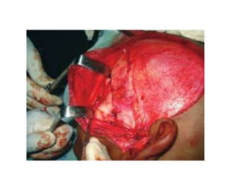
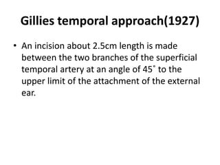
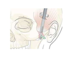
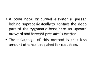
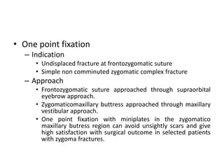
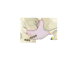
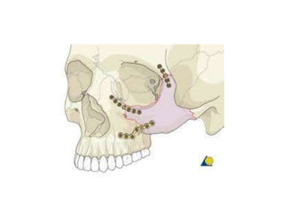
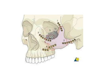
This document discusses the classification, clinical features, investigation, management, and complications of zygomatic bone fractures. It describes 8 types of zygomatic bone fractures classified by Row and Killey or Knight and Northwood. Clinical features include midface deformities, ocular symptoms like diplopia, neurological symptoms, oral symptoms like trismus, and nasal symptoms like epistaxis. Management involves surgical approaches like bicoronal or Gillies temporal to reduce the fracture using indirect or direct methods, then fixing with miniplates in 1-4 points. Complications can include infraorbital numbness, enophthalmos, diplopia, or superior orbital fissure syndrome.
Zygoma: Strong buttress of lateral midface lying between zygomatic process of frontal bone and maxilla.
The high incidence of zygomatic complex fracture relates to its prominent position within the facial skeleton.
Zygoma: Strong buttress of lateral midface lying between zygomatic process of frontal bone and maxilla.
The high incidence of zygomatic complex fracture relates to its prominent position within the facial skeleton.
Temporomandibular disorders (TMDs) are considered the major cause of orofacial pain. Internal derangement (ID) of the temporomandibular joint (TMJ), which is classified as disc displacement with or without reduction, is one of the disorders of the TMJ that is frequently seen.
Displacement of the articular disc can result in decreased joint space, joint noise (clicking, popping, or crepitation), arthritis, condylar resorption, inflammation, and compression of the bilaminar tissue, all of which can cause various degrees of pain and dysfunction.
Maxillofacial Surgery 1
Dental Students Fifth Year First semester
Lecture Name maxillofacial trauma Part 3
Al Azhar University Gaza Palestine
Dr. Lama El Banna
A seminar prepared during my omfs posting hours. Short points are added for easiness to study and bihart. Reference taken from Balaji and Neelima Anil Malik
Telehealth Psychology Building Trust with Clients.pptxThe Harvest Clinic
Telehealth psychology is a digital approach that offers psychological services and mental health care to clients remotely, using technologies like video conferencing, phone calls, text messaging, and mobile apps for communication.
Deep Leg Vein Thrombosis (DVT): Meaning, Causes, Symptoms, Treatment, and Mor...The Lifesciences Magazine
Deep Leg Vein Thrombosis occurs when a blood clot forms in one or more of the deep veins in the legs. These clots can impede blood flow, leading to severe complications.
Navigating Challenges: Mental Health, Legislation, and the Prison System in B...Guillermo Rivera
This conference will delve into the intricate intersections between mental health, legal frameworks, and the prison system in Bolivia. It aims to provide a comprehensive overview of the current challenges faced by mental health professionals working within the legislative and correctional landscapes. Topics of discussion will include the prevalence and impact of mental health issues among the incarcerated population, the effectiveness of existing mental health policies and legislation, and potential reforms to enhance the mental health support system within prisons.
CRISPR-Cas9, a revolutionary gene-editing tool, holds immense potential to reshape medicine, agriculture, and our understanding of life. But like any powerful tool, it comes with ethical considerations.
Unveiling CRISPR: This naturally occurring bacterial defense system (crRNA & Cas9 protein) fights viruses. Scientists repurposed it for precise gene editing (correction, deletion, insertion) by targeting specific DNA sequences.
The Promise: CRISPR offers exciting possibilities:
Gene Therapy: Correcting genetic diseases like cystic fibrosis.
Agriculture: Engineering crops resistant to pests and harsh environments.
Research: Studying gene function to unlock new knowledge.
The Peril: Ethical concerns demand attention:
Off-target Effects: Unintended DNA edits can have unforeseen consequences.
Eugenics: Misusing CRISPR for designer babies raises social and ethical questions.
Equity: High costs could limit access to this potentially life-saving technology.
The Path Forward: Responsible development is crucial:
International Collaboration: Clear guidelines are needed for research and human trials.
Public Education: Open discussions ensure informed decisions about CRISPR.
Prioritize Safety and Ethics: Safety and ethical principles must be paramount.
CRISPR offers a powerful tool for a better future, but responsible development and addressing ethical concerns are essential. By prioritizing safety, fostering open dialogue, and ensuring equitable access, we can harness CRISPR's power for the benefit of all. (2998 characters)
Health Education on prevention of hypertensionRadhika kulvi
Hypertension is a chronic condition of concern due to its role in the causation of coronary heart diseases. Hypertension is a worldwide epidemic and important risk factor for coronary artery disease, stroke and renal diseases. Blood pressure is the force exerted by the blood against the walls of the blood vessels and is sufficient to maintain tissue perfusion during activity and rest. Hypertension is sustained elevation of BP. In adults, HTN exists when systolic blood pressure is equal to or greater than 140mmHg or diastolic BP is equal to or greater than 90mmHg. The
Defecation
Normal defecation begins with movement in the left colon, moving stool toward the anus. When stool reaches the rectum, the distention causes relaxation of the internal sphincter and an awareness of the need to defecate. At the time of defecation, the external sphincter relaxes, and abdominal muscles contract, increasing intrarectal pressure and forcing the stool out
The Valsalva maneuver exerts pressure to expel faeces through a voluntary contraction of the abdominal muscles while maintaining forced expiration against a closed airway. Patients with cardiovascular disease, glaucoma, increased intracranial pressure, or a new surgical wound are at greater risk for cardiac dysrhythmias and elevated blood pressure with the Valsalva maneuver and need to avoid straining to pass the stool.
Normal defecation is painless, resulting in passage of soft, formed stool
CONSTIPATION
Constipation is a symptom, not a disease. Improper diet, reduced fluid intake, lack of exercise, and certain medications can cause constipation. For example, patients receiving opiates for pain after surgery often require a stool softener or laxative to prevent constipation. The signs of constipation include infrequent bowel movements (less than every 3 days), difficulty passing stools, excessive straining, inability to defecate at will, and hard feaces
IMPACTION
Fecal impaction results from unrelieved constipation. It is a collection of hardened feces wedged in the rectum that a person cannot expel. In cases of severe impaction the mass extends up into the sigmoid colon.
DIARRHEA
Diarrhea is an increase in the number of stools and the passage of liquid, unformed feces. It is associated with disorders affecting digestion, absorption, and secretion in the GI tract. Intestinal contents pass through the small and large intestine too quickly to allow for the usual absorption of fluid and nutrients. Irritation within the colon results in increased mucus secretion. As a result, feces become watery, and the patient is unable to control the urge to defecate. Normally an anal bag is safe and effective in long-term treatment of patients with fecal incontinence at home, in hospice, or in the hospital. Fecal incontinence is expensive and a potentially dangerous condition in terms of contamination and risk of skin ulceration
HEMORRHOIDS
Hemorrhoids are dilated, engorged veins in the lining of the rectum. They are either external or internal.
FLATULENCE
As gas accumulates in the lumen of the intestines, the bowel wall stretches and distends (flatulence). It is a common cause of abdominal fullness, pain, and cramping. Normally intestinal gas escapes through the mouth (belching) or the anus (passing of flatus)
FECAL INCONTINENCE
Fecal incontinence is the inability to control passage of feces and gas from the anus. Incontinence harms a patient’s body image
PREPARATION AND GIVING OF LAXATIVESACCORDING TO POTTER AND PERRY,
An enema is the instillation of a solution into the rectum and sig
Antibiotic Stewardship by Anushri Srivastava.pptxAnushriSrivastav
Stewardship is the act of taking good care of something.
Antimicrobial stewardship is a coordinated program that promotes the appropriate use of antimicrobials (including antibiotics), improves patient outcomes, reduces microbial resistance, and decreases the spread of infections caused by multidrug-resistant organisms.
WHO launched the Global Antimicrobial Resistance and Use Surveillance System (GLASS) in 2015 to fill knowledge gaps and inform strategies at all levels.
ACCORDING TO apic.org,
Antimicrobial stewardship is a coordinated program that promotes the appropriate use of antimicrobials (including antibiotics), improves patient outcomes, reduces microbial resistance, and decreases the spread of infections caused by multidrug-resistant organisms.
ACCORDING TO pewtrusts.org,
Antibiotic stewardship refers to efforts in doctors’ offices, hospitals, long term care facilities, and other health care settings to ensure that antibiotics are used only when necessary and appropriate
According to WHO,
Antimicrobial stewardship is a systematic approach to educate and support health care professionals to follow evidence-based guidelines for prescribing and administering antimicrobials
In 1996, John McGowan and Dale Gerding first applied the term antimicrobial stewardship, where they suggested a causal association between antimicrobial agent use and resistance. They also focused on the urgency of large-scale controlled trials of antimicrobial-use regulation employing sophisticated epidemiologic methods, molecular typing, and precise resistance mechanism analysis.
Antimicrobial Stewardship(AMS) refers to the optimal selection, dosing, and duration of antimicrobial treatment resulting in the best clinical outcome with minimal side effects to the patients and minimal impact on subsequent resistance.
According to the 2019 report, in the US, more than 2.8 million antibiotic-resistant infections occur each year, and more than 35000 people die. In addition to this, it also mentioned that 223,900 cases of Clostridoides difficile occurred in 2017, of which 12800 people died. The report did not include viruses or parasites
VISION
Being proactive
Supporting optimal animal and human health
Exploring ways to reduce overall use of antimicrobials
Using the drugs that prevent and treat disease by killing microscopic organisms in a responsible way
GOAL
to prevent the generation and spread of antimicrobial resistance (AMR). Doing so will preserve the effectiveness of these drugs in animals and humans for years to come.
being to preserve human and animal health and the effectiveness of antimicrobial medications.
to implement a multidisciplinary approach in assembling a stewardship team to include an infectious disease physician, a clinical pharmacist with infectious diseases training, infection preventionist, and a close collaboration with the staff in the clinical microbiology laboratory
to prevent antimicrobial overuse, misuse and abuse.
to minimize the developme
Empowering ACOs: Leveraging Quality Management Tools for MIPS and BeyondHealth Catalyst
Join us as we delve into the crucial realm of quality reporting for MSSP (Medicare Shared Savings Program) Accountable Care Organizations (ACOs).
In this session, we will explore how a robust quality management solution can empower your organization to meet regulatory requirements and improve processes for MIPS reporting and internal quality programs. Learn how our MeasureAble application enables compliance and fosters continuous improvement.
3. INTRODUCTION
• Zygomatic bone is intimately associated with
the maxilla, frontal and temporal bones
zygomatic complex.
• The zygomatic bone fractures in the region of
the zygomatico-frontal suture, the
zygomaticotemporal suture and the
zygomatico-maxillary suture
4. CLASSIFICATION
I. Row and Killey classification(1968)
Type I – no significant displacement
Type II – Fracture of zygomatic arch
Type III – rotation around horizontal axis (inward or outward
displacement)
Type IV – rotation around vertical axis(medial or lateral
displacement)
Type V – displacement of complex enblock
Type VI – displacement of orbitoantral partition
Type VII – displacement of orbital rim segment
Type VIII – isolated fracture of orbital wall
5. II. KNIGHT & NORTHWOOD CLASSIFICATION
ON THE BASIS OF OCCIPITOMENTAL VIEW:
1. No significant displacement
2. Fracture of zygomatic arch only
3. Unrotational body fracture
4. Medial rotational body fracture
5. Lateral rotational body fracture
6. Complete rotational body fracture
6. CLINICAL FEATURES
• SKELETAL DEFORMITIES
– Asymmetry of the mid face
– Depression or flattening of malar prominence
– Flattening , hollowing or broadening over the
zygomatic arch
– Step deformity of orbital margins
7. • OCULAR /OPHTHALMIC SYMPTOMS
– Periorbital edema
– Pseudoptosis
– Increased visibility of sclera
– Downward slant of palpebral fissure
– Malposition of the lateral canthus
– Vertical shortening of the lower eye lid
9. – Subcutaneous periorbital air emphysema
– Pneumoexophthalmos
– Amaurosis
– Superior orbital fissure syndrome
– Diplopia
10. • Test for diplopia
1. Finger gaze:-
Finger moved infront of eye in all nine
directions of gaze at a distance of 30cm.
2. Forced duction test:-
Tissue holding forceps are used to hold
tendon of inferior fornix . The globe is
manipulated through its entire range of motion.
Inability to rotate the globe superiorly signifies
entraptment of muscle in orbital floor.
11.
12. • NEUROLOGICAL SYMPTOMS
– Paresthesia of infraorbital nerve
– Parethesia of supra orbital and supra trochlear
nerve
– Paresthesia of zygomatico temporal and
zygomatico facial nerve
– Paresis of facial nerve
– Paresis of extraocular muscles
13. • ORAL SYMPTOMS
– Ecchymosis in the buccal sulcus of maxillary arch
– Deformity of zygomatic buttress of maxilla
– Trismus
– Pain
– Impacted /flattened zygomatic arch
• NASAL SYMPTOMS
– Ipsilateral epistaxis
– Ipsilateral hematosinus
14. INVESTIGATION
• Plain radiographs
water’s view or paranasal view of
zygomaticomaxillary complex fracture,floor of
orbit,infra orbital rim
submentovertex- Arch fracture
• CT scan
15. MANAGEMENT
• Surgical approach:-
A. Extra oral approach
Bicoronal/hemicoronal
Gillies temporal approach
Superolateral
Supraorbital approach;lateral eyebrow
Upper eyelid
Lower eyelid
Infra orbital
Subtarsal
Subcilliary
Transconjunctival
percutaneous
17. Bicoronal/hemicoronal approach
• The zygoma fracture reduction is complete if
the sphenozygomatic suture is reduced. This
suture can be visualized only by this
approach. Moreover, this approach is ideal in
zygomatic complex fracture involving the
frontal bone,orbital roof reconstruction ,arch
fracture requiring fixation and laterally
displaced zygoma fracture requiring 3 or 4
point fixation.
18.
19. Gillies temporal approach(1927)
• An incision about 2.5cm length is made
between the two branches of the superficial
temporal artery at an angle of 45˚ to the
upper limit of the attachment of the external
ear.
20.
21. • elevator is also used in this approach for the
reduction of the zygomatic fracture.
• Dissection is carried out till the temporal fascia. A
Bristow’s elevator is passed down through this
incision beneath the zygomatic bone which is
then gradually reduced to its position.
• The incision is then closed in layers.
• Rowe pattern zygomatic
22. • Bristow’s elevator has adisadvantage of using
the temporal bone as fulcrum causing risk of
fracturing the temporal bone during the
procedure. This was overcome by the design
in Rowe zygoma elevator
23. Transoral/keen’s approach
• Also known as buccal sulcus incision /lateral
maxillary vestibular incision
• A bone hook can be passed from a transverse
incision made in the region of buccal sulcus
and the fractured segment can be reduced.
• An incision 1cm in length is made in the buccal
sulcus behind the zygomatic buttress.
24.
25. • A bone hook or curved elevator is passed
behind supraperiosteally,to contact the deep
part of the zygomatic bone.here an upward
outward and forward pressure is exerted.
• The advantage of this method is that less
amount of force is required for reduction.
27. • Fixation
– 1 point fixation
– 2 point fixation
– 3 point fixation
– 4 point fixation
28. • One point fixation
– Indication
• Undisplaced fracture at frontozygomatic suture
• Simple non comminuted zygomatic complex fracture
– Approach
• Frontozygomatic suture approached through supraorbital
eyebrow approach.
• Zygomaticomaxillary buttress approached through maxillary
vestibular approach.
• One point fixation with miniplates in the zygomatico
maxillary butress region can avoid unsightly scars and give
high satisfaction with surgical outcome in selected patients
with zygoma fractures.
29.
30. • Two point fixation
– Indication
• Displaced fracture unstable after reduction
• Fracture at frontozygomatic suture,infraorbital rim and
buttress.
– Approach
• Exposure of frontozygomatic suture through lower
eyelid incision or maxillary vestibular incision.
• A 2 point fixation using low profile plate at
zygomaticomaxillary buttress or at the infra orbital rim
suffice.
31.
32. • Three point fixation
– Fixation is done at frontozygomatic
suture,zygomaticomaxillary buttress and the
infraorbital rim.
– Good reduction of these 3 sites mostly reduces
the arch fracture which is not fixed.
33.
34. • Four point fixation
– Unique from 3 point technique in that the surgeon
visualizes the zygomatic arch. The order of
placement of the plates will be dependant on the
least damaged landmarks. The zygomatic arch is
an excellent reference to restore proper
anteroposterior projection of the midface
35.
36. • Fixation is again of two types:
i. Direct fixation
• Transosseous wiring
ii. Indirect fixation
• Internal pin fixation
• Transfixation with kirshner wire
37. COMPLICATIONS
• Infraorbital paraesthesia
• Malunion and assymmetry
• Diplopia
• Traumatic hyphema
• Enopthalmos
• Traumatic optic neuropathy- mild visual deficit to complete loss of
vision
• Superior orbital fissure syndrome-include ptosis, ophthalmoplegia,
forehead anesthesia, and a fixed dilated pupil. Proptosis may be
present. Treatment may include reduction of fractures, steroids,
orbital apex exploration, and aspiration of retrobulbar hematoma if
present
• Trismus
• Retrobulbar haemorrhage
38. REFERENCE
1. Textbook of oral and maxillofacial surgery;2nd
edition- S.M Balaji
2. Textbook of oral and maxillofacial surgery;3rd
edition- Neelima Mallik
3. Textbook of oral and maxillofacial surgery-
Chithra chakravarthi
4. Oral and maxillofacial surgery-Laskin