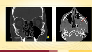
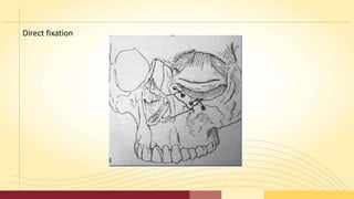
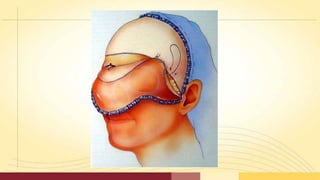
ZYGOMATICOMAXILLARY
COMPLEX (ZMC)
FRACTURE
Contents
• Introduction
• Anatomy
• Classification
• Signs and Symptoms
• Clinical Examinations
• Radiological Examinations
• Management
• Complications
Introduction
• Second most common facial fracture, after nasal fracture
• The high incidence relates to zygoma’s prominent position within the facial skeleton
• Male predilection, 4:1 ratio
• Peak incidence - 2nd to 3rd decades of life
• Mostly due to altercations followed by motor vehicle accidents
• During altercations left zygoma is most commonly affected
• Zygoma plays an important role in facial contour. Disruption of the zygomatic position
creates impairement of ocular and mandibular function
• Zygoma or the malar complex forms the central support of the cheek an is a strong
buttress of the lateral and middle third of the facial skeleton.
ANATOMY
• Zygoma is roughly quadrilateral in shape, with
an outer convex (cheek) surface and an inner
concave (temporal) surface.
• It forms the point of greatest prominence
of the cheek.
• Resembles a four sided pyramid, which
has temporal, orbital, maxillary and
frontal processes.
• Zygoma articulates with four bones- the
frontal, sphenoid, maxillary and temporal.
• Body of the zygoma extensively articulates with
the maxilla along the anterior maxilla and along
the orbital floor.
• It forms the superolateral aspect and part of
the supero-anterior aspect of the maxillary
sinus.
• The zygomatico-temporal articulation is a
very thin ,delicate connection, which
fractures frequently.
• The zygoma provides origin to a major portion
of the masseter muscle along the body and
temporal surface.
• The temporal fascia also attaches along
the arch and the temporal process.
The inferior orbital fissure is the key to remember the usual lines of
ZMC fracture
Three fracture lines extend from the inferior orbital fissure in an
antero-medial, a supero-lateral, and an inferior direction.
FRACTURE PATTERNS
1. One fracture extends from the
inferior orbital fissure antero-
medially along the orbital floor
mostly through the orbital process of
maxilla.
2. Second line of fracture from the
inferior orbital fissure runs inferiorly
through the posterior (infra
temporal) aspect of maxilla and joins
the fracture from the anterior aspect
of maxilla, under the zygomatico
maxillary buttress.
3. Third line of fracture extends
superiorly from the inferior
orbital fissure along the lateral
orbital wall posterior to the rim,
usually separating the
zygomatico-sphenoid suture. A
ZMC fracture that follows this
pattern usually has one
additional fracture line through
the zygomatic arch.
Classification
Knight and North (1961)
Rowe and Killey (1968)
Yanagisawa (1973)
Larsen and Thomson (1978)
Rowe and Williams (1985)
Poswillo (1988)
Knight and North (1961)
Based on the direction of displacement
On a Water 's view radiograph,
• Group I - Non displaced fractures
• Group II - Arch fractures
• Group Ill - Un-rotated fractures
• Group IV - Medially rotated fractures
• Group V - Laterally rotated fractures
• Group VI - Complex fractures
ROWE AND KILLEY (1968)
Type I : No significant displacement
Type II : Fracture of the zygomatic arch
Type III : Rotation around vertical axis
- Inward displacement of orbital rim
- Outward displacement of orbital rim
Type IV : Rotation around longitudinal axis
- Medial displacement of frontal process
- Lateral displacement of frontal process
Type V : Displacement of the complex en bloc
- Medial
- Inferior
- lateral (Rare)
Type VI : Displacement of orbitoantral partition
- Inferiorly
- Superiorly
Type VII : Displacement of orbital rim segments
Type VIII : Complex comminuted fractures.
Type I : no significant displacement
Type II . Fracture of the zygomatic arch
Outward Displacement
Inward Displacement
Type III. Rotation around vertical axis
Type IV. Rotation around longitudinal axis
Type V. Displacement of the complex en bloc
Type VI. Displacement of orbitoantral partition
Type VII. Displacement of orbital rim segments
Type VIII. Complex comminuted fractures
Yanagisawa ( 1973 )
GROUPS I & II - Unchanged
GROUP Ill - Medial or lateral rotation around a vertical axis
GROUP IV - Medial or lateral rotation around a longitudinal axis
GROUP V - Medial or lateral displacement without rotation
GROUP VI - Isolated arch fracture
GROUP VII - All complex fractures
Larsen and Thomson (1978)
Group I – Non displaced fractures requiring no treatment
Group II – All fractures requiring treatment
ROWE AND WILLIAMS (1985)
•Fractures stable after elevation
a) Arch only (medially displaced)
b) Rotation around the vertical axis
i) medially
ii) laterally
•Fractures unstable after elevation
a) Arch only (inferiorly displaced)
b) Rotation around horizontal axis
i) Medially
ii) Laterally
c) Dislocations en bloc
i) inferiorly
ii) medially
iii) postero - laterally
d) Communited fractures
Poswillo’s classification (1988)
Inward and downward displacement
Inward and posterior displacement
Outward displacement of the zygomatic complex
Communition
Fracture of the arch alone
Signs and Symptoms
Deformity at the zygomatic buttress of the maxilla
Deformity of the orbital region
Trismus
Abnormal nerve sensibility
Epistaxis
Crepitation from air emphysema
RADIOLOGIC EXAMINATION
• Plain films and Computed Tomography have their place in
determining the type,location, magnitude, and direction of
displacement of zygomatic fractures.
• This includes,
Water 's view, Submentovertex view, Computed Tomography.
• A single Water 1
s view is an important adjunct to clinical examination.
Submento vertex : “Jug handle view”
• If fractures are noted, CT should be the procedure of
choice.
• Two dimensional CT is now considered the best and most
useful means of radiologic assessment of the facial
skeleton.
• CT scans allow complete assessment orbital
floor and walls.
• The axial scan is helpful in evaluating the medial and
lateral walls, and the coronal scan defines the extent of
injury to the orbital floor
Treatment
• Historical review:
• Various authors have given various treatment modalities and
techniques for the
management of ZMC fractures.
• Dating back to 1751, Duverney stressed the role of contraction
of temporal muscle in realigning the medial displacement of
the zygomatic arch.
• Ferrier in 1825,attempted to reduce fracture of
zygomatic arch through an incision above the arch.
• Dupuytren in 1847,discovered the important
relationship of the temporal fascia and the muscle as a
pathway to the zygomatic arch.
• Gillies in 1927 emphasized the cosmetic value of placing the incision
within the hair line.
• Stroymeyer in 1844 described the percutaneous
hook technique.
• Cheyne and Burghard in 1901 discussed the intraoral digital
manipulation technique.
• Smith and Yanagisawa in 1961 stressed the importance of cosmetic
aspects of the treatment.
GENERAL PRINCIPLES OF TREATMENT
• No treatment
• Indirect reduction with,
1. No fixation
2. Temporary support
3. Direct fixation
4. Indirect fixation
• Direct reduction and fixation
Surgical Approaches
Indirect reduction with
No fixation
1. the temporal fossa (gillies)
2. the upper buccal sulcus (intraoral),
keens and balasubramanium
3. the cheek (percutaneous),
4. the nose (transantral)
5. the eyebrow (lateral brow), dingman
& natwig
Temporary support- packing into the
antrum
Direct fixation – transosseous wiring,
miniplates
Indirect fixation
Zygomatico-zygomatic (Trans-maxillary)
Naso-zygomatic
Zygomatico-palatal
Maxillo-zygomatic
Fronto-zygomatic
Cranio-zygomatic
Direct reduction and fixation
Extra oral
a. Upper eyelid
b. Supra orbital eyebrow
c. Lower eyelid
i. Subciliary
ii. Infra orbital
iii. Trans conjunctival
d. Coronal
Intra oral
a. Maxillary vestibular
No treatment
No Treatment
Cases with a minimal degree of displacement, which following
union, are considered unlikely to result any
• cosmetic deformity,
• disturbance of vision,
• persistent paresthesia or
• impairment of mandibular movement.
Surgical Approaches
Indirect reduction with
No fixation
1. the temporal fossa (gillies)
2. the upper buccal sulcus (intraoral),
keens and balasubramanium
3. the cheek (percutaneous),
4. the nose (transantral)
5. the eyebrow (lateral brow), dingman
& natwig
Temporary support- packing into the
antrum
Direct fixation – transosseous wiring,
miniplates
Indirect fixation
Zygomatico-zygomatic (Trans-maxillary)
Naso-zygomatic
Zygomatico-palatal
Maxillo-zygomatic
Fronto-zygomatic
Cranio-zygomatic
Direct reduction and fixation
Extra oral
a. Upper eyelid
b. Supra orbital eyebrow
c. Lower eyelid
i. Subciliary
ii. Infra orbital
iii. Trans conjunctival
d. Coronal
Intra oral
a. Maxillary vestibular
No treatment
INDIRECT REDUCTION
NO FIXATION
Includes procedures which do not involve exposure of the fracture sites.
The principle is to disimpact and reduce the fracture by direct application of an
instrument, through an indirect approach remote from the fracture line.
The techniques which have been developed for this operative
approach, are based upon the introduction of an instrument
through,
1. the temporal fossa,
2. the upper buccal sulcus (intraoral),
3. the cheek (percutaneous),
4. the nose (transantral)
5. the eyebrow (lateral brow)
Temporal fossa approach
• Temporal fossa approach:
• This method was introduced by Gillies et al (1927) for elevation of the zygomatic
arch.
• Incision of about 2.5 cm long, made above and parallel to the anterior branch of
the temporal artery.
Upper buccal sulcus - Balasubramaniam (1967)
• The advantages of this technique have been discussed by Balasubramaniam (1967)
who considers that '' less force is required by the intraoral approach than by the
extraoral, because the force is exerted where it should be, i.e., more at the centre of
the fractured fragment''.
Upper buccal sulcus – Keen’s approach
Access is gained by an incision of about 1cm in length at the reflection of the
upper buccal sulcus immediately behind the zygomatic buttress.
Quinn in 1977 described a modification –
Which employs a lateral coronoid approach through an incision situated over
the anterior border of ramus.
Percutaneous approach: (Stroymeyer 1844)
• This method consists of inserting a hook through the
skin below and behind the zygomatic bone so that it
engages the deep aspect and allows reduction by strong
outward traction on the handle of the
instrument.
• Poswillo advises that the exact location of the initial
stab wound for insertion is found at the intersection of
a perpendicular line dropped from the outer canthus of
the eye and a horizontal line extended posteriorly from
the alar margin of the nostril.
Percutaneous approach: (Poswillo)
Intra nasal transantral approach: (Lathrop's approach 1906)
• Not common in use.
• An opening is made into the antrum below the inferior meatus, and a
curved instrument (urethral sound) introduced and manipulated so
that its tip lies on the antral aspect of the zygomatic bone. Firm
outward and upward pressure is applied to reposition the bone.
Lateral brow approach: (Dingman & Natwig 1964)
• The advantage of this technique is that the
fracture at the orbital rim is visualized
directly.
• The fronto-zygomatic area of the lateral
orbital rim is exposed by the eyebrow
incision.
• The instrument is inserted to lift the
zygoma anteriorly, laterally and
superiorly.
Surgical Approaches
Indirect reduction with
No fixation
1. the temporal fossa (gillies)
2. the upper buccal sulcus (intraoral),
keens and balasubramanium
3. the cheek (percutaneous),
4. the nose (transantral)
5. the eyebrow (lateral brow), dingman
& natwig
Temporary support- packing into the
antrum
Direct fixation – transosseous wiring,
miniplates
Indirect fixation
Zygomatico-zygomatic (Trans-maxillary)
Naso-zygomatic
Zygomatico-palatal
Maxillo-zygomatic
Fronto-zygomatic
Cranio-zygomatic
Direct reduction and fixation
Extra oral
a. Upper eyelid
b. Supra orbital eyebrow
c. Lower eyelid
i. Subciliary
ii. Infra orbital
iii. Trans conjunctival
d. Coronal
Intra oral
a. Maxillary vestibular
No treatment
• Temporary support is a concept which is primarily based upon the
introduction of a pack or other material into the antrum so as to exert
counter-pressure against those forces which tend to bring about a
relapse of the position achieved by indirect reduction.
TEM PORARY SUPPORT
This may be indicated,as a supplementary measure, under the following
circumstances:
• When the zygomatic complex is unstable following reduction,
• When there is gross comminution of the zygomatic bone.
• When there is comminution without bone loss of the orbital floor.
Surgical Approaches
Indirect reduction with
No fixation
1. the temporal fossa (gillies)
2. the upper buccal sulcus (intraoral),
keens and balasubramanium
3. the cheek (percutaneous),
4. the nose (transantral)
5. the eyebrow (lateral brow), dingman
& natwig
Temporary support- packing into the
antrum
Direct fixation – transosseous wiring,
miniplates
Indirect fixation
Zygomatico-zygomatic (Trans-maxillary)
Naso-zygomatic
Zygomatico-palatal
Maxillo-zygomatic
Fronto-zygomatic
Cranio-zygomatic
Direct reduction and fixation
Extra oral
a. Upper eyelid
b. Supra orbital eyebrow
c. Lower eyelid
i. Subciliary
ii. Infra orbital
iii. Trans conjunctival
d. Coronal
Intra oral
a. Maxillary vestibular
No treatment
DIRECT FIXATION
• Indirect reduction, combined with direct fixation following
exposure of the fracture site, provides an excellent
method of treatment.
• Direct fixation is needed when the fractures remain unstable
after indirect reduction.
Transosseous wiring or osteosynthesis
Incisions on the face should be placed parallel to or within the skin creases.
It is preferable to incise the skin through the outer end of the eyebrow. The incision
should not be at right angles to the skin, but directed downwards at the same angle as
the emerging hairs.
Direct fixation
Surgical Approaches
Indirect reduction with
No fixation
1. the temporal fossa (gillies)
2. the upper buccal sulcus (intraoral),
keens and balasubramanium
3. the cheek (percutaneous),
4. the nose (transantral)
5. the eyebrow (lateral brow), dingman
& natwig
Temporary support- packing into the
antrum
Direct fixation – transosseous wiring,
miniplates
Indirect fixation
Zygomatico-zygomatic (Trans-maxillary)
Naso-zygomatic
Zygomatico-palatal
Maxillo-zygomatic
Fronto-zygomatic
Cranio-zygomatic
Direct reduction and fixation
Extra oral
a. Upper eyelid
b. Supra orbital eyebrow
c. Lower eyelid
i. Subciliary
ii. Infra orbital
iii. Trans conjunctival
d. Coronal
Intra oral
a. Maxillary vestibular
No treatment
INDIRECT FIXATION
• Indirect fixation implies that the zygomatic bone will be
rigidly secured to some point elsewhere on the facial
skeleton until union occurs.
• The required degree of firmness can only be achieved by
means of internal (intramedullary) pins or wires or external
pins and rods which are linked together.
• Indirect fixation has only limited application at the present
time in view of the greater efficiency and comfort obtained by
internal fixation techniques.
The indirect fixation can be achieved by the following methods:
1. Zygomatico-zygomatic (Trans-maxillary)
2. Naso-zygomatic
3. Zygomatico-palatal
4. Maxillo-zygomatic
5. Fronto-zygomatic
6. Cranio-zygomatic
Surgical Approaches
Indirect reduction with
No fixation
1. the temporal fossa (gillies)
2. the upper buccal sulcus (intraoral),
keens and balasubramanium
3. the cheek (percutaneous),
4. the nose (transantral)
5. the eyebrow (lateral brow), dingman
& natwig
Temporary support- packing into the
antrum
Direct fixation – transosseous wiring,
miniplates
Indirect fixation
Zygomatico-zygomatic (Trans-maxillary)
Naso-zygomatic
Zygomatico-palatal
Maxillo-zygomatic
Fronto-zygomatic
Cranio-zygomatic
Direct reduction and fixation
Extra oral
a. Upper eyelid
b. Supra orbital eyebrow
c. Lower eyelid
i. Subciliary
ii. Infra orbital
iii. Trans conjunctival
d. Coronal
Intra oral
a. Maxillary vestibular
No treatment
Intra-oral
Gingival Buccal Sulcus Incision
• The transoral approach was
popularized by Keen in 1909
with later modifications by
Goldthwaite and Quinn.
Direct reduction and fixation
Surgical Approaches
Indirect reduction with
No fixation
1. the temporal fossa (gillies)
2. the upper buccal sulcus (intraoral),
keens and balasubramanium
3. the cheek (percutaneous),
4. the nose (transantral)
5. the eyebrow (lateral brow), dingman
& natwig
Temporary support- packing into the
antrum
Direct fixation – transosseous wiring,
miniplates
Indirect fixation
Zygomatico-zygomatic (Trans-maxillary)
Naso-zygomatic
Zygomatico-palatal
Maxillo-zygomatic
Fronto-zygomatic
Cranio-zygomatic
Direct reduction and fixation
Extra oral
a. Upper eyelid
b. Supra orbital eyebrow
c. Lower eyelid
i. Subciliary
ii. Infra orbital
iii. Trans conjunctival
d. Coronal
Intra oral
a. Maxillary vestibular
No treatment
Direct reduction and fixation – Extra oral
2-For Infra - Orbital Rim Fracture.
Infraorbital approach.
Subciliary incision.
Transcongunctival Approach.
3-For the fractured Maxillary Buttress
Gingival buccal sulcus approach. (transoral
approach).
1-For ZF Frcature.
Lateral eyebrow approach.
Upper blepharoplasty incision.
4-For comminuted Fracture.
Coronal approach.
Plate Fixation
The first two screws should be
placed in the plate holes
closest to the fracture, one on
each side of the fracture. Make
sure that the fracture is
adequately spanned so that
each screw is placed in solid
bone.
Approaches to
Infraorbital
Rim
•Transconjunctival approach.
• Subciliary incision.
•Infraorbital approach.
Transconjunctival Approach
 Retroseptal method:
In this method an
incision is sited 2mm
below the tarsal plate
to reach the orbital
rim.
 Preseptal method: In
this method incision is
made at the edge of
the tarsal plate to
create a space infront
of the orbital septum to
reach the orbital rim.
Tranconjunctival approaches
• is that they produce excellent cosmetic
results
• no skin or muscle dissection is necessary.
Advantage:
 limited medial extension by the lacrimal drainage
system.
Disadvantage:
Subciliary Approach
1
2
1
•2nd Incision:
•periorbital fat to
herniate into the wound.
•The skin and muscle
flap, maintains a better
blood supply to the skin,
and pigmentation of the
lower lid has not been
seen.
3
1•1ST Incision:
•"buttonhole" dehiscence.
•slight darkening of the skin in this
area after healing.
• An increase in the incidence of
ectropion has also been noted by
some investigators with this
approach.
3rd Incision:
•the pretarsal fibers of the
orbicularis oculi can be kept
attached to the tarsal plate,
presumably assisting in
maintaninig the position of
the eyelid and its contact
with the globe
postoperatively.
Approaches to
ZF Suture
Approaches to ZF suture
• Upper eye lid
Approach.
• Lateral Brow
Approach.
• Hemicoronal
Approach.
• also called upper
blepharoplasty, upper
eyelid crease, and
supratarsal fold
approach.
Upper eye lid Approach
• Technique:
1-globe protection.
2-Identification of and marking
Incision Line.
3-incision.
4-Disection
5-closure.
Lateral eye brow Approach
• Technique:
1-Vasoconstriction.
2-Skin Incision.
3-Periosteal Incision.
4-Subperiosteal
Dissection of Lateral
Orbital Rim and
Lateral Orbit.
5. Closure.
Lateral eye brow
Approach
•Gives simple and rapid access to the
frontozygomatic
• area.
•If the incision is made almost entirely within
the confines of the eyebrow, the scar is usually
imperceptible.
Advantage:
•extremely limited access.
•Occasionally, some hair loss occurs, making the
scar perceptible.
•Incisions made along the lateral orbital rim
outside of the eyebrow are very conspicuous in
such individuals, and another type of incision
may be indicated.
Disadvantage:
Lateral eye brow
Approach
Bicoronal Approach
 surgical approach to
the upper and middle
regions of the facial
skeleton, including the
zygomatic arch.
 It provides excellent
access to these areas with
minimal complications
and scar.
Skin Layer
•S = skin
•C = subcutaneous tissue
•A = aponeurosis and muscle
•L = loose areolar tissue
•P = pericranium (periosteum)
Anatomical Relation
A thick layer arises from the
superior temporal line, where it
fuses with the pericranium .
 At the level of the superior
orbital rim, the temporalis fascia
splitts into the superficial & deep
layer.
Temporalis Fascia
Superficial Temporal Fat Pad
Buccal Fat Pad
TECHNIQUE
TECHNIQUE
COMPLICATIONS
• lnfraorbita l nerve disorders
• Implant extrusion,displacement and infection
• Maxilla ry sinusitis
• Persistent diplopia
• Enophthalmos
• Ankylosis of zygoma to coronoid process
• Malunion of the zygoma
CONCLUSION
• Thus, the zygomatic complex fractures are common injuries,
second in frequency after the nasal bone fractures.
• There being a wide range of treatment modalities and
techniques for the management of zygomaticomaxiIlary
complex fractures.
• It is the a patient judgement and knowledge of the
surgical anatomy on the part of the surgeon enabling him
to effectively manage the ZMC fractures with the desired
outcome.
REFERENCES
• Rowe and Williams volume – 1
• Fonseca trauma volume – 2
• Maxillofacial surgery - Peter ward booth – 2nd edition
• Peterson’s Principles of oral and maxillofacial surgery – 3rd edition
• Surgical Approaches to Facial Skeleton, 2nd edition
Surgical Approaches
Indirect
Extra oral
a. Temporal
b. Percutaneous
Intra oral
a. Keen
b. Quin
Direct
Extra oral
a. Upper eyelid
b. Supra orbital eyebrow
c. Lower eyelid
i. Subciliary
ii. Infra orbital
iii. Trans conjunctival
d. Coronal
Intra oral
a. Maxillary
vestibular

Zmc fracture

  • 1.
  • 2.
    Contents • Introduction • Anatomy •Classification • Signs and Symptoms • Clinical Examinations • Radiological Examinations • Management • Complications
  • 3.
    Introduction • Second mostcommon facial fracture, after nasal fracture • The high incidence relates to zygoma’s prominent position within the facial skeleton • Male predilection, 4:1 ratio • Peak incidence - 2nd to 3rd decades of life • Mostly due to altercations followed by motor vehicle accidents • During altercations left zygoma is most commonly affected • Zygoma plays an important role in facial contour. Disruption of the zygomatic position creates impairement of ocular and mandibular function • Zygoma or the malar complex forms the central support of the cheek an is a strong buttress of the lateral and middle third of the facial skeleton.
  • 4.
    ANATOMY • Zygoma isroughly quadrilateral in shape, with an outer convex (cheek) surface and an inner concave (temporal) surface. • It forms the point of greatest prominence of the cheek. • Resembles a four sided pyramid, which has temporal, orbital, maxillary and frontal processes.
  • 5.
    • Zygoma articulateswith four bones- the frontal, sphenoid, maxillary and temporal. • Body of the zygoma extensively articulates with the maxilla along the anterior maxilla and along the orbital floor. • It forms the superolateral aspect and part of the supero-anterior aspect of the maxillary sinus.
  • 6.
    • The zygomatico-temporalarticulation is a very thin ,delicate connection, which fractures frequently. • The zygoma provides origin to a major portion of the masseter muscle along the body and temporal surface. • The temporal fascia also attaches along the arch and the temporal process.
  • 7.
    The inferior orbitalfissure is the key to remember the usual lines of ZMC fracture Three fracture lines extend from the inferior orbital fissure in an antero-medial, a supero-lateral, and an inferior direction. FRACTURE PATTERNS
  • 8.
    1. One fractureextends from the inferior orbital fissure antero- medially along the orbital floor mostly through the orbital process of maxilla.
  • 9.
    2. Second lineof fracture from the inferior orbital fissure runs inferiorly through the posterior (infra temporal) aspect of maxilla and joins the fracture from the anterior aspect of maxilla, under the zygomatico maxillary buttress.
  • 10.
    3. Third lineof fracture extends superiorly from the inferior orbital fissure along the lateral orbital wall posterior to the rim, usually separating the zygomatico-sphenoid suture. A ZMC fracture that follows this pattern usually has one additional fracture line through the zygomatic arch.
  • 11.
    Classification Knight and North(1961) Rowe and Killey (1968) Yanagisawa (1973) Larsen and Thomson (1978) Rowe and Williams (1985) Poswillo (1988)
  • 12.
    Knight and North(1961) Based on the direction of displacement On a Water 's view radiograph, • Group I - Non displaced fractures • Group II - Arch fractures • Group Ill - Un-rotated fractures • Group IV - Medially rotated fractures • Group V - Laterally rotated fractures • Group VI - Complex fractures
  • 13.
    ROWE AND KILLEY(1968) Type I : No significant displacement Type II : Fracture of the zygomatic arch Type III : Rotation around vertical axis - Inward displacement of orbital rim - Outward displacement of orbital rim Type IV : Rotation around longitudinal axis - Medial displacement of frontal process - Lateral displacement of frontal process Type V : Displacement of the complex en bloc - Medial - Inferior - lateral (Rare) Type VI : Displacement of orbitoantral partition - Inferiorly - Superiorly Type VII : Displacement of orbital rim segments Type VIII : Complex comminuted fractures.
  • 14.
    Type I :no significant displacement
  • 15.
    Type II .Fracture of the zygomatic arch
  • 16.
    Outward Displacement Inward Displacement TypeIII. Rotation around vertical axis
  • 17.
    Type IV. Rotationaround longitudinal axis
  • 18.
    Type V. Displacementof the complex en bloc
  • 19.
    Type VI. Displacementof orbitoantral partition
  • 20.
    Type VII. Displacementof orbital rim segments
  • 21.
    Type VIII. Complexcomminuted fractures
  • 22.
    Yanagisawa ( 1973) GROUPS I & II - Unchanged GROUP Ill - Medial or lateral rotation around a vertical axis GROUP IV - Medial or lateral rotation around a longitudinal axis GROUP V - Medial or lateral displacement without rotation GROUP VI - Isolated arch fracture GROUP VII - All complex fractures
  • 23.
    Larsen and Thomson(1978) Group I – Non displaced fractures requiring no treatment Group II – All fractures requiring treatment
  • 24.
    ROWE AND WILLIAMS(1985) •Fractures stable after elevation a) Arch only (medially displaced) b) Rotation around the vertical axis i) medially ii) laterally •Fractures unstable after elevation a) Arch only (inferiorly displaced) b) Rotation around horizontal axis i) Medially ii) Laterally c) Dislocations en bloc i) inferiorly ii) medially iii) postero - laterally d) Communited fractures
  • 25.
    Poswillo’s classification (1988) Inwardand downward displacement Inward and posterior displacement Outward displacement of the zygomatic complex Communition Fracture of the arch alone
  • 26.
  • 28.
    Deformity at thezygomatic buttress of the maxilla Deformity of the orbital region Trismus Abnormal nerve sensibility Epistaxis Crepitation from air emphysema
  • 29.
    RADIOLOGIC EXAMINATION • Plainfilms and Computed Tomography have their place in determining the type,location, magnitude, and direction of displacement of zygomatic fractures. • This includes, Water 's view, Submentovertex view, Computed Tomography.
  • 30.
    • A singleWater 1 s view is an important adjunct to clinical examination.
  • 31.
    Submento vertex :“Jug handle view”
  • 32.
    • If fracturesare noted, CT should be the procedure of choice. • Two dimensional CT is now considered the best and most useful means of radiologic assessment of the facial skeleton.
  • 33.
    • CT scansallow complete assessment orbital floor and walls. • The axial scan is helpful in evaluating the medial and lateral walls, and the coronal scan defines the extent of injury to the orbital floor
  • 35.
    Treatment • Historical review: •Various authors have given various treatment modalities and techniques for the management of ZMC fractures. • Dating back to 1751, Duverney stressed the role of contraction of temporal muscle in realigning the medial displacement of the zygomatic arch.
  • 36.
    • Ferrier in1825,attempted to reduce fracture of zygomatic arch through an incision above the arch. • Dupuytren in 1847,discovered the important relationship of the temporal fascia and the muscle as a pathway to the zygomatic arch.
  • 37.
    • Gillies in1927 emphasized the cosmetic value of placing the incision within the hair line. • Stroymeyer in 1844 described the percutaneous hook technique. • Cheyne and Burghard in 1901 discussed the intraoral digital manipulation technique. • Smith and Yanagisawa in 1961 stressed the importance of cosmetic aspects of the treatment.
  • 38.
    GENERAL PRINCIPLES OFTREATMENT • No treatment • Indirect reduction with, 1. No fixation 2. Temporary support 3. Direct fixation 4. Indirect fixation • Direct reduction and fixation
  • 39.
    Surgical Approaches Indirect reductionwith No fixation 1. the temporal fossa (gillies) 2. the upper buccal sulcus (intraoral), keens and balasubramanium 3. the cheek (percutaneous), 4. the nose (transantral) 5. the eyebrow (lateral brow), dingman & natwig Temporary support- packing into the antrum Direct fixation – transosseous wiring, miniplates Indirect fixation Zygomatico-zygomatic (Trans-maxillary) Naso-zygomatic Zygomatico-palatal Maxillo-zygomatic Fronto-zygomatic Cranio-zygomatic Direct reduction and fixation Extra oral a. Upper eyelid b. Supra orbital eyebrow c. Lower eyelid i. Subciliary ii. Infra orbital iii. Trans conjunctival d. Coronal Intra oral a. Maxillary vestibular No treatment
  • 40.
    No Treatment Cases witha minimal degree of displacement, which following union, are considered unlikely to result any • cosmetic deformity, • disturbance of vision, • persistent paresthesia or • impairment of mandibular movement.
  • 41.
    Surgical Approaches Indirect reductionwith No fixation 1. the temporal fossa (gillies) 2. the upper buccal sulcus (intraoral), keens and balasubramanium 3. the cheek (percutaneous), 4. the nose (transantral) 5. the eyebrow (lateral brow), dingman & natwig Temporary support- packing into the antrum Direct fixation – transosseous wiring, miniplates Indirect fixation Zygomatico-zygomatic (Trans-maxillary) Naso-zygomatic Zygomatico-palatal Maxillo-zygomatic Fronto-zygomatic Cranio-zygomatic Direct reduction and fixation Extra oral a. Upper eyelid b. Supra orbital eyebrow c. Lower eyelid i. Subciliary ii. Infra orbital iii. Trans conjunctival d. Coronal Intra oral a. Maxillary vestibular No treatment
  • 42.
    INDIRECT REDUCTION NO FIXATION Includesprocedures which do not involve exposure of the fracture sites. The principle is to disimpact and reduce the fracture by direct application of an instrument, through an indirect approach remote from the fracture line.
  • 43.
    The techniques whichhave been developed for this operative approach, are based upon the introduction of an instrument through, 1. the temporal fossa, 2. the upper buccal sulcus (intraoral), 3. the cheek (percutaneous), 4. the nose (transantral) 5. the eyebrow (lateral brow)
  • 44.
    Temporal fossa approach •Temporal fossa approach: • This method was introduced by Gillies et al (1927) for elevation of the zygomatic arch. • Incision of about 2.5 cm long, made above and parallel to the anterior branch of the temporal artery.
  • 46.
    Upper buccal sulcus- Balasubramaniam (1967) • The advantages of this technique have been discussed by Balasubramaniam (1967) who considers that '' less force is required by the intraoral approach than by the extraoral, because the force is exerted where it should be, i.e., more at the centre of the fractured fragment''.
  • 47.
    Upper buccal sulcus– Keen’s approach Access is gained by an incision of about 1cm in length at the reflection of the upper buccal sulcus immediately behind the zygomatic buttress. Quinn in 1977 described a modification – Which employs a lateral coronoid approach through an incision situated over the anterior border of ramus.
  • 48.
    Percutaneous approach: (Stroymeyer1844) • This method consists of inserting a hook through the skin below and behind the zygomatic bone so that it engages the deep aspect and allows reduction by strong outward traction on the handle of the instrument. • Poswillo advises that the exact location of the initial stab wound for insertion is found at the intersection of a perpendicular line dropped from the outer canthus of the eye and a horizontal line extended posteriorly from the alar margin of the nostril. Percutaneous approach: (Poswillo)
  • 49.
    Intra nasal transantralapproach: (Lathrop's approach 1906) • Not common in use. • An opening is made into the antrum below the inferior meatus, and a curved instrument (urethral sound) introduced and manipulated so that its tip lies on the antral aspect of the zygomatic bone. Firm outward and upward pressure is applied to reposition the bone.
  • 50.
    Lateral brow approach:(Dingman & Natwig 1964) • The advantage of this technique is that the fracture at the orbital rim is visualized directly. • The fronto-zygomatic area of the lateral orbital rim is exposed by the eyebrow incision. • The instrument is inserted to lift the zygoma anteriorly, laterally and superiorly.
  • 51.
    Surgical Approaches Indirect reductionwith No fixation 1. the temporal fossa (gillies) 2. the upper buccal sulcus (intraoral), keens and balasubramanium 3. the cheek (percutaneous), 4. the nose (transantral) 5. the eyebrow (lateral brow), dingman & natwig Temporary support- packing into the antrum Direct fixation – transosseous wiring, miniplates Indirect fixation Zygomatico-zygomatic (Trans-maxillary) Naso-zygomatic Zygomatico-palatal Maxillo-zygomatic Fronto-zygomatic Cranio-zygomatic Direct reduction and fixation Extra oral a. Upper eyelid b. Supra orbital eyebrow c. Lower eyelid i. Subciliary ii. Infra orbital iii. Trans conjunctival d. Coronal Intra oral a. Maxillary vestibular No treatment
  • 52.
    • Temporary supportis a concept which is primarily based upon the introduction of a pack or other material into the antrum so as to exert counter-pressure against those forces which tend to bring about a relapse of the position achieved by indirect reduction. TEM PORARY SUPPORT
  • 53.
    This may beindicated,as a supplementary measure, under the following circumstances: • When the zygomatic complex is unstable following reduction, • When there is gross comminution of the zygomatic bone. • When there is comminution without bone loss of the orbital floor.
  • 54.
    Surgical Approaches Indirect reductionwith No fixation 1. the temporal fossa (gillies) 2. the upper buccal sulcus (intraoral), keens and balasubramanium 3. the cheek (percutaneous), 4. the nose (transantral) 5. the eyebrow (lateral brow), dingman & natwig Temporary support- packing into the antrum Direct fixation – transosseous wiring, miniplates Indirect fixation Zygomatico-zygomatic (Trans-maxillary) Naso-zygomatic Zygomatico-palatal Maxillo-zygomatic Fronto-zygomatic Cranio-zygomatic Direct reduction and fixation Extra oral a. Upper eyelid b. Supra orbital eyebrow c. Lower eyelid i. Subciliary ii. Infra orbital iii. Trans conjunctival d. Coronal Intra oral a. Maxillary vestibular No treatment
  • 55.
    DIRECT FIXATION • Indirectreduction, combined with direct fixation following exposure of the fracture site, provides an excellent method of treatment. • Direct fixation is needed when the fractures remain unstable after indirect reduction.
  • 56.
    Transosseous wiring orosteosynthesis Incisions on the face should be placed parallel to or within the skin creases. It is preferable to incise the skin through the outer end of the eyebrow. The incision should not be at right angles to the skin, but directed downwards at the same angle as the emerging hairs.
  • 57.
  • 58.
    Surgical Approaches Indirect reductionwith No fixation 1. the temporal fossa (gillies) 2. the upper buccal sulcus (intraoral), keens and balasubramanium 3. the cheek (percutaneous), 4. the nose (transantral) 5. the eyebrow (lateral brow), dingman & natwig Temporary support- packing into the antrum Direct fixation – transosseous wiring, miniplates Indirect fixation Zygomatico-zygomatic (Trans-maxillary) Naso-zygomatic Zygomatico-palatal Maxillo-zygomatic Fronto-zygomatic Cranio-zygomatic Direct reduction and fixation Extra oral a. Upper eyelid b. Supra orbital eyebrow c. Lower eyelid i. Subciliary ii. Infra orbital iii. Trans conjunctival d. Coronal Intra oral a. Maxillary vestibular No treatment
  • 59.
    INDIRECT FIXATION • Indirectfixation implies that the zygomatic bone will be rigidly secured to some point elsewhere on the facial skeleton until union occurs. • The required degree of firmness can only be achieved by means of internal (intramedullary) pins or wires or external pins and rods which are linked together. • Indirect fixation has only limited application at the present time in view of the greater efficiency and comfort obtained by internal fixation techniques.
  • 60.
    The indirect fixationcan be achieved by the following methods: 1. Zygomatico-zygomatic (Trans-maxillary) 2. Naso-zygomatic 3. Zygomatico-palatal 4. Maxillo-zygomatic 5. Fronto-zygomatic 6. Cranio-zygomatic
  • 62.
    Surgical Approaches Indirect reductionwith No fixation 1. the temporal fossa (gillies) 2. the upper buccal sulcus (intraoral), keens and balasubramanium 3. the cheek (percutaneous), 4. the nose (transantral) 5. the eyebrow (lateral brow), dingman & natwig Temporary support- packing into the antrum Direct fixation – transosseous wiring, miniplates Indirect fixation Zygomatico-zygomatic (Trans-maxillary) Naso-zygomatic Zygomatico-palatal Maxillo-zygomatic Fronto-zygomatic Cranio-zygomatic Direct reduction and fixation Extra oral a. Upper eyelid b. Supra orbital eyebrow c. Lower eyelid i. Subciliary ii. Infra orbital iii. Trans conjunctival d. Coronal Intra oral a. Maxillary vestibular No treatment
  • 63.
    Intra-oral Gingival Buccal SulcusIncision • The transoral approach was popularized by Keen in 1909 with later modifications by Goldthwaite and Quinn. Direct reduction and fixation
  • 65.
    Surgical Approaches Indirect reductionwith No fixation 1. the temporal fossa (gillies) 2. the upper buccal sulcus (intraoral), keens and balasubramanium 3. the cheek (percutaneous), 4. the nose (transantral) 5. the eyebrow (lateral brow), dingman & natwig Temporary support- packing into the antrum Direct fixation – transosseous wiring, miniplates Indirect fixation Zygomatico-zygomatic (Trans-maxillary) Naso-zygomatic Zygomatico-palatal Maxillo-zygomatic Fronto-zygomatic Cranio-zygomatic Direct reduction and fixation Extra oral a. Upper eyelid b. Supra orbital eyebrow c. Lower eyelid i. Subciliary ii. Infra orbital iii. Trans conjunctival d. Coronal Intra oral a. Maxillary vestibular No treatment
  • 66.
    Direct reduction andfixation – Extra oral 2-For Infra - Orbital Rim Fracture. Infraorbital approach. Subciliary incision. Transcongunctival Approach. 3-For the fractured Maxillary Buttress Gingival buccal sulcus approach. (transoral approach). 1-For ZF Frcature. Lateral eyebrow approach. Upper blepharoplasty incision. 4-For comminuted Fracture. Coronal approach.
  • 67.
    Plate Fixation The firsttwo screws should be placed in the plate holes closest to the fracture, one on each side of the fracture. Make sure that the fracture is adequately spanned so that each screw is placed in solid bone.
  • 68.
    Approaches to Infraorbital Rim •Transconjunctival approach. •Subciliary incision. •Infraorbital approach.
  • 69.
    Transconjunctival Approach  Retroseptalmethod: In this method an incision is sited 2mm below the tarsal plate to reach the orbital rim.
  • 70.
     Preseptal method:In this method incision is made at the edge of the tarsal plate to create a space infront of the orbital septum to reach the orbital rim.
  • 71.
    Tranconjunctival approaches • isthat they produce excellent cosmetic results • no skin or muscle dissection is necessary. Advantage:
  • 72.
     limited medialextension by the lacrimal drainage system. Disadvantage:
  • 73.
    Subciliary Approach 1 2 1 •2nd Incision: •periorbitalfat to herniate into the wound. •The skin and muscle flap, maintains a better blood supply to the skin, and pigmentation of the lower lid has not been seen. 3 1•1ST Incision: •"buttonhole" dehiscence. •slight darkening of the skin in this area after healing. • An increase in the incidence of ectropion has also been noted by some investigators with this approach. 3rd Incision: •the pretarsal fibers of the orbicularis oculi can be kept attached to the tarsal plate, presumably assisting in maintaninig the position of the eyelid and its contact with the globe postoperatively.
  • 74.
  • 75.
    Approaches to ZFsuture • Upper eye lid Approach. • Lateral Brow Approach. • Hemicoronal Approach.
  • 76.
    • also calledupper blepharoplasty, upper eyelid crease, and supratarsal fold approach. Upper eye lid Approach
  • 77.
    Upper eye lidApproach • Technique: 1-globe protection. 2-Identification of and marking Incision Line. 3-incision. 4-Disection 5-closure.
  • 78.
    Lateral eye browApproach • Technique: 1-Vasoconstriction. 2-Skin Incision. 3-Periosteal Incision. 4-Subperiosteal Dissection of Lateral Orbital Rim and Lateral Orbit. 5. Closure.
  • 79.
    Lateral eye brow Approach •Givessimple and rapid access to the frontozygomatic • area. •If the incision is made almost entirely within the confines of the eyebrow, the scar is usually imperceptible. Advantage:
  • 80.
    •extremely limited access. •Occasionally,some hair loss occurs, making the scar perceptible. •Incisions made along the lateral orbital rim outside of the eyebrow are very conspicuous in such individuals, and another type of incision may be indicated. Disadvantage: Lateral eye brow Approach
  • 81.
    Bicoronal Approach  surgicalapproach to the upper and middle regions of the facial skeleton, including the zygomatic arch.  It provides excellent access to these areas with minimal complications and scar.
  • 82.
    Skin Layer •S =skin •C = subcutaneous tissue •A = aponeurosis and muscle •L = loose areolar tissue •P = pericranium (periosteum)
  • 83.
  • 84.
    A thick layerarises from the superior temporal line, where it fuses with the pericranium .  At the level of the superior orbital rim, the temporalis fascia splitts into the superficial & deep layer. Temporalis Fascia Superficial Temporal Fat Pad Buccal Fat Pad
  • 85.
  • 86.
  • 90.
    COMPLICATIONS • lnfraorbita lnerve disorders • Implant extrusion,displacement and infection • Maxilla ry sinusitis • Persistent diplopia • Enophthalmos • Ankylosis of zygoma to coronoid process • Malunion of the zygoma
  • 91.
    CONCLUSION • Thus, thezygomatic complex fractures are common injuries, second in frequency after the nasal bone fractures. • There being a wide range of treatment modalities and techniques for the management of zygomaticomaxiIlary complex fractures. • It is the a patient judgement and knowledge of the surgical anatomy on the part of the surgeon enabling him to effectively manage the ZMC fractures with the desired outcome.
  • 92.
    REFERENCES • Rowe andWilliams volume – 1 • Fonseca trauma volume – 2 • Maxillofacial surgery - Peter ward booth – 2nd edition • Peterson’s Principles of oral and maxillofacial surgery – 3rd edition • Surgical Approaches to Facial Skeleton, 2nd edition
  • 93.
    Surgical Approaches Indirect Extra oral a.Temporal b. Percutaneous Intra oral a. Keen b. Quin Direct Extra oral a. Upper eyelid b. Supra orbital eyebrow c. Lower eyelid i. Subciliary ii. Infra orbital iii. Trans conjunctival d. Coronal Intra oral a. Maxillary vestibular