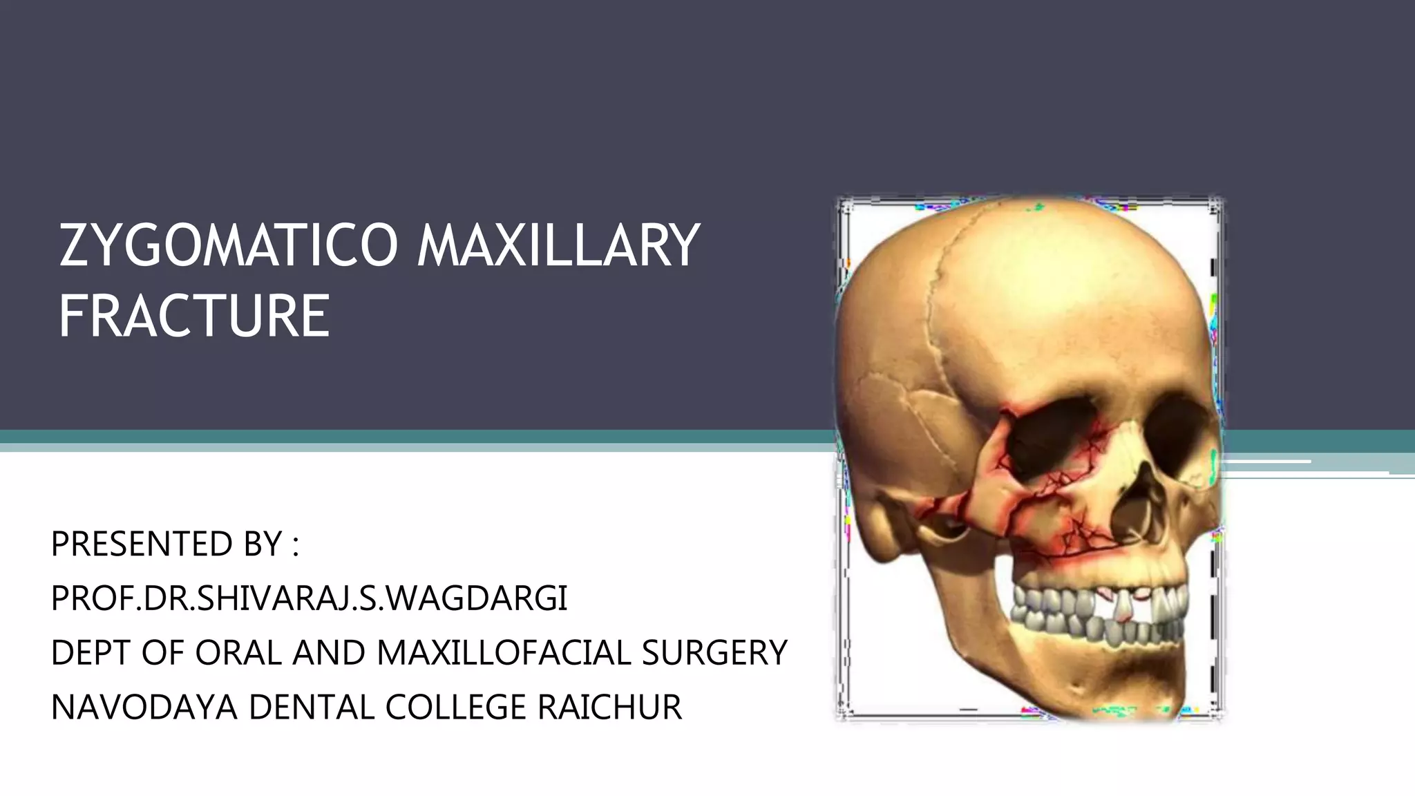
1) The document discusses zygomaticomaxillary fractures, providing classifications, examination techniques, signs and symptoms, and treatment approaches.
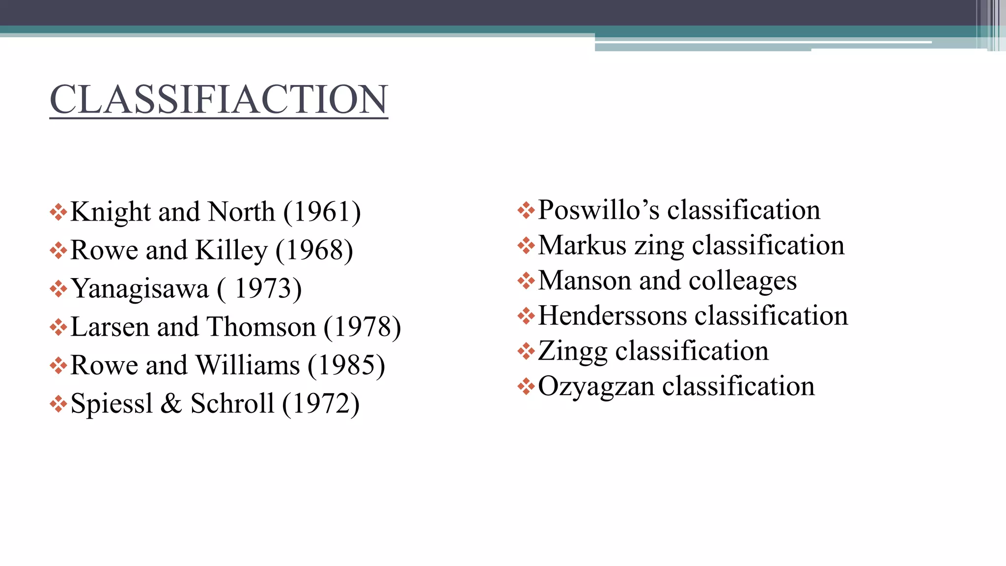
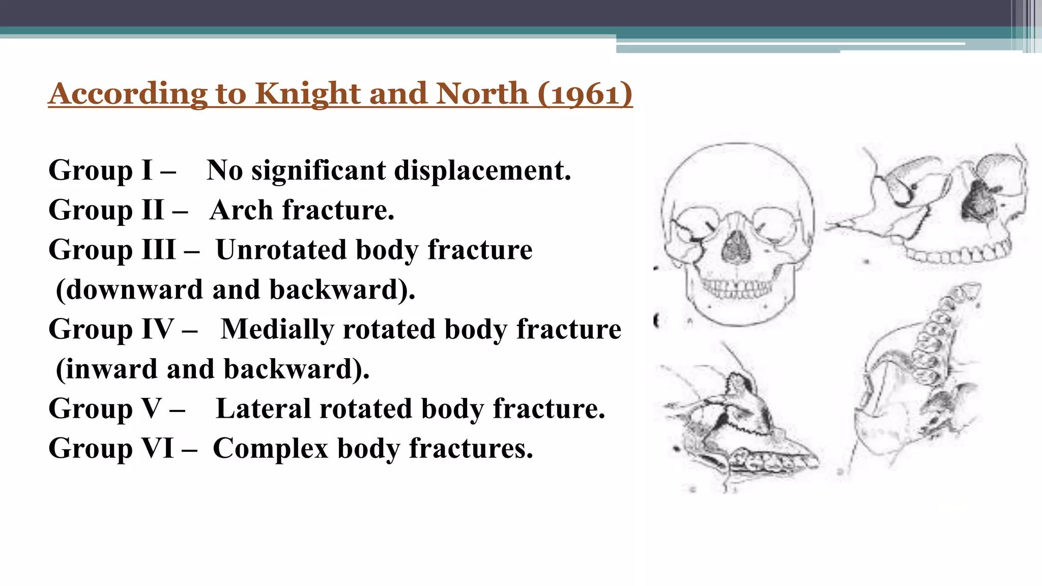
2) Common classification systems include the Knight and North system that categorizes fractures based on displacement patterns. Treatment depends on factors like displacement and stability.
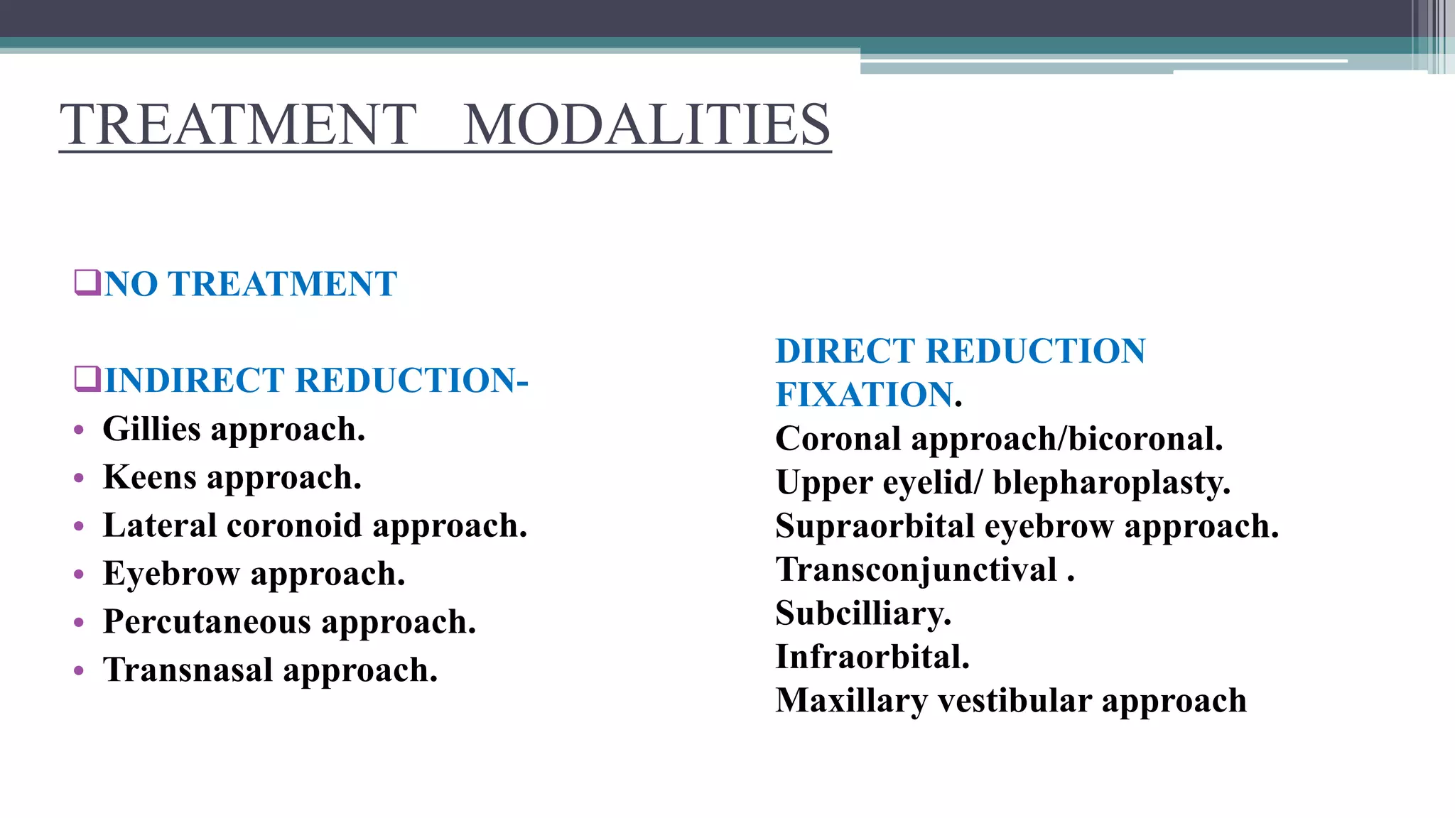
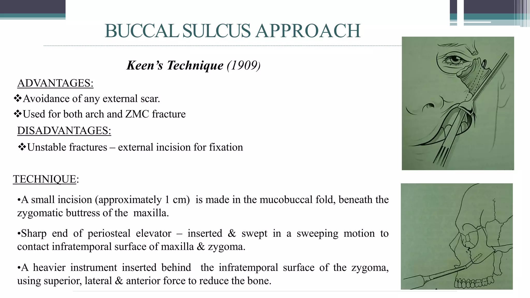
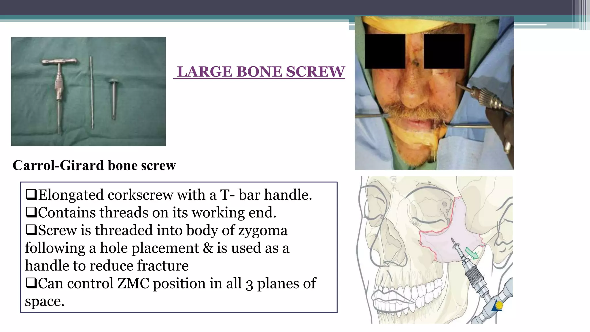
3) Approaches to treatment include no treatment for non-displaced fractures, indirect reduction using techniques like the temporal or buccal sulcus approaches, and direct open reduction using approaches like the coronal or maxillary vestibular approaches with fixation if needed.
Zygoma: Strong buttress of lateral midface lying between zygomatic process of frontal bone and maxilla.
The high incidence of zygomatic complex fracture relates to its prominent position within the facial skeleton.
Zygoma: Strong buttress of lateral midface lying between zygomatic process of frontal bone and maxilla.
The high incidence of zygomatic complex fracture relates to its prominent position within the facial skeleton.
Dr. Ahmed M. Adawy, Professor Emeritus, Dep. Oral & Maxillofacial Surgery. Former Dean, Faculty of Dental Medicine, Al-Azhar University. Mandibular angle fractures account for 23% to 42% of all facial fractures. Fracture of mandibular angle can be classified as (A) Vertical favorable or unfavorable, (B) Horizontally favorable of unfavorable. Traditionally, mandibular angle fractures have been treated with either closed reduction and inter-maxillary fixation or open reduction and internal fixation with or without inter-maxillary fixation. Patients treated with inter-maxillary fixation have a restricted airway and loose excess weight. Rigid internal fixation and early return to function have eliminated the use of wire osteosenthysis and prolonged use of inter-maxillary fixation. The principal of rigid fixation, however, have inherent set of disadvantages including damage to the inferior alveolar nerve and the marginal mandibular branch of facial nerve. Postoperative malocclusion rates are also high. With the introduction of semi-rigid technique fracture of the mandibular angle could be treated according to Champy’s Ideal lines of osteosenthysis. The technique involves placement of a single monocortial miniplate on the superior border of the mandible. However, some studies suggested using a second miniplate along the inferior border. Wether one or two miniplates should be used is still debatable. The application of 3D plates may provide additional stability in 3 dimension and good resistance against torque forces.
Dentist in pune.(BDS. MDS) - Dr. Amit T. Suryawanshi..Zygomaticomaxillary com...All Good Things
Dentist in pune. (BDS. MDS) - Dr. Amit T. Suryawanshi. Seminar-Canine Impaction.
Email ID- amitsuryawanshi999@gmail.com
Contact -Ph no.-9405622455
Subscribe our channel on youtube - https://www.youtube.com/channel/UC_gylEXTrjmEbbOTSXjuZ4Q/videos?view_as=public
Follow us on slideshare
The Indian Dental Academy is the Leader in continuing dental education , training dentists in all aspects of dentistry and
offering a wide range of dental certified courses in different formats.for more details please visit
www.indiandentalacademy.com
A seminar prepared during my omfs posting hours. Short points are added for easiness to study and bihart. Reference taken from Balaji and Neelima Anil Malik
ARTIFICIAL INTELLIGENCE IN HEALTHCARE.pdfAnujkumaranit
Artificial intelligence (AI) refers to the simulation of human intelligence processes by machines, especially computer systems. It encompasses tasks such as learning, reasoning, problem-solving, perception, and language understanding. AI technologies are revolutionizing various fields, from healthcare to finance, by enabling machines to perform tasks that typically require human intelligence.
Title: Sense of Smell
Presenter: Dr. Faiza, Assistant Professor of Physiology
Qualifications:
MBBS (Best Graduate, AIMC Lahore)
FCPS Physiology
ICMT, CHPE, DHPE (STMU)
MPH (GC University, Faisalabad)
MBA (Virtual University of Pakistan)
Learning Objectives:
Describe the primary categories of smells and the concept of odor blindness.
Explain the structure and location of the olfactory membrane and mucosa, including the types and roles of cells involved in olfaction.
Describe the pathway and mechanisms of olfactory signal transmission from the olfactory receptors to the brain.
Illustrate the biochemical cascade triggered by odorant binding to olfactory receptors, including the role of G-proteins and second messengers in generating an action potential.
Identify different types of olfactory disorders such as anosmia, hyposmia, hyperosmia, and dysosmia, including their potential causes.
Key Topics:
Olfactory Genes:
3% of the human genome accounts for olfactory genes.
400 genes for odorant receptors.
Olfactory Membrane:
Located in the superior part of the nasal cavity.
Medially: Folds downward along the superior septum.
Laterally: Folds over the superior turbinate and upper surface of the middle turbinate.
Total surface area: 5-10 square centimeters.
Olfactory Mucosa:
Olfactory Cells: Bipolar nerve cells derived from the CNS (100 million), with 4-25 olfactory cilia per cell.
Sustentacular Cells: Produce mucus and maintain ionic and molecular environment.
Basal Cells: Replace worn-out olfactory cells with an average lifespan of 1-2 months.
Bowman’s Gland: Secretes mucus.
Stimulation of Olfactory Cells:
Odorant dissolves in mucus and attaches to receptors on olfactory cilia.
Involves a cascade effect through G-proteins and second messengers, leading to depolarization and action potential generation in the olfactory nerve.
Quality of a Good Odorant:
Small (3-20 Carbon atoms), volatile, water-soluble, and lipid-soluble.
Facilitated by odorant-binding proteins in mucus.
Membrane Potential and Action Potential:
Resting membrane potential: -55mV.
Action potential frequency in the olfactory nerve increases with odorant strength.
Adaptation Towards the Sense of Smell:
Rapid adaptation within the first second, with further slow adaptation.
Psychological adaptation greater than receptor adaptation, involving feedback inhibition from the central nervous system.
Primary Sensations of Smell:
Camphoraceous, Musky, Floral, Pepperminty, Ethereal, Pungent, Putrid.
Odor Detection Threshold:
Examples: Hydrogen sulfide (0.0005 ppm), Methyl-mercaptan (0.002 ppm).
Some toxic substances are odorless at lethal concentrations.
Characteristics of Smell:
Odor blindness for single substances due to lack of appropriate receptor protein.
Behavioral and emotional influences of smell.
Transmission of Olfactory Signals:
From olfactory cells to glomeruli in the olfactory bulb, involving lateral inhibition.
Primitive, less old, and new olfactory systems with different path
263778731218 Abortion Clinic /Pills In Harare ,sisternakatoto
263778731218 Abortion Clinic /Pills In Harare ,ABORTION WOMEN’S CLINIC +27730423979 IN women clinic we believe that every woman should be able to make choices in her pregnancy. Our job is to provide compassionate care, safety,affordable and confidential services. That’s why we have won the trust from all generations of women all over the world. we use non surgical method(Abortion pills) to terminate…Dr.LISA +27730423979women Clinic is committed to providing the highest quality of obstetrical and gynecological care to women of all ages. Our dedicated staff aim to treat each patient and her health concerns with compassion and respect.Our dedicated group ABORTION WOMEN’S CLINIC +27730423979 IN women clinic we believe that every woman should be able to make choices in her pregnancy. Our job is to provide compassionate care, safety,affordable and confidential services. That’s why we have won the trust from all generations of women all over the world. we use non surgical method(Abortion pills) to terminate…Dr.LISA +27730423979women Clinic is committed to providing the highest quality of obstetrical and gynecological care to women of all ages. Our dedicated staff aim to treat each patient and her health concerns with compassion and respect.Our dedicated group of receptionists, nurses, and physicians have worked together as a teamof receptionists, nurses, and physicians have worked together as a team wwww.lisywomensclinic.co.za/
These lecture slides, by Dr Sidra Arshad, offer a quick overview of physiological basis of a normal electrocardiogram.
Learning objectives:
1. Define an electrocardiogram (ECG) and electrocardiography
2. Describe how dipoles generated by the heart produce the waveforms of the ECG
3. Describe the components of a normal electrocardiogram of a typical bipolar leads (limb II)
4. Differentiate between intervals and segments
5. Enlist some common indications for obtaining an ECG
Study Resources:
1. Chapter 11, Guyton and Hall Textbook of Medical Physiology, 14th edition
2. Chapter 9, Human Physiology - From Cells to Systems, Lauralee Sherwood, 9th edition
3. Chapter 29, Ganong’s Review of Medical Physiology, 26th edition
4. Electrocardiogram, StatPearls - https://www.ncbi.nlm.nih.gov/books/NBK549803/
5. ECG in Medical Practice by ABM Abdullah, 4th edition
6. ECG Basics, http://www.nataliescasebook.com/tag/e-c-g-basics
NVBDCP.pptx Nation vector borne disease control programSapna Thakur
NVBDCP was launched in 2003-2004 . Vector-Borne Disease: Disease that results from an infection transmitted to humans and other animals by blood-feeding arthropods, such as mosquitoes, ticks, and fleas. Examples of vector-borne diseases include Dengue fever, West Nile Virus, Lyme disease, and malaria.
New Drug Discovery and Development .....NEHA GUPTA
The "New Drug Discovery and Development" process involves the identification, design, testing, and manufacturing of novel pharmaceutical compounds with the aim of introducing new and improved treatments for various medical conditions. This comprehensive endeavor encompasses various stages, including target identification, preclinical studies, clinical trials, regulatory approval, and post-market surveillance. It involves multidisciplinary collaboration among scientists, researchers, clinicians, regulatory experts, and pharmaceutical companies to bring innovative therapies to market and address unmet medical needs.
Lung Cancer: Artificial Intelligence, Synergetics, Complex System Analysis, S...Oleg Kshivets
RESULTS: Overall life span (LS) was 2252.1±1742.5 days and cumulative 5-year survival (5YS) reached 73.2%, 10 years – 64.8%, 20 years – 42.5%. 513 LCP lived more than 5 years (LS=3124.6±1525.6 days), 148 LCP – more than 10 years (LS=5054.4±1504.1 days).199 LCP died because of LC (LS=562.7±374.5 days). 5YS of LCP after bi/lobectomies was significantly superior in comparison with LCP after pneumonectomies (78.1% vs.63.7%, P=0.00001 by log-rank test). AT significantly improved 5YS (66.3% vs. 34.8%) (P=0.00000 by log-rank test) only for LCP with N1-2. Cox modeling displayed that 5YS of LCP significantly depended on: phase transition (PT) early-invasive LC in terms of synergetics, PT N0—N12, cell ratio factors (ratio between cancer cells- CC and blood cells subpopulations), G1-3, histology, glucose, AT, blood cell circuit, prothrombin index, heparin tolerance, recalcification time (P=0.000-0.038). Neural networks, genetic algorithm selection and bootstrap simulation revealed relationships between 5YS and PT early-invasive LC (rank=1), PT N0—N12 (rank=2), thrombocytes/CC (3), erythrocytes/CC (4), eosinophils/CC (5), healthy cells/CC (6), lymphocytes/CC (7), segmented neutrophils/CC (8), stick neutrophils/CC (9), monocytes/CC (10); leucocytes/CC (11). Correct prediction of 5YS was 100% by neural networks computing (area under ROC curve=1.0; error=0.0).
CONCLUSIONS: 5YS of LCP after radical procedures significantly depended on: 1) PT early-invasive cancer; 2) PT N0--N12; 3) cell ratio factors; 4) blood cell circuit; 5) biochemical factors; 6) hemostasis system; 7) AT; 8) LC characteristics; 9) LC cell dynamics; 10) surgery type: lobectomy/pneumonectomy; 11) anthropometric data. Optimal diagnosis and treatment strategies for LC are: 1) screening and early detection of LC; 2) availability of experienced thoracic surgeons because of complexity of radical procedures; 3) aggressive en block surgery and adequate lymph node dissection for completeness; 4) precise prediction; 5) adjuvant chemoimmunoradiotherapy for LCP with unfavorable prognosis.
Flu Vaccine Alert in Bangalore Karnatakaaddon Scans
As flu season approaches, health officials in Bangalore, Karnataka, are urging residents to get their flu vaccinations. The seasonal flu, while common, can lead to severe health complications, particularly for vulnerable populations such as young children, the elderly, and those with underlying health conditions.
Dr. Vidisha Kumari, a leading epidemiologist in Bangalore, emphasizes the importance of getting vaccinated. "The flu vaccine is our best defense against the influenza virus. It not only protects individuals but also helps prevent the spread of the virus in our communities," he says.
This year, the flu season is expected to coincide with a potential increase in other respiratory illnesses. The Karnataka Health Department has launched an awareness campaign highlighting the significance of flu vaccinations. They have set up multiple vaccination centers across Bangalore, making it convenient for residents to receive their shots.
To encourage widespread vaccination, the government is also collaborating with local schools, workplaces, and community centers to facilitate vaccination drives. Special attention is being given to ensuring that the vaccine is accessible to all, including marginalized communities who may have limited access to healthcare.
Residents are reminded that the flu vaccine is safe and effective. Common side effects are mild and may include soreness at the injection site, mild fever, or muscle aches. These side effects are generally short-lived and far less severe than the flu itself.
Healthcare providers are also stressing the importance of continuing COVID-19 precautions. Wearing masks, practicing good hand hygiene, and maintaining social distancing are still crucial, especially in crowded places.
Protect yourself and your loved ones by getting vaccinated. Together, we can help keep Bangalore healthy and safe this flu season. For more information on vaccination centers and schedules, residents can visit the Karnataka Health Department’s official website or follow their social media pages.
Stay informed, stay safe, and get your flu shot today!
micro teaching on communication m.sc nursing.pdfAnurag Sharma
Microteaching is a unique model of practice teaching. It is a viable instrument for the. desired change in the teaching behavior or the behavior potential which, in specified types of real. classroom situations, tends to facilitate the achievement of specified types of objectives.
Prix Galien International 2024 Forum ProgramLevi Shapiro
June 20, 2024, Prix Galien International and Jerusalem Ethics Forum in ROME. Detailed agenda including panels:
- ADVANCES IN CARDIOLOGY: A NEW PARADIGM IS COMING
- WOMEN’S HEALTH: FERTILITY PRESERVATION
- WHAT’S NEW IN THE TREATMENT OF INFECTIOUS,
ONCOLOGICAL AND INFLAMMATORY SKIN DISEASES?
- ARTIFICIAL INTELLIGENCE AND ETHICS
- GENE THERAPY
- BEYOND BORDERS: GLOBAL INITIATIVES FOR DEMOCRATIZING LIFE SCIENCE TECHNOLOGIES AND PROMOTING ACCESS TO HEALTHCARE
- ETHICAL CHALLENGES IN LIFE SCIENCES
- Prix Galien International Awards Ceremony
Couples presenting to the infertility clinic- Do they really have infertility...Sujoy Dasgupta
Dr Sujoy Dasgupta presented the study on "Couples presenting to the infertility clinic- Do they really have infertility? – The unexplored stories of non-consummation" in the 13th Congress of the Asia Pacific Initiative on Reproduction (ASPIRE 2024) at Manila on 24 May, 2024.
3. INTRODUCTION
The zygoma or malar complex forms the central support of the cheek and is a strong
buttress of the lateral and middle third of the facial skeleton
• Zygomatic or malar fracture are the terms commonly used to described fractures that
involve the lateral one third of the middle face. Other names for this fracture are:
Zygomaticomaxillary complex
Zygomaticomaxillary compound
zygomatico orbital
Zygomatic complex
Malar
Trimalar
Tripod
4. CLASSIFIACTION
Knight and North (1961)
Rowe and Killey (1968)
Yanagisawa ( 1973)
Larsen and Thomson (1978)
Rowe and Williams (1985)
Spiessl & Schroll (1972)
Poswillo’s classification
Markus zing classification
Manson and colleages
Henderssons classification
Zingg classification
Ozyagzan classification
5. According to Knight and North (1961)
Group I – No significant displacement.
Group II – Arch fracture.
Group III – Unrotated body fracture
(downward and backward).
Group IV – Medially rotated body fracture
(inward and backward).
Group V – Lateral rotated body fracture.
Group VI – Complex body fractures.
6. According to Larsen and Thomsen (1968)
Stable fractures that shows - minimal or no displacement ( no
intervention)
Stable fracture – with displacement ( no fixation)
Unstable fracture with great displacement and disruption at
frontozygomatic suture and comminuted fractures.
Fractures of zygomatic arch are classified again into:
Minimum or no displacement.
V type in fracture.
Comminuted fracture.
7. ROWE’S AND KILLEY (1968)
Type I : No significant displacement
Type II : Fracture of the zygomatic arch
Type III : Rotation around horizontal axis
- Inward displacement of orbital rim
- Outward displacement of orbital rim
Type IV : Rotation around longitudinal axis
- Medial displacement of frontal process
- Lateral displacement of frontal process
Type V : Displacement of the complex
enbloc - Medial
-Inferior
- lateral (Rare)
Type VI : Displacement of orbitoantral
partition - Inferiorly
-Superiorly
Type VII : Displacement of orbital rim
segments
Type VIII : Complex comminuted fractures
8. ROWE & WILLIAMS CLASSIFICATION
Fractures unstable after
elevation
a)Arch only (inferiorly displaced)
b)Rotation around horizontal axis
i)medially
ii)Laterally
c)Dislocations en bloc
i) inferiorly
ii)medially
iii)Postero-laterally
Fractures stable after elevation
a)Arch only (medially displaced)
b)Rotation around the vertical axis
i)medially
ii)Laterally
9. SPIESSLAND SCHROLL (1972)
• TYPE 1- zygomatic arch fracture.
• TYPE 2- zygomatic - complex fracture with no significant displacement.
• TYPE 3- zygomatic complex fracture - partial medial displacement. (Kinking
at F-Z suture)
• TYPE 4 - Z.C with total medial displacement(complex fracture of F-Z suture).
• TYPE 5- Z.C dorsal displacement (2 fracture sites in zygomatic arch).
• TYPE 6- Z.C, inferior displacement.
• TYPE 7- Z.C, comminuted fracture.
10. YANAGISAWA(1973)
• GROUP 1- non displaced fracture- no treatment.
• GROUP 2- arch fracture(pure)
• GROUP 3- medial/lateral rotation around vertical axis.
• GROUP 4- medial/ lateral rotation around longitudinal axis.
• GROUP 5-medial/ lateral displacement without rotation.
• GROUP 6- isolated rim fracture.
• GROUP 7- all complex fracture.
11. EBERHARD KRUGER(1986)
FRACTURE OF ZYGOMA
• No displacement.
• Partial medial
• Total medial
• Dorsal displacement.
• Inferior displacement.
• Comminuted fracture.
12. MANSON& COLLEAGUES(1990)
Based on
- Amount of energy dissipated by the facial bones.
- Secondary to the traumatic force.
- Findings in CT scans
High energy fractures Lower energy fractures
• Extreme displacement. Displacement
• Comminution of articulations. NIL
• Segmentation of bones.
Treatment- Treatment-
• Require extensive exposure Less agressive
and Aggressive fixation.
13. ANATOMY
PRINCIPAL STRUCTURE OF MIDFACE
MAJOR ROLE IN FACIAL CONTOUR
4 PROCESSES -ORBITAL
-FRONTAL
-TEMPORAL
-MAXILLARY
4 ARTICULATIONS -FRONTAL
-TEMPORAL
-MAXILLARY
-SPHENOID
MUSCLE ATTACHMENTS - MASSETER
-TEMPORAL FASCIA
16. FRACTURE PATTERNS
• Depends on- DIRECTION & MAGNITUDE OF FORCE
• Fracture lines pass through lines of greatest weakness
• Malar bone represent strong bone on fragile suppport
• Force appiled on zygoma - distributed through four processes
to adjacent articulating bone –weaker than zygoma
• Three lines of fracture extend from IOF
Anteromedial – orbital floor
Superolateral -
Inferior direction
17. Clinical examination
1.NEUROLOGICAL STATUS.
• First step is to assess neurological
status…….
• Associated neurologic injury was
encountered in 57% of patients.
2.VISUAL STATUS
3.EXAMINATION OF ZYGOMA
18. INSPECTION
Symmetry of arches.
Pupillary level.
Presence of orbital oedema.
Sub-conjunctival ecchymosis.
Ant & lat projection of zygoma.
Intra-oral.
• PALPATION
-Systematic & thorough.
-Compare one side with another.
19. SIGNS AND SYMPTOMS
• Periorbital ecchymosis and edema.
• Flattening of the malar prominence.
• Flattening of the zygomatic arch.
• Pain.
• Ecchymosis of the maxillary buccal sulcus.
• Deformity of the zygomatic buttress of the maxilla.
• Deformity of the infra-orbital margin.
• Trismus.
• Abnormal nerve sensibility.
• Epistasis.
• Subconjunctival ecchymosis.
• Crepitation from air emphysema.
• Displacement of palpebral fissure.
• Diplopia.
• Enopthalmos.
20. Orbital signs and symptoms
Periorbital
Oedema
Circumorbital
ecchymosis
Subconjunctival
haemorrhage
Orbital
Emphysema Diplopia
Unequal
pupillary
levels
21. Flattening of malar prominence
• Characteristic sign
• Presence of edema flattening may be difficult to discern
Flattening of zygomatic arch
• Loss of normal convex curvature
23. ORBITAL EXAMINATION
FORCED DUCTION TEST
• To determine presence of
mechanical obstruction to
globe movement.
• Test done to assess the cause
of reduced movement of
extraocular muscles
following trauma to mid-face
region.
• It helps to determine if the
cause is mechanical/
neurological.
DIPLOPIA
24.
25. RADIOGRAPHIC EXAMINATION
Postero-anterior oblique view (OM/PNS view): excellent assessment of
sinuses and their walls, zygoma and its processes and rims of orbit
Submentovertex view is specific for zygomatic arch fractures
28. STEPS IN TREATING ZMC FRACTURE
1. Prophylactic antibiotics.
2. Anesthesia.
3. Clinical examination and forced duction test.
4. Protection of the globe.
5. Antiseptic preparation.
6. Reduction of the fracture.
7. Assessment of reduction.
8. Determination of necessity for fixation.
29. 9. Application of fixation device.
10. Assessment of ocular motility.
11. Bone graft for extra orbital osseous defects.
12. Post surgical ocular examinations.
13. Post surgical images.
30. NEED FOR FIXATION
Indications for fixation
1. Comminuted fracture fragments.
2. Doubt regarding the stability
Role of MASSETER in displacement:
• Albright and McFarland recommended IMF following fracture reduction
helps to reduce the pull of the masseter muscle on the repositioned ZMC.
• Ellis et al reviewed series of isolated ZMC fractures treated by different
approaches and fixation schemes and found no evidence of post reduction
instability
33. NO TREATMENT
NON-DISPLACED FRACTURES-9-50 % OF ZMC #
MEDICAL CONTRAINDICATIONS
MINIMAL DEGREE OF DISPLACEMENT.
UNLIKELY TO RESULT IN
COSMETIC DEFORMITY
DISTURBANCE OF VISION
PARESTHESIA
IMPAIRED MANDIBULAR MOVEMENTS.
34. DECISION TO INTERVENE
DEPENDS ON
SIGNS & SYMPTOMS.
FUNCTIONAL IMPAIRMENT.
PRESENCE OF OPHTHALMIC INJURIES.
PROGRESSIVE PROPTOSIS.
VISUAL ACUITY DETERIORATION.
VISUAL INTEGRITY ON UNAFFECTED SITE.
OTHER RELATED FACIAL INJURIES.
MEDICAL CONDITION OF PATIENT.
35. DELAYED TREATMENT
1. MANIPULATION OF ORBITAL BONES
2. RISK OF EYEBALL INJURY.
3. EXISTING HAEMORRGHAGE.
4. PROGRESSIVE PROPTOSIS.
5. PRE-EXISTING BLINDNESS IN OTHER EYE.
6. GENERAL MEDICAL CONDITION.
7. NEUROLOGICAL STATUS QUESTIONABLE.
8. GROSS OEDEMA.
9. CRITICAL PERIOD - 5-10th DAY
36. DELAYED TREATMENT
ADVANTAGES
OEDEMA RESOLVES- DETAILED EXAMINATION OF
EYE.
ANTRUM CLEARS - BETTER RADIOGRAPH.
HAEMATOMA STILL NOT ORGANISED –DISSECTION
EASIER.
>5-10 DAYS
DIFFICULTY IN DISIMPACTING & REDUCING.
PHYSIOLOGICAL RESORPTION OF # MARGINS.
INTERDIGITATION LESS ACCURATE.
INELASTICITY OF FIBROUS TISSUE AT SITE OF
MALUNION.
37. IMMEDIATE TREATMENT
• IN THE ABSENCE OF COMPLICATIONS.
• NO OEDEMA.
ADVANTAGES
EXCELLENT REDUCTION -NOT COMPROMISED
NO SOFT TISSUE SCARRING
NO CHANGE IN MORPHOLOGY
SUPERIOR FACIAL SOFT TISSUE CONTOUR.
39. TEMPORAL APPROACH
Popular approach
First described by Gillies & coworkers in 1927 – arch fractures
ADVANTAGES :
Allows application of greater amount of controlled force to
disimpact even the most difficult zygomatic fracture.
For treatment of fractures which are consolidated already
Quick and simple method
DISADVANTAGE:
Encountered temporal vessels- hemorrhage
40. Incision – 2.5cm, through skin & subcutaneous tissue
White glistening surface Temporalis muscle bulge
42. BUCCALSULCUS APPROACH
Keen’s Technique (1909)
ADVANTAGES:
Avoidance of any external scar.
Used for both arch and ZMC fracture
DISADVANTAGES:
Unstable fractures – external incision for fixation
TECHNIQUE:
•A small incision (approximately 1 cm) is made in the mucobuccal fold, beneath the
zygomatic buttress of the maxilla.
•Sharp end of periosteal elevator – inserted & swept in a sweeping motion to
contact infratemporal surface of maxilla & zygoma.
•A heavier instrument inserted behind the infratemporal surface of the zygoma,
using superior, lateral & anterior force to reduce the bone.
43. LATERAL CORONOID APPROACH
Quinnin1977–described
Usedonlyforarchfractures
TECHNIQUE :
•3 to 4 cm incision -anterior border of the ramus.
•Insision made - to the depth of the temporal muscle
insertion (not to the bone)
•Instrument between the temporal muscle and the
zygomatic arch - readily palpable.
•A flat-bladed instrument- inserted into the pocket&
arch is elevated while palpating the arch extraorally.
44. ELEVATION FROM EYEBROWAPPROACH
•Used for both ZMC & arch fracture
ADVANTAGE:
•Fracture at the orbital rim is visualized
directly
•Fixation can be done with same incision.
DISADVANTAGE:
•Difficult to generate a large amount of force,
especially in the superior direction.
DINGMAN ZYGOMATIC
ELEVATOR
45. PERCUTANEOUS APPROACH
•Most simple of all techniques as no soft tissue dissection is necessary
•Direct route to elevate the depressed zygoma is through the skin
surface of the face overlying the zygoma.
ADVANTAGE:
Produces forces anteriorly, laterally, and superiorly in a very direct
manner
DISADVANTAGE:
Scar on the face in a very noticeable location.
46. ELEVATION OF THE ZYGOMA WITH A
BONE HOOK
•Strohmeyer in 1844
•Poswillo`s
intersecting lines.
•Stab incision made
and hook inserted.
•Apply strong traction.
Stacey bone hook
47. Carrol-Girard bone screw
Elongated corkscrew with a T- bar handle.
Contains threads on its working end.
Screw is threaded into body of zygoma
following a hole placement & is used as a
handle to reduce fracture
Can control ZMC position in all 3 planes of
space.
LARGE BONE SCREW
49. MAXILLARY VESTIBULAR APPROACH
• Most useful when performing any of
wide variety of procedures in the
midface.
• Safe access to the entire facial surface
of the midfacial skeleton- from the
zygomatic arch to the infraorbital rim
to the frontal process of the maxilla.
ADVANTAGE:
• Hidden intraoral scar.
• Rapid and simple approach
• Complications are few.
50. TECHNIQUE
Subperiosteal Dissection
V –Y Closure
Incision through the mucosa, submucosa, facial
musculature,and periosteum (3-5mm superior to
mucogingival junction)
Infraorbital rim, anterior
maxilla & ZMC Buttress -
treated
51. LATERAL BROW APPROACH
Popular approach - Access to the lateral
orbital rim and the frontozygomatic suture
ADVANTAGE:
Simple, safe and rapid approach
Scar is usually hidden within the confines of
the eyebrow
No important neurovascular structures are
involved in this approach.
52. DISADVANTAGE :
In individual who has no eyebrows extending laterally and
inferiorly along the orbital margin, this approach is undesirable.
Incisions made along the lateral orbital rim outside of the
eyebrow are very conspicuous in such individuals, and another
type of incision may be indicated.
Extremely limited access.
53. TECHNIQUE
•Incision – parallel to the eyebrow hair.
•The incision is made through skin and subcutaneous tissue
to the level of the periosteum in one stroke. (2cm)
•Incision through periosteum along lateral orbital rim and
subperiosteal dissection into lacrimal fossa.
•Because of the concavity just behind the orbital rim in this area,
the periosteal elevator is oriented laterally as dissection proceeds
posteriorly.
Closure:
The incision is closed in two layers, the periosteum and the skin.
54. UPPER EYELID APPROACH
The upper eyelid approach to the superolateral orbital rim is also called UPPER
BLEPHAROPLASTY, UPPER EYELID CREASE, & SUPRATARSAL FOLD
APPROACH.
In this approach, a natural skin crease in the upper eyelid is used to make the
incision.
Advantage: ADVANTAGE:
Inconspicuous scar it creates - best
approaches to the region.
55. TECHNIQUE
Closure:The wound is closed in two layers, periosteum and skin/muscle.
To facilitate retraction of the skin/muscle flap, it can be widely undermined
laterally and retracted with small retractors.
Periosteum is divided 2-3mm posterior to the orbital rim with a scalpel.
Sagittal section through orbit and globe showing dissection
between orbicularis oculi muscle and the orbital septum
Incision – begins 10mm superior to upper lid margin
& lies 6mm above lateral canthus as it extends laterally.
The initial incision is made through skin and muscle.
56. LOWER EYELID APPROACH
SUBTARSAL approach:
• Frequently employed – access to infraorbital rim & orbital floor
• Along natural skin crease at/below the level of tarsus, approx half
distance between lash margin and orbital rim
ADVANTAGE:
• Relatively easy
• Scar is imperceptible
• Minimal complication
57. SUBCILIARY approach:
• Also called INFRACILIARY APPROACH/ BLEPHAROPLASTY
• Incision is - approx 2 mm below the eyelashes and can be extended laterally as necessary
(top dashed line) along the natural crease inferior to lateral canthal ligament.
ADVANTAGE:
• Imperceptible scar
• Simultaneously exposes infraorbital
& F-Z areas.
DISADVANTAGE:
• Technically difficult
• Risk of postoperative ectropion
58. TECHNIQUE
•Skin incision
•Dissect b/w skin & muscle
until orbital rim is reached
•2nd incision – through muscle
& periosteum to bone
•Incision – skin & muscle at
same level
•Dissect down anterior to
orbital septum to orbital rim
•Combination of these – STEPPED
incision
•Subcutaneous dissection toward
rim – few mm
•Followed by muscle incision at
lower level & then following orbital
septum to rim.
•Fine scissors are used during dissection –spreading motion.
•Incision through periosteum – 3-4mm below rim
59. TRANSCONJUNCTIVALAPPROACH
Originally described by Bourguet in 1928.
Also called INFERIOR FORNIX APPROACH.
2 types:
Preseptal (Tessier)
Retroseptal (Tenzel&Miller) approaches.
Converse & colleagues added a lateral canthotomy to
transconjunctival retroseptal incision for improved lateral
exposure.
61. TECHNIQUE
Sagital section through orbit showing preseptal and
retroseptal placement of incision.
Initial incision for lateral canthotomy
Initial canthopexy incision to dissect in the
subconjunctival plane. The dissection should be just
below the tarsal plate and extend no farther medially
than the lacrimal punctum.
Closure of transconjunctival incision and inferior
canthopexy
62. ADVANTAGE :
Produce excellent cosmetic results because the scar is
hidden in the conjunctiva.
If a canthotomy is performed in conjunction with the
approach, the only visible scar is the lateral extension,
which heals with an inconspicuous scar.
Rapid and no skin or muscle dissection is necessary.
DISADVANTAGE :
• medial extent of the incision is limited by the lacrimal
drainage system.
63. CORONALAPPROACH
The coronal or bi-frontal incision is a
versatile surgical approach to the upper
and middle regions of the facial skeleton,
including the zygomatic arch
ADVANTAGE:
•The surgical scar is hidden within the hairline.
•Excellent acess to orbits, zygoma & arch.
64. TECHNIQUE
Incision placement:
•Hairline of patient
•Amount of inferior access
•Incision – skin, subcutaneous tissue & galea
•Lateral aspect of skull- superficial layer of temporal fascia incised 2cm supr
to zygomatic arch
•Dissect inferiorly - zygomatic arch
•Periosteal incision – along the superior aspect of arch
•Pericranium is incised along lateral orbital rim – FZ suture
•Wound is closed in layers – fascia , galea, skin.
66. MODIFICATION OF HEMICORONAL
APPROACH
•The anterior arm of the incision is
curved downward toward the superior
wall of the orbit before it reaches the
vertex of the skull within the hairline.
•The ‘backcut’ provides excellent
exposure of the entire zygomatic
complex and the arch.
• Aesthetic and is less invasive thereby
being quite acceptable by patients.
Journal of Maxillofacial and Oral Surgery 2010
Volume 9, Number 3, 270-272
67. Common methods include
wire osteosynthesis and
rigid fixation by plates
Less common methods include
external pin fixation and
maxillary antral support
IMMOBILIZATION
68. PIN FIXATION
External pin fixation
Can be used for fractures that demonstrate
an intact body of the zygoma but severe
communition at the junction with the
surrounding bones
Internal pin fixation
Was introduced by Fryer and results in
stable entity and relatively free of
complications
Techniques make use of K-wire placement
69. SINUS PACKING SUPPORT
Gauze or balloon can be used to
provide inferior support to the
zygoma
Lateral wall is approached
through a Caldwell-Luc
½ inch gauze dipped in antibiotic
of choice is placed along the
floor anteroposteriorly
Antral balloon can be used by it is
relatively imprecise and cannot
adapt to the topography
71. INDIRECT FIXATION
• Zygomatic bone - rigidly secured to facial skeleton.
INDICATION
• Gross loss of bone in FZ region and infra orbital rim.
ZYGOMATICO-ZYGOMATIC
NASO-ZYGOMATIC
ZYGOMATICO-PALATAL
MAXILLO-ZYGOMATIC
FRONTO-ZYGOMATIC
CRANIO-ZYGOMATIC
73. EXTERNAL FIXATION
Accomplished with wires suspended from plaster head caps,
head frames and by pins connected to one another with
universal joints and cold cure acrylic.
ADVANTAGES-
1. Three dimensional stability.
2. Minimal scarring.
3. Adjustability of the reduction.
DISADVANTAGES-
1. Patient comfort is compromised.
2. Need for specific hardware.
3. Lack of usefulness in comminuted fractures.
75. FIXATION TECHNIQUES - PRINCIPLES
1. Use self-threading bone screws.
2. Use hardware that will not scatter postoperative CT scans.
3. Place at least two screws through the plate on each side of the fracture.
4. Avoid important anatomic structures.
•Use Y,L,T shaped plates where fracture line
in the zmc buttress region is low.
•Prevents damage to the roots and nerve
bundle.
76. 5. Use a thin plate as possible in the periorbital areas.
6. Place as many bone plates in as many locations as
necessary for ensuring stability.
7. If concomitant fractures of other midfacial bones exist,
it will be necessary to apply fixation devices more
liberally.
8. In areas of comminution or bone loss, span the gap
with the bone plate.
77. BONE PLATES
FOUR POINT FIXATION-
COMMINUTED ZMC FRACTURES
SITES OF FIXATION-
1. F-Z SUTURE.
2. INFRAORBITAL RIM.
3. ZYGOMATIC ARCH.
4. MAXILLARY BUTTRESS.
78. THREE POINT FIXATION-
NON-COMMINUTED ZMC FRACTURES
SITES OF FIXATION-
•F-Z SUTURE
• INFRAORBITAL RIM
• ZYGOMATIC ARCH
(OR)
MAXILLARY BUTTRESS.
79. TWO POINT FIXATION-
SIMPLE NON-COMMINUTED ZMC
FRACTURES
SITES OF FIXATION-
F-Z SUTURE
INFRAORBITAL RIM
OR
MAXILLARY BUTTRESS
83. Complications of periorbital incision
1. Minor - dehiscence
hematoma /seroma
lymphedema
2. Vertical shortening of lower lid
prevention - superior support of lower lid for several days( best
achieved with frost sutures).
3. Ectropion – associated with subciliary incision and
trans conjunctival incision(mild /moderate/severe)
4. Entropion – inward curl of lower eyelid
-occurs less commonly but more
distressing (subciliary incision )
84. 5.Infraorbital nerve injury
Either direct injury to nerve due to trauma or
iatrogenic
Mostly these injuries are temporary(neuropraxia) due to
stretching or compression of infraorbital nerve.
Markedly displaced fractures - neurotmesis can occur
Patient may complain of numbness , different
sensation and pain on heat /cold / light touch .
ZMC fracture which are treated with rigid fixation
– early recovery of neurosensory deficit .
85. 6.Persistent Diplopia
Binocular diplopia initially present with ZMC fracture should
resolve within 5-7 days after fracture treatment
Result of
edema or hematoma of one or more
extraocular muscles or their nerves
Introrbital edema
Ocassionally muscle entrapment
If persists, it may be due to scar contracture and adhesions either
within the ocular muscles or between them.
86. 7.Enopthalmos
Most commonly caused by increased volume of orbit
Difficult to correct secondarily, however
improvement is possible.
Surgery can be done to reduce orbital volume by
– reconstructing the internal orbit
- by placing a space occupying material behind
the globe
( glass beads , silicon sheets , sponges , teflon beads ,
cartilage graft, hydroxyapatite, metallic mesh or plate)
87. 8.Blindness
Occasionally reported after ZMC fracture
Causes- direct damage to optic nerve
- hemorrhage into optic sheath
- intraocular edema
- retrobulbar hemorrhage
9.Maxillary sinusitis
Caused by inflammation of sinus membrane and occlusion of
ostium.
Usually respond to antibiotic and decongestant therapy.
88. 10.Ankylosis of zygoma to coronoid process
- very rare
- when noted usually fibrous.
Causes-
- improper reduction of zygoma leaving arch in close
proximity to coronoid process.
- untreated zygomatic fracture
- post-operative infection
89. 11. Malunion of the zygoma
Signs and symptoms –
•Flattening of malar prominence
• Enopthalmos
•Altered pupillary level
• Limitation of mandibular movements
Treatment:
- camouflaging the defect with implant or transplant
- repositioning of malpositioned bone
90. CONCLUSION
Surgical intervention is an effective treatment modality
of depressed ZMC fracture, whereas a nonsurgical
approach is often used for nondisplaced fractures.
Most of them can be treated solely by an intraoral
approach & rigid fixation at ZM buttress.
Further exposure of FZ junction or infra orbital rim is
necessary for severely displaced fractures, requiring
additional fixation.
91. REFERENCES
• ORAL AND MAXILLOFACIAL TRAUMA-FONSECA
• PETERSON’S PRINCIPLES OF ORAL AND MAXILLOFACIAL SURGERY
• ROWE AND WILLIAMS MAXILLOFACIAL TRAUMATOLOGY
• TEXTBOOK OF ORAL AND MAXILLOFACIAL SURGERY