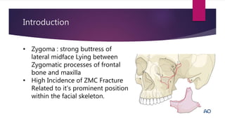
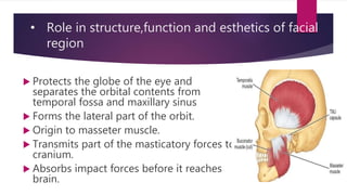
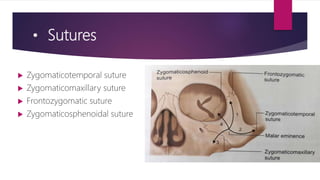
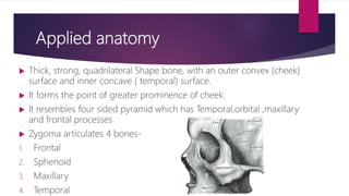
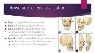
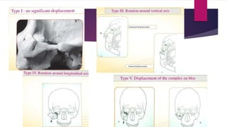
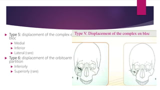
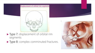
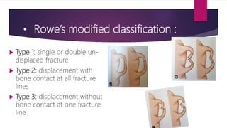
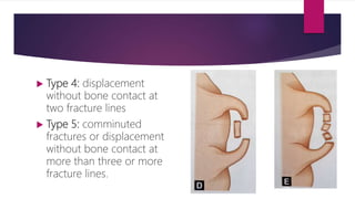
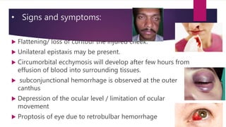
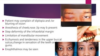
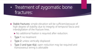
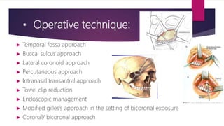
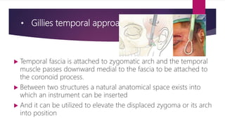
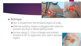
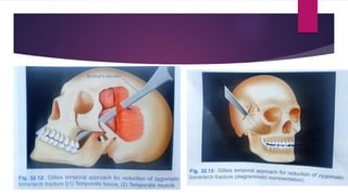
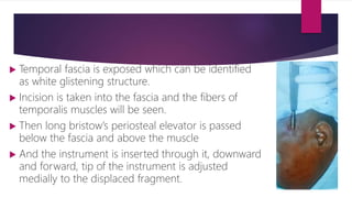
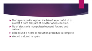
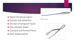
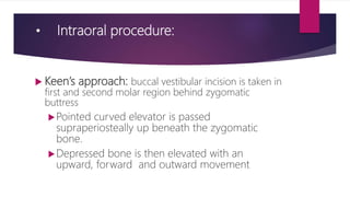
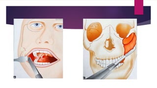
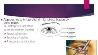
Fractures of zygomatic bone
Dr Deepak Kumar
Introduction
• Zygoma : strong buttress of
lateral midface Lying between
Zygomatic processes of frontal
bone and maxilla
• High Incidence of ZMC Fracture
Related to it’s prominent position
within the facial skeleton.
• Role in structure,function and esthetics of facial
region
 Protects the globe of the eye and
separates the orbital contents from
temporal fossa and maxillary sinus
 Forms the lateral part of the orbit.
 Origin to masseter muscle.
 Transmits part of the masticatory forces to
cranium.
 Absorbs impact forces before it reaches
brain.
• Sutures
 Zygomaticotemporal suture
 Zygomaticomaxillary suture
 Frontozygomatic suture
 Zygomaticosphenoidal suture
Applied anatomy
 Thick, strong, quadrilateral Shape bone, with an outer convex (cheek)
surface and inner concave ( temporal) surface.
 It forms the point of greater prominence of cheek.
 It resembles four sided pyramid which has Temporal,orbital ,maxillary
and frontal processes
 Zygoma articulates 4 bones-
1. Frontal
2. Sphenoid
3. Maxillary
4. Temporal
Rowe and killey classification:
 Type 1: no significant displacement
 Type 2: fractures of zygomatic arch
 Type 3: rotation around vertical axis
 Inward displacement of orbital rim
 Outward displacement of orbital rim
 Type 4: rotation around the longitudinal axis
 Medial displacement of the frontal process
 Lateral displacement of frontal process
 Type 5: displacement of the complex en
bloc
 Medial
 Inferior
 Lateral (rare)
 Type 6: displacement of the orbitoantral
partition
 Inferiorly
 Superiorly (rare)
 Type 7: displacement of orbital rim
segments
 Type 8: complex comminuted fractures.
• Rowe’s modified classification :
 Type 1: single or double un-
displaced fracture
 Type 2: displacement with
bone contact at all fracture
lines
 Type 3: displacement without
bone contact at one fracture
line
 Type 4: displacement
without bone contact at
two fracture lines
 Type 5: comminuted
fractures or displacement
without bone contact at
more than three or more
fracture lines.
• Signs and symptoms:
 Flattening/ loss of contour the injured cheek.
 Unilateral epistaxis may be present.
 Circumorbital ecchymosis will develop after few hours from
effusion of blood into surrounding tissues.
 subconjunctional hemorrhage is observed at the outer
canthus
 Depression of the ocular level / limitation of ocular
movement
 Proptosis of eye due to retrobulbar hemorrhage
 Patient may complain of diplopia and /or
blurring of vision
 Anesthesia of cheek,nose ,lip may b present
 Step deformity of the infraorbital margin
 Limitation of mandibular movement
 Ecchymosis and tenderness in the upper buccal
sulcus,change in sensation of the teeth and
gums.
 Enophthalmos may be seen
• Treatment of zygomatic bone
fractures:
 Stable fractures: simple elevation will be sufficient,because of
high degree of stability due to integrity of temporal fascia and
interdigitation of the fracture lines.
 No additional fixation is required after reduction.
1. Type 1: no treatment
2. Type 2: unless vertically displaced
3. Type 3 and type 4(a): open reduction may be required and
transosseous wiring is advisable
 Unstable fractures: require open reduction and transosseous
wiring or bone plating .
 Type 4(b)
 Type 5,6 and 7,8
• Operative technique:
 Temporal fossa approach
 Buccal sulcus approach
 Lateral coronoid approach
 Percutaneous approach
 Intranasal transantral approach
 Towel clip reduction
 Endoscopic management
 Modified gilles’s approach in the setting of bicoronal exposure
 Coronal/ bicoronal approach
• Gillies temporal approach:
 Temporal fascia is attached to zygomatic arch and the temporal
muscle passes downward medial to the fascia to be attached to
the coronoid process.
 Between two structures a natural anatomical space exists into
which an instrument can be inserted
 And it can be utilized to elevate the displaced zygoma or its arch
into position
 Technique:
 Hair is shaved from the temporal region of scalp.
 External auditory meatus is plugged with cotton to
prevent any fluid or blood getting inside.
 Incision about 2- 2.5cm in length and inclined
forward at 45° to zygomatic arch, well in temporal
region.
 Temporal fascia is exposed which can be identified
as white glistening structure.
 Incision is taken into the fascia and the fibers of
temporalis muscles will be seen.
 Then long bristow’s periosteal elevator is passed
below the fascia and above the muscle
 And the instrument is inserted through it, downward
and forward, tip of the instrument is adjusted
medially to the displaced fragment.
 Thick gauze pad is kept on the lateral aspect of skull to
protect it from pressure of elevator while reduction.
 Tip of elevator is manipulated upward, forward and
outward
 Snap sound is heard as reduction procedure is complete
 Wound is closed in layers
 Shave Temporal region
 Incision and dissection
 Incision of temporal Fascia
 Pass elevator blade
 Outward and forward force
 And closed wound
• Intraoral procedure:
 Keen’s approach: buccal vestibular incision is taken in
first and second molar region behind zygomatic
buttress
Pointed curved elevator is passed
supraperiosteally up beneath the zygomatic
bone.
Depressed bone is then elevated with an
upward, forward and outward movement
 Approaches to infraorbital rim for direct fixation by
bone plates:
 Existing skin laceration
 Infraorbital rim incision
 Subtarsal incision
 Subciliary incision
 Transconjuctival incision
Thank you

fractures of zygomatic bone

  • 1.
    Fractures of zygomaticbone Dr Deepak Kumar
  • 2.
    Introduction • Zygoma :strong buttress of lateral midface Lying between Zygomatic processes of frontal bone and maxilla • High Incidence of ZMC Fracture Related to it’s prominent position within the facial skeleton.
  • 3.
    • Role instructure,function and esthetics of facial region  Protects the globe of the eye and separates the orbital contents from temporal fossa and maxillary sinus  Forms the lateral part of the orbit.  Origin to masseter muscle.  Transmits part of the masticatory forces to cranium.  Absorbs impact forces before it reaches brain.
  • 4.
    • Sutures  Zygomaticotemporalsuture  Zygomaticomaxillary suture  Frontozygomatic suture  Zygomaticosphenoidal suture
  • 5.
    Applied anatomy  Thick,strong, quadrilateral Shape bone, with an outer convex (cheek) surface and inner concave ( temporal) surface.  It forms the point of greater prominence of cheek.  It resembles four sided pyramid which has Temporal,orbital ,maxillary and frontal processes  Zygoma articulates 4 bones- 1. Frontal 2. Sphenoid 3. Maxillary 4. Temporal
  • 6.
    Rowe and killeyclassification:  Type 1: no significant displacement  Type 2: fractures of zygomatic arch  Type 3: rotation around vertical axis  Inward displacement of orbital rim  Outward displacement of orbital rim  Type 4: rotation around the longitudinal axis  Medial displacement of the frontal process  Lateral displacement of frontal process
  • 8.
     Type 5:displacement of the complex en bloc  Medial  Inferior  Lateral (rare)  Type 6: displacement of the orbitoantral partition  Inferiorly  Superiorly (rare)
  • 9.
     Type 7:displacement of orbital rim segments  Type 8: complex comminuted fractures.
  • 10.
    • Rowe’s modifiedclassification :  Type 1: single or double un- displaced fracture  Type 2: displacement with bone contact at all fracture lines  Type 3: displacement without bone contact at one fracture line
  • 11.
     Type 4:displacement without bone contact at two fracture lines  Type 5: comminuted fractures or displacement without bone contact at more than three or more fracture lines.
  • 12.
    • Signs andsymptoms:  Flattening/ loss of contour the injured cheek.  Unilateral epistaxis may be present.  Circumorbital ecchymosis will develop after few hours from effusion of blood into surrounding tissues.  subconjunctional hemorrhage is observed at the outer canthus  Depression of the ocular level / limitation of ocular movement  Proptosis of eye due to retrobulbar hemorrhage
  • 13.
     Patient maycomplain of diplopia and /or blurring of vision  Anesthesia of cheek,nose ,lip may b present  Step deformity of the infraorbital margin  Limitation of mandibular movement  Ecchymosis and tenderness in the upper buccal sulcus,change in sensation of the teeth and gums.  Enophthalmos may be seen
  • 14.
    • Treatment ofzygomatic bone fractures:  Stable fractures: simple elevation will be sufficient,because of high degree of stability due to integrity of temporal fascia and interdigitation of the fracture lines.  No additional fixation is required after reduction. 1. Type 1: no treatment 2. Type 2: unless vertically displaced 3. Type 3 and type 4(a): open reduction may be required and transosseous wiring is advisable
  • 15.
     Unstable fractures:require open reduction and transosseous wiring or bone plating .  Type 4(b)  Type 5,6 and 7,8
  • 16.
    • Operative technique: Temporal fossa approach  Buccal sulcus approach  Lateral coronoid approach  Percutaneous approach  Intranasal transantral approach  Towel clip reduction  Endoscopic management  Modified gilles’s approach in the setting of bicoronal exposure  Coronal/ bicoronal approach
  • 17.
    • Gillies temporalapproach:  Temporal fascia is attached to zygomatic arch and the temporal muscle passes downward medial to the fascia to be attached to the coronoid process.  Between two structures a natural anatomical space exists into which an instrument can be inserted  And it can be utilized to elevate the displaced zygoma or its arch into position
  • 18.
     Technique:  Hairis shaved from the temporal region of scalp.  External auditory meatus is plugged with cotton to prevent any fluid or blood getting inside.  Incision about 2- 2.5cm in length and inclined forward at 45° to zygomatic arch, well in temporal region.
  • 20.
     Temporal fasciais exposed which can be identified as white glistening structure.  Incision is taken into the fascia and the fibers of temporalis muscles will be seen.  Then long bristow’s periosteal elevator is passed below the fascia and above the muscle  And the instrument is inserted through it, downward and forward, tip of the instrument is adjusted medially to the displaced fragment.
  • 21.
     Thick gauzepad is kept on the lateral aspect of skull to protect it from pressure of elevator while reduction.  Tip of elevator is manipulated upward, forward and outward  Snap sound is heard as reduction procedure is complete  Wound is closed in layers
  • 22.
     Shave Temporalregion  Incision and dissection  Incision of temporal Fascia  Pass elevator blade  Outward and forward force  And closed wound
  • 23.
    • Intraoral procedure: Keen’s approach: buccal vestibular incision is taken in first and second molar region behind zygomatic buttress Pointed curved elevator is passed supraperiosteally up beneath the zygomatic bone. Depressed bone is then elevated with an upward, forward and outward movement
  • 25.
     Approaches toinfraorbital rim for direct fixation by bone plates:  Existing skin laceration  Infraorbital rim incision  Subtarsal incision  Subciliary incision  Transconjuctival incision
  • 26.