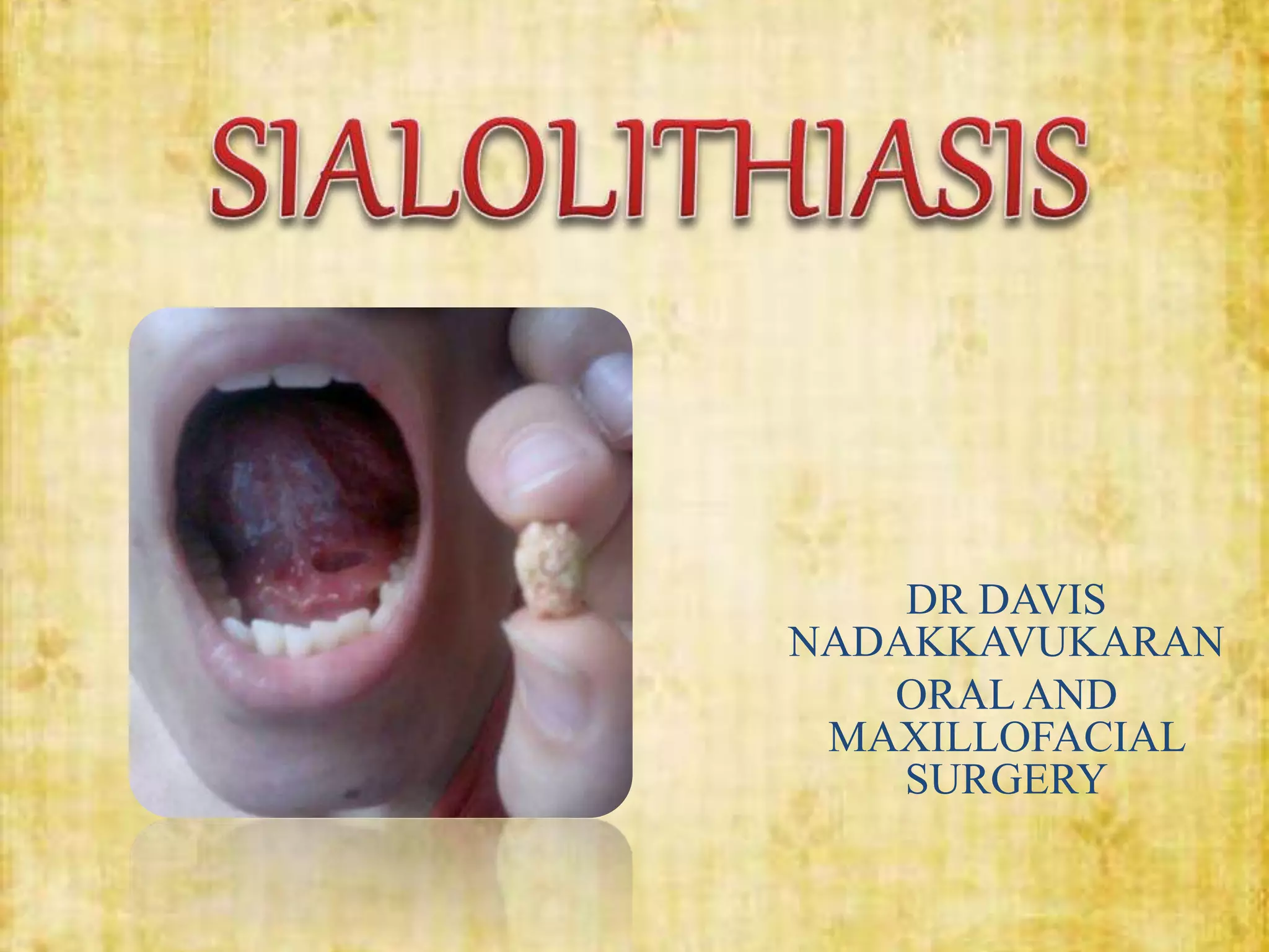
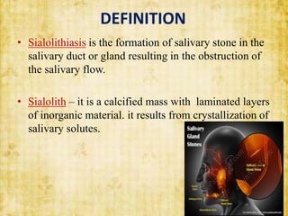
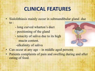
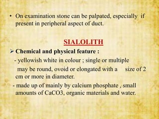
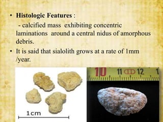
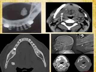
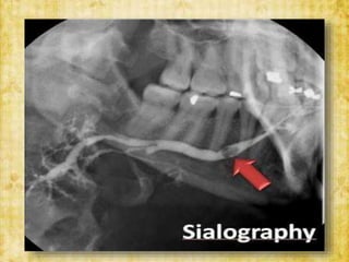
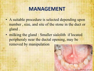
DR DAVIS
NADAKKAVUKARAN
ORAL AND
MAXILLOFACIAL
SURGERY
CONTENTS
• INTRODUCTION
• DEFINITION
• ETIOLOGY
• CLINICAL FEATURES
• INVESTIGATIONS
• COMPLICATIONS
• MANAGEMENT
INTRODUCTION
• Salivary glands can be classified as major and minor
salivary glands
• major Salivary glands are parotid , submandibular
and sublingual glands.
• Minor salivary glands are widely distributed in oral
cavity.
• Function : produce saliva
DEFINITION
• Sialolithiasis is the formation of salivary stone in the
salivary duct or gland resulting in the obstruction of
the salivary flow.
• Sialolith – it is a calcified mass with laminated layers
of inorganic material. it results from crystallization of
salivary solutes.
ETIOLOGY
• Inflammation stagnation of saliva
• Irritation
formation of inorganic
• Drugs nidus and its calcification
• Metabolic cause – not established yet
CLINICAL FEATURES
• Sialolithiasis mainly occur in submandibular gland due
to :
- long curved wharton’s duct
- positioning of the gland
- tenacity of saliva due to its high
mucin content.
-alkalinity of saliva
• Can occur at any age – in middle aged persons
• Patients complaints of pain and swelling during and after
eating of food.
• On examination stone can be palpated, especially if
present in peripheral aspect of duct.
SIALOLITH
 Chemical and physical feature :
- yellowish white in colour ; single or multiple
may be round, ovoid or elongated with a size of 2
cm or more in diameter.
- made up of mainly by calcium phosphate , small
amounts of CaCO3, organic materials and water.
• Histologic Features :
- calcified mass exhibiting concentric
laminations around a central nidus of amorphous
debris.
• It is said that sialolith grows at a rate of 1mm
/year.
INVESTIGATIONS
• Radiographs :- AnteroPosterior view
- lateral oblique
- occlusal view
• Sialography : specialized radiographic method
involving filling of ducts and parenchyma with a
radiopaque agent and make them visible on a
radiograph.
COMPLICATIONS
• Sialolithiasis is both a cause and consequence of
sialadinitis and it is frequently a cause of acute
suppurative sialadinitis.
• Sialoangiectasis : dilatation of the gland and duct
system can happen because of stasis of saliva.
• Retention of saliva – formation of mucoceles.
• Complete obstruction of the duct may result in
atrophy of gland.
MANAGEMENT
• A suitable procedure is selected depending upon
number , size, and site of the stone in the duct or
gland .
• milking the gland : Smaller sialolith if located
peripheraly near the ductal opening, may be
removed by manipulation
• Surgical removal – larger sialoliths
 transoral sialolithotomy of submandibular duct
• carried out under local anesthesia
• The exact site of stone is located by X-rays
• a suture is placed behind the stone to prevent
its backward movement
• Tongue is lifted and held with the help of a guaze
• Insicion is made in the mucosa parallel to the duct
taking care not to injure structures like lingual nerve
and sublingual glands
• After this , a blunt dissection is carried out. The
tissues are displaced to locate the gland.
• Part of the duct lodging the stone is identified, then a
longitudinal incision is made over the stone
• Stone is removed using forceps . If the stone is large,
it is crushed using forceps.
• A cannula is passed to aspirate the pieces of stone.
A probe is passed to ensure the patency of duct
anterior to surgical area
• Sutures are placed at the level of mucosa.
• Sometimes stones which are not impacted, may
be extracted through intubation of the duct with
fine soft plastic catheter and application of
suction to the tube.
• Modern techniques like piezoelectric shockwave
lithotripsy to fragment the salivary stone can also
be used.the fragments pass through the duct as
salivary flow is stimulated by use of sialogogues.
REFERANCE
• TEXT BOOK OF ORAL AND
MAXILLOFACIAL SURGERY – NEELIMA
ANIL MALIK , third edition
• SHAFER’S TEXT BOOK OF ORAL
PATHOLOGY - SHAFER, HINE , LEVY ,
seventh edition
THANK YOU

SIALOLITHIASIS - OMFS.pptx

  • 1.
  • 2.
    CONTENTS • INTRODUCTION • DEFINITION •ETIOLOGY • CLINICAL FEATURES • INVESTIGATIONS • COMPLICATIONS • MANAGEMENT
  • 3.
    INTRODUCTION • Salivary glandscan be classified as major and minor salivary glands • major Salivary glands are parotid , submandibular and sublingual glands. • Minor salivary glands are widely distributed in oral cavity. • Function : produce saliva
  • 4.
    DEFINITION • Sialolithiasis isthe formation of salivary stone in the salivary duct or gland resulting in the obstruction of the salivary flow. • Sialolith – it is a calcified mass with laminated layers of inorganic material. it results from crystallization of salivary solutes.
  • 5.
    ETIOLOGY • Inflammation stagnationof saliva • Irritation formation of inorganic • Drugs nidus and its calcification • Metabolic cause – not established yet
  • 6.
    CLINICAL FEATURES • Sialolithiasismainly occur in submandibular gland due to : - long curved wharton’s duct - positioning of the gland - tenacity of saliva due to its high mucin content. -alkalinity of saliva • Can occur at any age – in middle aged persons • Patients complaints of pain and swelling during and after eating of food.
  • 7.
    • On examinationstone can be palpated, especially if present in peripheral aspect of duct. SIALOLITH  Chemical and physical feature : - yellowish white in colour ; single or multiple may be round, ovoid or elongated with a size of 2 cm or more in diameter. - made up of mainly by calcium phosphate , small amounts of CaCO3, organic materials and water.
  • 8.
    • Histologic Features: - calcified mass exhibiting concentric laminations around a central nidus of amorphous debris. • It is said that sialolith grows at a rate of 1mm /year.
  • 9.
    INVESTIGATIONS • Radiographs :-AnteroPosterior view - lateral oblique - occlusal view • Sialography : specialized radiographic method involving filling of ducts and parenchyma with a radiopaque agent and make them visible on a radiograph.
  • 12.
    COMPLICATIONS • Sialolithiasis isboth a cause and consequence of sialadinitis and it is frequently a cause of acute suppurative sialadinitis. • Sialoangiectasis : dilatation of the gland and duct system can happen because of stasis of saliva. • Retention of saliva – formation of mucoceles. • Complete obstruction of the duct may result in atrophy of gland.
  • 13.
    MANAGEMENT • A suitableprocedure is selected depending upon number , size, and site of the stone in the duct or gland . • milking the gland : Smaller sialolith if located peripheraly near the ductal opening, may be removed by manipulation
  • 14.
    • Surgical removal– larger sialoliths  transoral sialolithotomy of submandibular duct • carried out under local anesthesia • The exact site of stone is located by X-rays • a suture is placed behind the stone to prevent its backward movement • Tongue is lifted and held with the help of a guaze
  • 15.
    • Insicion ismade in the mucosa parallel to the duct taking care not to injure structures like lingual nerve and sublingual glands • After this , a blunt dissection is carried out. The tissues are displaced to locate the gland. • Part of the duct lodging the stone is identified, then a longitudinal incision is made over the stone • Stone is removed using forceps . If the stone is large, it is crushed using forceps.
  • 16.
    • A cannulais passed to aspirate the pieces of stone. A probe is passed to ensure the patency of duct anterior to surgical area • Sutures are placed at the level of mucosa.
  • 18.
    • Sometimes stoneswhich are not impacted, may be extracted through intubation of the duct with fine soft plastic catheter and application of suction to the tube. • Modern techniques like piezoelectric shockwave lithotripsy to fragment the salivary stone can also be used.the fragments pass through the duct as salivary flow is stimulated by use of sialogogues.
  • 20.
    REFERANCE • TEXT BOOKOF ORAL AND MAXILLOFACIAL SURGERY – NEELIMA ANIL MALIK , third edition • SHAFER’S TEXT BOOK OF ORAL PATHOLOGY - SHAFER, HINE , LEVY , seventh edition
  • 21.