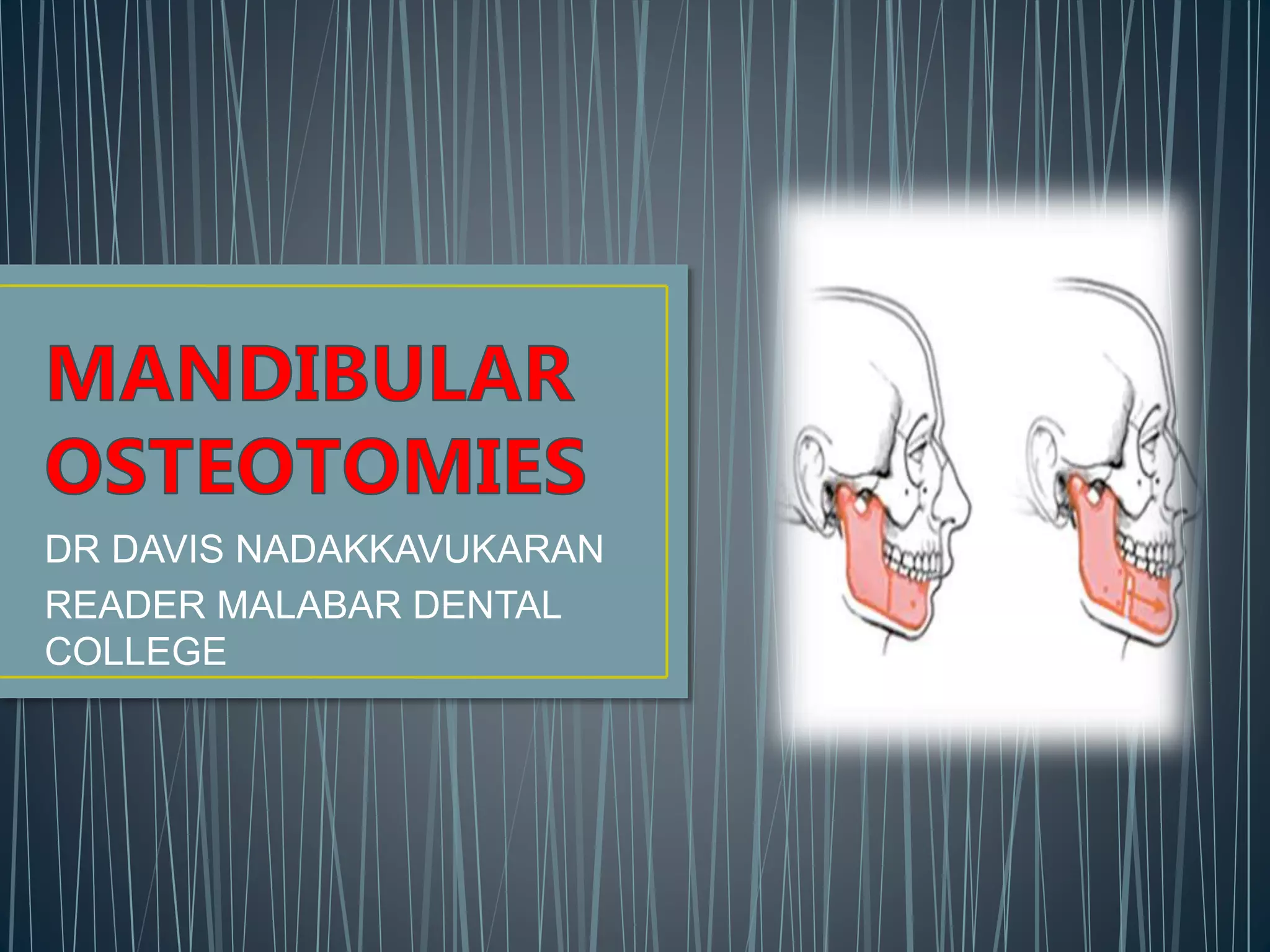
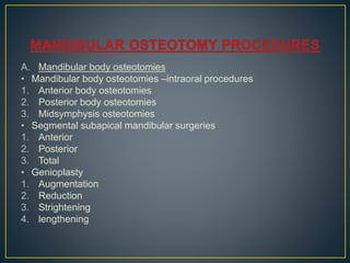
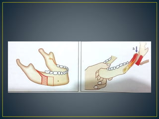
DR DAVIS NADAKKAVUKARAN
READER MALABAR DENTAL
COLLEGE
1. INTRODUCTION
2. CLASSIFICATION
3. MANDIBULAR BODY OSTEOTOMY
4. MANDIBULAR RAMUS OSTEOTOMY
5. COMPLICATION
• Definition of orthognatic surgery is art and science of
diagnosis , treatment planning and execution of treatment
by combining orthodontics and oral and maxillofacial
surgery to correct the muskuloskeltal , dentooseeous and
soft tissue deformities of the jaws and associated
structures . In the severe skeltal deformities orthodontics
may compromise the esthetics and stability and surgery
alone ,may compramise the function and stability
A. Mandibular body osteotomies
• Mandibular body osteotomies –intraoral procedures
1. Anterior body osteotomies
2. Posterior body osteotomies
3. Midsymphysis osteotomies
• Segmental subapical mandibular surgeries
1. Anterior
2. Posterior
3. Total
• Genioplasty
1. Augmentation
2. Reduction
3. Strightening
4. lengthening
B.Mandibular ramus osteotomies
• Subcondylar ramus osteotomies
1. Extra oral
2. Intraoral
3. Arching ramus
• Intraoral modified sagittal split osteotomy
Anterior body osteotomy
• Performed anterior to the mental foramen
Indication
1. Mandibular prognathism with functional posterir
occlusion
2. Class III malocclusion with or without anterior open bite
3. Posterior crossbite in dental in nature
Incision
Removal of the first or second premolars is required
Two small vestibular incision in Ist and 2 nd premolar
region
Procedure
• After putting incision mucoperiosteal tunneling is carried out
superiorly till the alveolar crest and inferiorly till inferir border of
area of extraction
• Periosteal elevator is inserted lingually through the extraction
site subperiosteally to protect the lingual soft tissue during
osteotomy cut
Osteotomy cut
• Modified retractor placed at the inferior border of the body of
mandible
• Osteotomy cut started in socket at the alveolar margin
involving both buccal and lingual cortices going towards the
inferior border
• Cuts are made parellel to each othe
• Same procrdure repeated on opposite side
• Occlusal splint tried and cut is modified till desired
position is achieved
• After fit of occlusal splint is achieved , fragments are
stabilized at superior border by passing figure of eight
wire around neck of canine and premolar and inferiorly
by using miniplate
• Wound is closed by single layer
• Modified step osteotomy
Posterior body osteotomy
• Osteotomy posterior to mental foramen
Indication
1. Missing posterior teeth
2. Class III deformity
3. Correction of crossbite
Incision
• Vestibular incision- one tooth anteriorly and one tooth
distal to osteotomy site and is extended posteriorly up to
the external oblique ridge for more relaxation
Osteotomy cut
• Channel retractor inserted at the site
• Cut started superior to neurovascular bundle and finished
through both the cortices
• At the level of bundle , small window is made by removal
of removal of only external cortex
• Neve hook is inserted to pull bundle towards buccal side
and lingual osteotomy finished
• Bundle retracted upward , inferior border cut can be
completed
• Same procedure on other side
• Occlusal splint is fitted by intraossous wiring or bone
plating
Mid symphysis osteotomy
• Used to widen or narrow the anterior
arch width
Incision
• Complete vestibular incision
Osteotomy cut
• Thin tapering saw or bur can be
used for making cut between two
mandibular incisor from the alveolar
crest to the inferior border
Anterior subapical mandibular osteotomy
Indication
1. Correction of mandibular dento alveolar proclination
2. Closing mild anterior open bite
3. Leveling an accentuated curve of spee
4. Correcting mndibular dental arch asymmetry
5. As adjunctive procedure with anterior maxillary
osteotomy
6. With mandibular advancement
7. Genioplasty procedure
Incision
• Circum vestibular incision from canine to canine
• It made into the lip and carried out tangentially down to the
bone
• Subperiosteal dissection is carried to the inferior body and the
symphysis region is degloved
Osteotomy cut
• Periosteal elevator is placed on the lingual suface of extraction
socket and vertical cut is made from the alveolar crest till the
level of premolar root apex through both the cortices .
• Same procude repeated on the same side
• Both vertical cut connected by the horizontal subapical
osteotomy cut made about 5mm below the anterior teeth
apices
• Fixation by miniplate and wound closed in two layer,mucosal
and muscular layer
Posterior subapical mandibular osteotomy procedure
Indication
1. Uprighting the posterior segment which is extreme
linguo –version or buccoversion
2. Closing a premolar or molar spaces
3. Leveling a supraerupted posterior teeth
Incision
• Horizontal vestibular incision
• Mucoperiosteal flap reflected downwards till the inferior
border of the mandible
Osteotomy cut
• Anterior vertical cut –area of missing first premolar or first
molar
• Second vertical cut -behind the last molar is exist
• Horizontal cut –below the apices of tooth
• Cut upto the level of neurovascular bundle
• Buccal cortex above the neurovascular bundle is removed
• Window extended posteriorly to the distal vertical cut
• Window is made to cut buccal cortex only and after
identification of nerve bundle lingual osteotomy cut is
completed
• Horizontal cut to connect the vertical cut and the entire
segment is mobilized
• Fixing the occlusal splint and placing circummandibular wiring
over splint
Total subapical mandibular osteotomy
Indications
1. To reposition entire mandibular dentoalveolar segment
anteriorly , posteriorly or superiorly
2. For lenthening of lower one third of the face
3. Advancing of mandibular dentoalveolar segment
Osteotomy cut
• Horizontal osteotomy – from anteriorly in the symphysis region
and proceed posteriorly and
• It is completed through lingual cortex by placing the guiding
finger on lingual side and bur is directed at an angle of 45
degree from buccal to lingual cortex .
• Vertical osteotomy is
made mm posterior to the
last molar
• Buccal cut made first upto
bundle and then lingual
cut is completed.
• In advancement cases ,
chance to overstrech the
nerve
Augmentation genioplasty
• To increase the chin projection
• Can be done by sliding
horizontal osteotomy or
autogeneous bone graft or
alloplastic material
Incision
• Vestibular incision to deglove
the entire inferior border of
symphysis
• Periosteal releasing incision
Inferior border is degloved after vestibular incision
Horizontal cut -at least 4-5 mm below the apices of canine
Cut completed through both buccal and lingual cortex
Segment is mobilized and forward with help of osteotome
Mobilized segment id pedicled over the geniohyoid muscle
Any bony interference is removed
Fix it at desired position by intraosseous wiring or miniplate
Reduction genioplasty
• Three different types of
procedures are desired
1. Horizontal osteotomy and set
back of the fragment
2. Vertical reduction amounts to
determining the movement .
3. Two horizontal osteotomy cut
–lower cut is first and then
superior cut and bony wedge
is removed
4. Vertical reduction and
posterior pushback is need in
some cases
• Subcondylar ramus osteotomies
1. Extra oral
2. Intraoral
3. Arching ramus
• Intraoral modified sagittal split osteotomy
Vertical subsigmoid osteotomy
Indication
1. To correct the mandibular
prognathism
2. To correct mild mandibular
retrognathism
Incision
1. Submandibular incision(extra orally)
2. Extended third molar incision (intra
oral)
Osteotomy cut
• Antilingula- prevent damage to inferior alveolar nerve
• Vertical bony cut – from mandibular notch to mandibular
foramen –reaching down to mandibular border
• Decortication of the part of ramus anterior to the
osteotomy cut can be made so that when the ramus is
pushed back to correct the prognathism
• Simple fixation using wires for to weeks
Inverted L osteotomy
• Indicated in retrognathic mandible
• Extraorally by submandibular incision
• Lateral asepect is exposed
• Osteotomy cut - first horizontal starting
from the anterior border of the ramus
base of the coroonoid process extending
above mandibular foramen
• Cut then vertically down to inferior border
of ramus
• Advancement done by using graft
• Fixation done by transosseous wiring or
bone plate
C osteotomy
• Modification of of L
osteotomy
• Difference is in osteotomy
cut
• Cut is stright down to the
inferior border of the
mandible brought forward
in a horizontal direction
towards the third molar
region
• described y obwegeser
• modification done by hunsuck and dalpont
Indication
• Prognathic mandible correction
• Retrognathic mandible correction
Incision
• Made third molar region just lateral to the crest of alveolus
• It extended anteriorly along external oblique ridge upto
planned vertical osteotomy cut
• Diastally extended along anterior border of ramus
• Lateral surface is exposed upto the lower border
• Medially subperiosteal disection done till the posterior border
of the ramus
Osteotomy cut
• Devided into three;-
1. Horizontal cut – on medial aspect over mandibular
foramen
2. Vertical cut-third molar region from exteranl oblique
ridge to the inferior border of mandible
3. A cut connecting them both runs along the external
oblique ridge
• Procedure done bilaterally
• Fixation done by wiring or single plate
Complication
1. Injury to inferior alveolar nerve
2. Troublsome bleeding
3. Unfavourable split
4. Avascular necrosis
5. Condylar resorption.
6. Malpositioned proximal segment
7. Shattering of the ramus in case of thin mandible
• Incision – preauricular
incision or
submandibular
incision
• Condylar neck is
sectioned obliquely
and mandible
positioned posteriorly
without damaging
mandibular foramen
1. Postoperative infection
2. Exposure of hardware
3. Devitalisation of teeth
4. Malunion
5. Malocclusion
6. Relapse
7. Injury to teeth
8. Periodontal problem
9. Respiratory decompensation
10.bleeding
• Text book of oral and maxillofacial surgery – Neelima Anil
Malik
• Textbook of oral and maxillofacial surgery –
Chitra Chakravarthy (2nd edition )
MANDIBULAR OSTEOTOMIES.pptx

MANDIBULAR OSTEOTOMIES.pptx

  • 1.
    DR DAVIS NADAKKAVUKARAN READERMALABAR DENTAL COLLEGE
  • 2.
    1. INTRODUCTION 2. CLASSIFICATION 3.MANDIBULAR BODY OSTEOTOMY 4. MANDIBULAR RAMUS OSTEOTOMY 5. COMPLICATION
  • 3.
    • Definition oforthognatic surgery is art and science of diagnosis , treatment planning and execution of treatment by combining orthodontics and oral and maxillofacial surgery to correct the muskuloskeltal , dentooseeous and soft tissue deformities of the jaws and associated structures . In the severe skeltal deformities orthodontics may compromise the esthetics and stability and surgery alone ,may compramise the function and stability
  • 4.
    A. Mandibular bodyosteotomies • Mandibular body osteotomies –intraoral procedures 1. Anterior body osteotomies 2. Posterior body osteotomies 3. Midsymphysis osteotomies • Segmental subapical mandibular surgeries 1. Anterior 2. Posterior 3. Total • Genioplasty 1. Augmentation 2. Reduction 3. Strightening 4. lengthening
  • 5.
    B.Mandibular ramus osteotomies •Subcondylar ramus osteotomies 1. Extra oral 2. Intraoral 3. Arching ramus • Intraoral modified sagittal split osteotomy
  • 6.
    Anterior body osteotomy •Performed anterior to the mental foramen Indication 1. Mandibular prognathism with functional posterir occlusion 2. Class III malocclusion with or without anterior open bite 3. Posterior crossbite in dental in nature Incision Removal of the first or second premolars is required Two small vestibular incision in Ist and 2 nd premolar region
  • 7.
    Procedure • After puttingincision mucoperiosteal tunneling is carried out superiorly till the alveolar crest and inferiorly till inferir border of area of extraction • Periosteal elevator is inserted lingually through the extraction site subperiosteally to protect the lingual soft tissue during osteotomy cut Osteotomy cut • Modified retractor placed at the inferior border of the body of mandible • Osteotomy cut started in socket at the alveolar margin involving both buccal and lingual cortices going towards the inferior border • Cuts are made parellel to each othe
  • 9.
    • Same procrdurerepeated on opposite side • Occlusal splint tried and cut is modified till desired position is achieved • After fit of occlusal splint is achieved , fragments are stabilized at superior border by passing figure of eight wire around neck of canine and premolar and inferiorly by using miniplate • Wound is closed by single layer • Modified step osteotomy
  • 10.
    Posterior body osteotomy •Osteotomy posterior to mental foramen Indication 1. Missing posterior teeth 2. Class III deformity 3. Correction of crossbite Incision • Vestibular incision- one tooth anteriorly and one tooth distal to osteotomy site and is extended posteriorly up to the external oblique ridge for more relaxation
  • 12.
    Osteotomy cut • Channelretractor inserted at the site • Cut started superior to neurovascular bundle and finished through both the cortices • At the level of bundle , small window is made by removal of removal of only external cortex • Neve hook is inserted to pull bundle towards buccal side and lingual osteotomy finished • Bundle retracted upward , inferior border cut can be completed • Same procedure on other side • Occlusal splint is fitted by intraossous wiring or bone plating
  • 13.
    Mid symphysis osteotomy •Used to widen or narrow the anterior arch width Incision • Complete vestibular incision Osteotomy cut • Thin tapering saw or bur can be used for making cut between two mandibular incisor from the alveolar crest to the inferior border
  • 14.
    Anterior subapical mandibularosteotomy Indication 1. Correction of mandibular dento alveolar proclination 2. Closing mild anterior open bite 3. Leveling an accentuated curve of spee 4. Correcting mndibular dental arch asymmetry 5. As adjunctive procedure with anterior maxillary osteotomy 6. With mandibular advancement 7. Genioplasty procedure
  • 15.
    Incision • Circum vestibularincision from canine to canine • It made into the lip and carried out tangentially down to the bone • Subperiosteal dissection is carried to the inferior body and the symphysis region is degloved Osteotomy cut • Periosteal elevator is placed on the lingual suface of extraction socket and vertical cut is made from the alveolar crest till the level of premolar root apex through both the cortices . • Same procude repeated on the same side • Both vertical cut connected by the horizontal subapical osteotomy cut made about 5mm below the anterior teeth apices • Fixation by miniplate and wound closed in two layer,mucosal and muscular layer
  • 16.
    Posterior subapical mandibularosteotomy procedure Indication 1. Uprighting the posterior segment which is extreme linguo –version or buccoversion 2. Closing a premolar or molar spaces 3. Leveling a supraerupted posterior teeth Incision • Horizontal vestibular incision • Mucoperiosteal flap reflected downwards till the inferior border of the mandible
  • 17.
    Osteotomy cut • Anteriorvertical cut –area of missing first premolar or first molar • Second vertical cut -behind the last molar is exist • Horizontal cut –below the apices of tooth • Cut upto the level of neurovascular bundle • Buccal cortex above the neurovascular bundle is removed • Window extended posteriorly to the distal vertical cut • Window is made to cut buccal cortex only and after identification of nerve bundle lingual osteotomy cut is completed • Horizontal cut to connect the vertical cut and the entire segment is mobilized • Fixing the occlusal splint and placing circummandibular wiring over splint
  • 18.
    Total subapical mandibularosteotomy Indications 1. To reposition entire mandibular dentoalveolar segment anteriorly , posteriorly or superiorly 2. For lenthening of lower one third of the face 3. Advancing of mandibular dentoalveolar segment Osteotomy cut • Horizontal osteotomy – from anteriorly in the symphysis region and proceed posteriorly and • It is completed through lingual cortex by placing the guiding finger on lingual side and bur is directed at an angle of 45 degree from buccal to lingual cortex .
  • 19.
    • Vertical osteotomyis made mm posterior to the last molar • Buccal cut made first upto bundle and then lingual cut is completed. • In advancement cases , chance to overstrech the nerve
  • 20.
    Augmentation genioplasty • Toincrease the chin projection • Can be done by sliding horizontal osteotomy or autogeneous bone graft or alloplastic material Incision • Vestibular incision to deglove the entire inferior border of symphysis • Periosteal releasing incision
  • 21.
    Inferior border isdegloved after vestibular incision Horizontal cut -at least 4-5 mm below the apices of canine Cut completed through both buccal and lingual cortex Segment is mobilized and forward with help of osteotome Mobilized segment id pedicled over the geniohyoid muscle Any bony interference is removed Fix it at desired position by intraosseous wiring or miniplate
  • 22.
    Reduction genioplasty • Threedifferent types of procedures are desired 1. Horizontal osteotomy and set back of the fragment 2. Vertical reduction amounts to determining the movement . 3. Two horizontal osteotomy cut –lower cut is first and then superior cut and bony wedge is removed 4. Vertical reduction and posterior pushback is need in some cases
  • 23.
    • Subcondylar ramusosteotomies 1. Extra oral 2. Intraoral 3. Arching ramus • Intraoral modified sagittal split osteotomy
  • 24.
    Vertical subsigmoid osteotomy Indication 1.To correct the mandibular prognathism 2. To correct mild mandibular retrognathism Incision 1. Submandibular incision(extra orally) 2. Extended third molar incision (intra oral)
  • 25.
    Osteotomy cut • Antilingula-prevent damage to inferior alveolar nerve • Vertical bony cut – from mandibular notch to mandibular foramen –reaching down to mandibular border • Decortication of the part of ramus anterior to the osteotomy cut can be made so that when the ramus is pushed back to correct the prognathism • Simple fixation using wires for to weeks
  • 26.
    Inverted L osteotomy •Indicated in retrognathic mandible • Extraorally by submandibular incision • Lateral asepect is exposed • Osteotomy cut - first horizontal starting from the anterior border of the ramus base of the coroonoid process extending above mandibular foramen • Cut then vertically down to inferior border of ramus • Advancement done by using graft • Fixation done by transosseous wiring or bone plate
  • 27.
    C osteotomy • Modificationof of L osteotomy • Difference is in osteotomy cut • Cut is stright down to the inferior border of the mandible brought forward in a horizontal direction towards the third molar region
  • 28.
    • described yobwegeser • modification done by hunsuck and dalpont Indication • Prognathic mandible correction • Retrognathic mandible correction Incision • Made third molar region just lateral to the crest of alveolus • It extended anteriorly along external oblique ridge upto planned vertical osteotomy cut • Diastally extended along anterior border of ramus • Lateral surface is exposed upto the lower border • Medially subperiosteal disection done till the posterior border of the ramus
  • 30.
    Osteotomy cut • Devidedinto three;- 1. Horizontal cut – on medial aspect over mandibular foramen 2. Vertical cut-third molar region from exteranl oblique ridge to the inferior border of mandible 3. A cut connecting them both runs along the external oblique ridge • Procedure done bilaterally • Fixation done by wiring or single plate
  • 32.
    Complication 1. Injury toinferior alveolar nerve 2. Troublsome bleeding 3. Unfavourable split 4. Avascular necrosis 5. Condylar resorption. 6. Malpositioned proximal segment 7. Shattering of the ramus in case of thin mandible
  • 33.
    • Incision –preauricular incision or submandibular incision • Condylar neck is sectioned obliquely and mandible positioned posteriorly without damaging mandibular foramen
  • 34.
    1. Postoperative infection 2.Exposure of hardware 3. Devitalisation of teeth 4. Malunion 5. Malocclusion 6. Relapse 7. Injury to teeth 8. Periodontal problem 9. Respiratory decompensation 10.bleeding
  • 35.
    • Text bookof oral and maxillofacial surgery – Neelima Anil Malik • Textbook of oral and maxillofacial surgery – Chitra Chakravarthy (2nd edition )