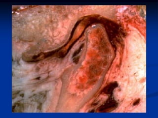
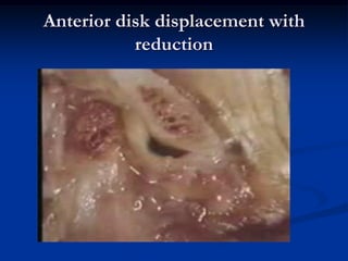
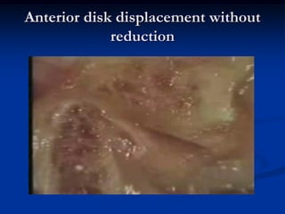
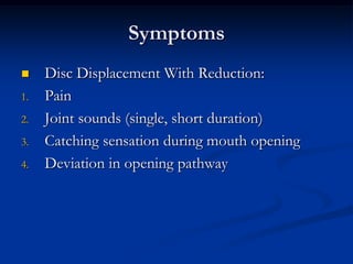
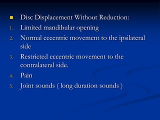
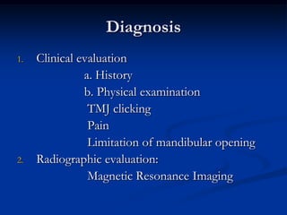
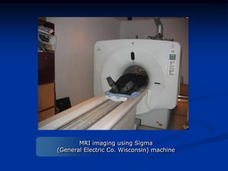
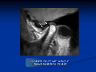
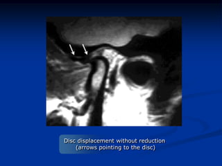
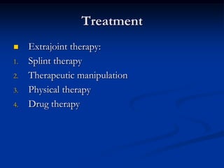
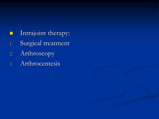
This document provides an overview of internal derangements of the temporomandibular joint (TMJ). It defines internal derangement as an abnormal relationship between the articular disc and condyle. The most common type is anterior disc displacement, which can be with or without reduction. Causes include trauma, functional overloading, joint laxity, and muscle spasms. Symptoms vary depending on the type but may include clicking, limited opening, and pain. Diagnosis involves clinical exams and MRI imaging. Treatment ranges from splint therapy to arthrocentesis or arthroscopy for lavage and relief of adhesions. Arthrocentesis is shown to improve opening and reduce pain by removing inflammatory factors from the
3. What to gain ???
1.
2.
3.
4.
5.
Understand Internal Derangements and its
underlying causes
Be able to diagnose Disc Displacements
Choose the correct line of treatment
Arthrocentesis, when, how, why ???
Future Directions
4. Definition
The Temporomandibular Joint (TMJ) is a
common site of complaint. Clicking sounds and
pain are indicators of a frequent condition called
internal derangement, most often affecting
females. As a general term, internal derangement
describes a structural abnormality within an
articulation.
5.
The internal derangement of the
temporomandibular joint (TMJ) is a specific
term defined as an abnormal positional and
functional relationship between the disk and
articulating surfaces.
6.
Emshoff R. and Rudisch A. (2003) defined
internal derangements of the
temporomandibular joint as an abnormal
relation of the articular disc to the mandibular
condyle and the articular eminence. Jaw pain,
clicking of the joint, irregular and limited
movement of the jaw are the characteristic
symptoms of this disorder.
7.
8.
9. Prevalence
Is TMJ Disorder a common disorder?
Internal derangement and associated
complications are the most common pathologic
entities affecting the jaw. Solberg W.K. (1979)
10.
Nebbe et al (2000) in his study on prevalence
of TMJ disc displacement found normal joints
in only 50% of boys and in 23%–29% of girls.
The rest of the study population presented with
different degrees of slight to full disk
displacement with or without a change in
morphology. In other studies, asymptomatic
disk displacement was documented in
approximately 30% of adolescents.
11.
82% of patients presenting with pain and
functional disturbance of their TMJ will have
displaced disks when examined with magnetic
resonance imaging. The overall prevalence of
symptomatic disk displacement or internal
derangement may range between 20% and 30%,
making them frequently encountered conditions.
12.
The National Institute of Dental and
Craniofacial Research indicates that 10.8 million
people in the United States suffer from TMJ
problems at any given time. Both men and
women experience TMJ problems; however, 90
percent of those seeking treatment are women
in their childbearing years.
13. Disc Displacements
Anterior disk displacement of the TMJ is a
malrelationship of the disk to the condylar head
and articular eminence. Although the disk may
displace medially, laterally, or (rarely) posteriorly
to the condyle, it generally displaces anteriorly.
First stage in the sequence of events leading to
osteoarthritis.
14.
TMJ morphology, have shown a path of
progression that includes changes not only in
the disc position, but also in its configuration.
The interpretation of the process leading up to a
dislocated disc as portrayed in the literature does
not always stand on firm evidence and at times
is contradictory.
15.
Disc displacement is considered to be associated
with clinically noticeable clicking noises on
opening and closing of the mouth as long as the
disc reduces to its normal position on opening.
When it becomes nonreducible, the clicking
noise disappears and instead there is a certain
degree of limitation in mouth opening.
16. Classification of Disc Displacements
Internal derangements can be divided into 2
categories: anterior disk displacement with reduction
and anterior disk displacement without reduction. The
condition in which the disk is located anteriorly
and slips back into its normal position during
opening of the mouth is called anterior disk
displacement with reduction; the opposite
condition is dubbed anterior disk displacement
without reduction.
20. Pathogenesis
TMJ disc displacement results from its inability
to slide smoothly due to increased friction or
degenerative changes in the joint surfaces.
The sequence of events, starting with increased
friction in the upper joint compartment and
culminating in disc displacement
Activation of various parafunctions, such as
clenching, compromises the lubrication system
in the upper TMJ compartment.
21.
The resulting increased friction prevents the disc
from sliding together with the condyle.
On jaw opening, the condyle is pulled away
from the disc by the inferior head of the lateral
pterygoid muscle. As a result, the ligaments
joining the disc to the condyle are gradually
stretched, and the ‘mobilized’ disc gravitates
slightly downward and forward.
22.
Subsequently, on clenching, the unstable disc is
propelled forward by pressure from the condyle.
At this point, the force on the slightly displaced
disc is shared between two vectors, one of
which is directed forward. Apparently, on
mouth closure, the superior belly of the lateral
pterygoid muscle pulls the disc anteriorly
23.
Subsequently, during mouth opening, the
condyle, which is now posterior to the loose
disc, gradually pushes it down the slope of the
eminence, displacing it further forward
24.
Since the lateral articular disc bears the bulk of
the shearing and compressive loads, persistent
loading tends to drive it in a medial direction,
which is the ‘path of least resistance’.
27.
1.
2.
3.
4.
5.
Disc Displacement Without Reduction:
Limited mandibular opening
Normal eccentric movement to the ipsilateral
side
Restricted eccentric movement to the
contralateral side.
Pain
Joint sounds ( long duration sounds )
34. Arthrocentesis
Nitzan et al (1991) described a technique of
irrigation of the upper compartment of the TMJ
with Ringer's lactate solution to treat limited
mouth opening due to internal derangement.
The authors called this technique
`arthrocentesis'.
35.
They reported an increase in mouth opening
from a range of 12±30 mm prior to the
procedure, to 35±50 mm following it. On a
visual analogue scale of 0±15, the pain decreased
from a mean rating of 8.75 to 2.3. This
technique marked an evolution towards less
surgical treatment.
36.
Arthrocentesis is the most recent surgical
approach for internal derangement of the TMJ.
In the past many cases of anterior displacement
of the disc or closed lock that did not improve
with medical treatment (bite plates, muscle
relaxants, diet and physical therapy) were initially
treated with surgical repositioning of the disc
and arthroplasty of the mandibular fossa.
37.
Arthrocentesis has an intermediate place
between the medical and the surgical forms of
treatment. Ease, lower cost of materials and
excellent published results so far include this
technique in the international protocol for the
treatment of TMJ dysfunction.
38.
Arthrocentesis is a simple yet effective treatment
of temporomandibular joint disorders, and it
requires minimal invasion. Significant
improvements in width of mouth opening have
been reported with proven long-term results. It
is speculated that the increase in mouth opening
results from the elimination of the vacuum
effect within the joint compartment.
39.
In 2003, Reston and Turkelson performed a
meta-analysis of surgical treatments for
temporomandibular articular disorders. They
concluded that among patients refractory to
nonsurgical therapies, surgical arthrocentesis and
arthroscopy were most effective for patients
with disc displacement without reduction.
40.
It is suspected that lavage under sufficient
hydraulic pressure could widen the narrowed
joint space and release adhesion in the joint
space. Arthrocentesis with sufficient pressure
could be effective for closed lock cases with
adhesions in the upper joint compartment.
41. Mechanism of Action
1.
Reduction in pain level:
Arthrocentesis reduces pain by removing
inflammatory mediators from the joint. The
combined treatment of arthrocentesis and
Sodium Hyaluronate injection may improve
the results due to the long-term lubricating
effect of Sodium Hyaluronate, which prevents
the onset of inflammatory mediators that are
responsible for pain.
42. 2.
Maximal Mouth Opening:
Arthrocentesis under high pressure is an
effective method to regain normal mouth
opening in closed lock cases. This effect is
usually due to elimination of the adhesions
around the disc. Also the lubricating effect of
Sodium Hyaluronate which either maintains
lubrication and minimizes wear and tear
mechanically, or plays a role in nutrition of the
avascular parts of the disc and condylar
cartilage.
46. “Relationship between the Canthal-Tragus
Distance and the Puncture Point in
Temporomandibular Joint Arthroscopy”
Wael Talaat Taha1, PhD, Thomas A. McGraw 1,
DMD,
and Bruce Klitzman1, PhD
Int J Oral Maxillofac Surg. 2010; 39: 57 - 60
47.
48.
49.
50.
51.
52.
53. Nearby Vital Structures
The frontal branch of the facial nerve is located
a mean distance of 20 mm from the anterior
margin of the bony external auditory canal as it
crosses over the posterior aspect of the
zygomatic arch (a range of 8 to 35mm). The
main trunk of the bifurcation of the facial nerve
is located a mean distance of 23 mm (a range of
15 to 28 mm) inferior to the lowest concavity of
the bony external auditory canal.
54.
Greene MW et al found the tympanic plate to be
located at a range of 6 to 9 mm anterior to the
posterior tragus and perpendicular to the skin at
a mean depth of 25.4 mm (range = 19 to 32
mm).
55. Future Directions
. In 2006, Betre et al designed a biologically based
drug delivery vehicle for intra-articular drug delivery
using elastin-like polypeptides (ELPs), a biopolymer
composed of repeating pentapeptides that undergo a
phase transition to form aggregates above their
transition temperature. The ELP drug delivery vehicle
was designed to aggregate upon intra-articular injection
at 37 °C, and form a drug ‘depot’ that could slowly
disaggregate and be cleared from the joint space over
time.