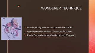
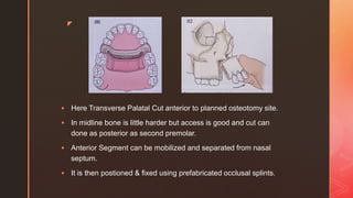
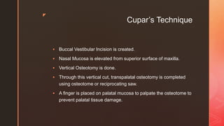
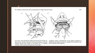
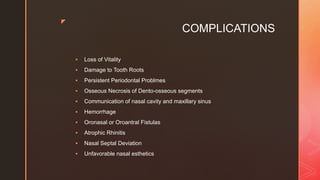
z
ANTERIOR
SEGMENTAL
MAXILLARY
OSTEOTOMY
PRESENTED BY
DINESH VENKATESAN
FINAL YEAR BDS
MAXILLARY OSTEOTOMY
z
HISTORICAL EVIDENCE
 1921 – Cohn Stock performed first maxillary ostetomy
 Wedge shaped maxillary Osteotomy through transverse palatal
incisions
 Succeeded by green stick fracture of Anterior maxillary segment
that was retracted
 Relapsed in 4 weeks
z
INDICATIONS
 In Correction of Protrusion of Maxillary Anterior Teeth with
normal axial inclination with alveolar bone.
 In Correction of Anterior Open Bite provided absence of
maxillary excess.
 Vertical Plane Premaxilla correction – Openbite / Deep Bite.
 Failure in Orthodontic Therapy due to Ankylois or Pathological
Resortion of Maxillary Anterior Teeth.
 To improve Facial Appearance in prognathic maxilla with
competent lips and adequate lip length.
z
Techniques in AMO
 Wassmund Technique
 Wunderer Technique
 Cupar’s Technique
z
WASSMUND TECHNIQUE
 Vertical Incision between Canine and Premolar extending to
nasal floor.
 Mucoperiosteum is reflected posteriorly.
 In apical region of canine, reflection is made till nasal pyriform
aperture.
 First Premolars are extracted (if planned) at this stage
z
 Midline Sagittal Incision is made anteroposteriorly on palate to
perform palatal osteotomy.
 Palatal Mucosa is reflected and care is taken not to injure anterior
palatine vessels.
z
 Bony Cut is made on buccal aspect of alveolus.
 Cut is taken vertically upwards and turned medially to pyriform
aperture so that 3-5 mm of bone remains intact in canine apex.
 Transpalatal osteotomy of palatal bone from alveolus of one side
to other side.
 As advancing to midline osteotomy becomes hard dure to
thickness of bone and access difficulty which is overcome by
Midpalatine insidion made early.
z
 Maxillary segment is now separated from nasal septum by finger
pressure sometimes.
 Usually Vertical incision is placed over anterior nasal spine;
mucoperiosteum is reflected from nasal spine & cartilaginous
nasal septum.
 Nasal Osteotome is used to separate anterior maxillary segment
and nasal septum.
 Folds of soft tissue are checked for proper blood supply and
closed using catgut 3-0
z
z
WUNDERER TECHNIQUE
 Used especially when second premolar is extracted
 Labial Approach is similar to Wassmund Technique.
 Palatal Surgery is started after Buccal part of Surgery.
z
 Here Transverse Palatal Cut anterior to planned osteotomy site.
 In midline bone is little harder but access is good and cut can
done as posterior as second premolar.
 Anterior Segment can be mobilized and separated from nasal
septum.
 It is then postioned & fixed using prefabricated occlusal splints.
z
z
Cupar’s Technique
 Buccal Vestibular Incision is created.
 Nasal Mucosa is elevated from superior surface of maxilla.
 Vertical Osteotomy is done.
 Through this vertical cut, transpalatal osteotomy is completed
using osteotome or reciprocating saw.
 A finger is placed on palatal mucosa to palpate the osteotome to
prevent palatal tissue damage.
z
z
z
AMO FIXATION
 OCCLUSAL STENT STABILIZATION
 OSSEOUS WIRING
 SKELETAL FIXATION
 REIGID INTERNAL FIXATION
z
ADVANTAGES
 Direct access to nasal structures
 Preservation of blood supply through excellent palatal pedicle.
 Ability to remove palatal none directly after down fracture of
anterior maxilla.
 Excellent access to superior maxilla.
 Easy placement of rigid fixation.
z
COMPLICATIONS
 Loss of Vitality
 Damage to Tooth Roots
 Persistent Periodontal Problmes
 Osseous Necrosis of Dento-osseous segments
 Communication of nasal cavity and maxillary sinus
 Hemorrhage
 Oronasal or Oroantral Fistulas
 Atrophic Rhinitis
 Nasal Septal Deviation
 Unfavorable nasal esthetics
z

ANTERIOR SEGMENTAL MAXILLARY OSTEOTOMY.pptx

  • 1.
  • 2.
    z HISTORICAL EVIDENCE  1921– Cohn Stock performed first maxillary ostetomy  Wedge shaped maxillary Osteotomy through transverse palatal incisions  Succeeded by green stick fracture of Anterior maxillary segment that was retracted  Relapsed in 4 weeks
  • 3.
    z INDICATIONS  In Correctionof Protrusion of Maxillary Anterior Teeth with normal axial inclination with alveolar bone.  In Correction of Anterior Open Bite provided absence of maxillary excess.  Vertical Plane Premaxilla correction – Openbite / Deep Bite.  Failure in Orthodontic Therapy due to Ankylois or Pathological Resortion of Maxillary Anterior Teeth.  To improve Facial Appearance in prognathic maxilla with competent lips and adequate lip length.
  • 4.
    z Techniques in AMO Wassmund Technique  Wunderer Technique  Cupar’s Technique
  • 5.
    z WASSMUND TECHNIQUE  VerticalIncision between Canine and Premolar extending to nasal floor.  Mucoperiosteum is reflected posteriorly.  In apical region of canine, reflection is made till nasal pyriform aperture.  First Premolars are extracted (if planned) at this stage
  • 6.
    z  Midline SagittalIncision is made anteroposteriorly on palate to perform palatal osteotomy.  Palatal Mucosa is reflected and care is taken not to injure anterior palatine vessels.
  • 7.
    z  Bony Cutis made on buccal aspect of alveolus.  Cut is taken vertically upwards and turned medially to pyriform aperture so that 3-5 mm of bone remains intact in canine apex.  Transpalatal osteotomy of palatal bone from alveolus of one side to other side.  As advancing to midline osteotomy becomes hard dure to thickness of bone and access difficulty which is overcome by Midpalatine insidion made early.
  • 8.
    z  Maxillary segmentis now separated from nasal septum by finger pressure sometimes.  Usually Vertical incision is placed over anterior nasal spine; mucoperiosteum is reflected from nasal spine & cartilaginous nasal septum.  Nasal Osteotome is used to separate anterior maxillary segment and nasal septum.  Folds of soft tissue are checked for proper blood supply and closed using catgut 3-0
  • 9.
  • 10.
    z WUNDERER TECHNIQUE  Usedespecially when second premolar is extracted  Labial Approach is similar to Wassmund Technique.  Palatal Surgery is started after Buccal part of Surgery.
  • 11.
    z  Here TransversePalatal Cut anterior to planned osteotomy site.  In midline bone is little harder but access is good and cut can done as posterior as second premolar.  Anterior Segment can be mobilized and separated from nasal septum.  It is then postioned & fixed using prefabricated occlusal splints.
  • 12.
  • 13.
    z Cupar’s Technique  BuccalVestibular Incision is created.  Nasal Mucosa is elevated from superior surface of maxilla.  Vertical Osteotomy is done.  Through this vertical cut, transpalatal osteotomy is completed using osteotome or reciprocating saw.  A finger is placed on palatal mucosa to palpate the osteotome to prevent palatal tissue damage.
  • 14.
  • 15.
  • 16.
    z AMO FIXATION  OCCLUSALSTENT STABILIZATION  OSSEOUS WIRING  SKELETAL FIXATION  REIGID INTERNAL FIXATION
  • 17.
    z ADVANTAGES  Direct accessto nasal structures  Preservation of blood supply through excellent palatal pedicle.  Ability to remove palatal none directly after down fracture of anterior maxilla.  Excellent access to superior maxilla.  Easy placement of rigid fixation.
  • 18.
    z COMPLICATIONS  Loss ofVitality  Damage to Tooth Roots  Persistent Periodontal Problmes  Osseous Necrosis of Dento-osseous segments  Communication of nasal cavity and maxillary sinus  Hemorrhage  Oronasal or Oroantral Fistulas  Atrophic Rhinitis  Nasal Septal Deviation  Unfavorable nasal esthetics
  • 19.