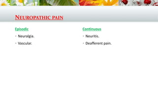
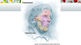
OROFACIAL PAIN
Dr. Saleh Bakry
Assistant Professor of Oral and Maxillofacial
Surgery
DEFINITION
 An unpleasant sensation caused by a noxious stimulus that is mediated only
along specific nerve pathway into the central nervous system, where it is
interpreted as pain.
EVALUATION OF
OROFACIAL
PAIN PATIENT
1. MEDICAL AND DENTAL HISTORY.
2. CHIEF COMPLAINT AND PAIN HISTORY :
 Pain quality: e.g. aching, throbbing, burning, shock like, paroxysmal or some
combination (quality).
 The duration of each episode of pain (duration).
 The site affected: ask the patient to point to the source of the pain and/or
outline the area affected by it. (Course).
 Initiating factors: anything that the patient remembers occurring immediately
before or at the same time as the start of their symptoms.
 Exacerbating factors: anything which makes the patient's symptoms worse.
 Relieving factors: Anything which relieves either partially or totally the
patient's symptom's e.g. nerve block anesthesia, anticonvilusent drug.
 Associated signs and symptoms: e.g. lacrimation, vomiting,
nausea, rhinorrhea, photophobia, phonophobia, fever.
 Previous investigations.
 Previous treatment.
3. PHYSICAL EXAMINATION
 Vital signs.
 Intraoral examination with oral cancer screening.
 Head & neck examination (lymph node, T.M.J, skin, and myofacial
examination).
 Cranial nerve examination (evaluate trigger zone, area of hyperesthesia, and
area of hypoesthesia or anesthesia).
 Diagnostic anesthetic testing → using V.C free L.A → define if a neuropathic
pain is due to peripheral causes (If pain is due to peripheral cause →
anesthesia will arrest pain).
4. IMAGING & SPECIAL INVESTIGATIONS:
 Cranial nerve screening examination.
 Laboratory blood analysis e.g. ESR.
 MRI.
 Bone scan.
PAIN
CLASSIFICATION BY
ORIGIN
 Somatic pain: Originating from the cells of the organ involved i.e. skin,
mucous membrane, bone, joint, muscles, etc…
 Neurogenic pain: Discomfort resulting within the nervous system.
Abnormality in the neural structures. No noxious stimulus
 Psychogenic pain: Resulting from psychic causes, No noxious stimulus, No
abnormality in neural structure
SOMATIC PAIN
Superfacial somatic
 Skin.
 Mucogingival.
Deep somatic
 Musculoskeletal
 Muscles.
 TMJ.
 Osseous.
 Periodontal.
 Visceral
 Pulp.
 Gland, Ear and eye.
 Neurovascular.
NEUROPATHIC PAIN
Episodic
 Neuralgia.
 Vascular.
Continuous
 Neuritis.
 Deafferent pain.
A- SOMATIC
Somatic pain is usually acute and localized, it also may be :
1. Superficial from the skin or mucous membrane due to noxious stimuli e.g. thermal or chemical
burns, mechanical, ulcerations, infection: bacterial, viral or Candidiasis (fungal).
Character: Burning, Pricking, Localized.
2. Deep from bone, muscles, joints and ligaments (Eagle’s syndrome which is due to calcification
of the stylohyoid ligament)
Character: dull aching, referred.
3. Inflammatory from collection of infected fluid e.g. Abscess, infected cyst, pericoronitis.
Character: throbbing with tenderness tends to be localized.
4. Referred from paraoral structures e.g. maxillary sinus, ear, eyes.
Character: deep
B-NEUROGENIC
1. Neuritis: inflammatory change of the nerves. (burning sensation)
2. Neuralgia: pain along the course of the nerve caused by vascular spasm and CNS
diseases
3. Vascular.
It’s usually poorly localized, chronic, preceded by minor electric shock like pain
CAUSES OF OROFACIAL PAIN
I- LOCAL CAUSES (SOMATIC):
 Diseases of teeth.
 Diseases of the periodontium.
 Diseases of oral mucosa.
 Disease of jaws.
 Diseases of the antrum.
 Diseases of the salivary glands.
 Diseases of the TMJ.
 Disease of the ears.
 Diseases of the eyes.
 Diseases of the sinuses and nasopharynx.
II-NEUROLOGICAL CAUSES (NEUROPATHIC)
 Trigeminal neuralgia.
 Glassopharyngeal neuralgia.
 Herps zoster.
 Post herpetic neuralgia.
 Geniculate herpes (Ramsay-hunt syndrome).
 Bell's palsy.
 Multiple sclerosis.
 Intracranial tumors.
 Causalgia.
CAUSES OF OROFACIAL PAIN
III-PSYCHOGENIC CAUSES
 Atypical odontologia.
 Atypical facial pain.
 Burning Mouth.
IV-VASCULAR CAUSES
 Migraine.
 Periodic migrainous neuralgia
(Sphenopalatine Neuralgia, Cluster
headache, alarm clock headache).
 Paroxysmal facial hemicrania.
 Giant cell (temporal, cranial)
arteritis.
 Referred pain, e.g. cardiac
ischemia.
I. NEUROLOGIC
PAIN
1. TRIGEMINAL
NEURALGIA
Definition:
A self limiting disorder characterized by instantaneous attacks, of sharp lancinating,
shooting pain confined to the area of distribution of the trigeminal nerve and
characterized by the presence of trigger zone.
Etiology:
1. Demyelination.
2. Vascular compression of the trigeminal ganglion.
3. Trauma or infection of the nerve.
4. Idiopathic.
INCIDENCE:
1. Involving areas supplied by the 2nd and 3rd divisions of trigeminal nerve
(teeth, jaws, face and associated structures).
2. Age: more than 40 years of age, in affected patients under 40 years, suspect
serious underlying pathology e.g. tumors or multiple sclerosis.
3. Sex: Females are affected twice more than males.
4. The right side is affected more commonly than the left side.
5. Mostly Unilateral, bilateral is relatively uncommon.
6. The 2nd division of trigeminal nerve (V2) is more commonly than the 3rd
division, on the other hand the ophthalmic nerve is involved only in 5% of
cases.
CLINICAL FEATURES:
Signs
 Spasmodic contraction of face muscles.
Symptoms
 Pain is limited to one of the three divisions of the
trigeminal nerve, most commonly the 2nd and 3rd divisions.
 The pain of trigeminal neuralgia never crosses the midline.
CLINICAL FEATURES:
 Pain is described as sharp and stabbing, electric shock, red hot needle type. It
is of rapid onset, short duration and with rapid recovery.
 Paroxysms occur most commonly in the first hours after awakening.
 The pain of trigeminal neuralgia is as clusters, patients having periods of daily
pain, then periods of remission. The remission may last days, weeks, months
or years.
 Trigeminal neuralgia does not affect sleep.
 This pain could be evoked by touch or even breeze to the trigger zone on the
face or mouth or it is evoked spontaneously.
TRIGGER ZONE:
 Represent primary site of origin for pain provocation.
 Half-inch finger signs: The patient points to the trigger area with his finger
without touching it, as this may precipitate the attack.
DIFFERENTIAL DIAGNOSIS:
1. Presence of trigger zone and periods of remissions.
2. Clinical examination of other cranial nerves to exclude other causes.
3. L.A nerve block of the trigger zone will arrest pain for the duration of LA.
4. Diagnostic aids:
 CT & MRI are used to exclude the presence of tumor.
 Tegretol can be used for diagnosis.
DIFFERENTIAL DIAGNOSIS
1. Multiple sclerosis: Occur at younger age + mainly bilateral while trigeminal
neuralgia is unilateral.
2. Cluster headache: headache occurs at night + No trigger zone.
3. Post-herpetic neuralgia: After herpes zoster of the 5th cranial Nerve +
history of skin lesion prior to pain aids in the diagnosis.
4. Psychogenic Neuralgia: the distribution of pain is unanatomical, it may cross
the midline with no trigger zone it is usually deep, vague, poorly localized.
DIFFERENTIAL DIAGNOSIS:
5. Neoplasia:
 Intracranial neoplasms may cause facial pain if they irritate or compress
the root or the ganglion of the trigeminal nerve.
 This may be indistinguishable from idiopathic trigeminal neuralgia and is
usually termed symptomatic trigeminal neuralgi.
6. Glossopharyngeal neuralgia: The pain is unilateral in the throat and base of
the tongue on one side, sometimes radiating to the ear.
7. Pain of dental origin: e.g. pulpitis, A.D.A.A. periodontitis, pericoronitis.
8. Pain of osseous origin (dry socket and acute osteomyelitis).
9. Pain originating in T.M.J.
TREATMENT:
I. Medical treatment:
1. Carbamazepine (Tegretol):
 Action as Dilantin.
 Usually begin with 200 mg, 2 times daily.
2. Second line drugs
 If the patient is unable to tolerate the side effects of carbamazepine or if the
carbamazepine has been ineffective after 4 weeks → the patient should be
started the second-line drugs.
 The second line drugs are antiepileptic medicines and tricyclic antidepressants.
TREATMENT:
II. PERIPHERAL PROCEDURES
 Trigeminal neuralgia can be modulated by interruption of any part of the
trigeminal pathway, from peripheral sensory nerves to the nerve root entry
zone.
 Thus local anesthetic blocks of peripheral nerves can be used as an emer-
gency measure.
 Peripheral nerve destruction usually by cryotherapy, alcohol injection, or
nerve avulsion is used.
 The supraorbital, infraorbital, or mental nerves are most commonly
approached.
TREATMENT:
III. GANGLION PROCEDURES
 Radiofrequency Thermocoagulation.
 Glycerol Injection.
 Balloon Compression.
 Radiosurgery (Gamma knife).
TREATMENT:
IV. SURGICAL TREATMENT (Open Procedures):
1. Trigeminal Root Section:
 It is an intra-cranial surgery in which the sensory roots of gasserian ganglion
are cut sparing the motor root.
2. Micro-vascular decompression "MVD"
 A loop of an artery (usually superior cerebellar artery) which is resting on the
trigeminal entry zone causing the nerve to produce the symptoms.
 In this operation the loop of the artery is dissected, elevated and then a small
prosthesis are put to separate the artery from the nerve (called Jannetta – S
operation).
2. PRETRIGEMINAL
NEURALGIA
 It is an aching dental pain in a region where physical and radiographic
examination reveals no abnormality.
 Local anesthetic block of the tooth arrests pain.
 Pre-TN responds to similar treatments as TN, beginning with anticonvulsant
therapy.
3. VAGO
GLOSSOPHARYNGEAL
NEURALGIA
DEFINITION:
It is sharp, paroxysmal electric shock pain radiate from the oropharynx or base
of the tongue to tonsils, larynx, soft palate, ear, the mandibular ramus or even
to region of TMJ.
Clinical picture:
1. Pain is unilateral and of short duration.
2. Swallowing, chewing, speaking, eating and drinking can trigger attacks.
3. Pain is stopped by anesthetizing the pharynx with topical anesthetic where
trigger point is located.
Incidence:
1. Middle-aged and the elderly are mainly affected.
2. Females > males.
3. Left side affected more than right side.
Etiology:
Vascular compression of the posterior inferior cerebellar artery on the root
entry at the medulla.
D.D:
1. Eagle syndrome: as similar pain distribution & intensity but Eagle syndrome
include dysphagia, foreign body sensation in throat, headache and pain on
turning the head to the other side.
Treatment:
1. Carbamazepine.
2. Surgical decompression.
4. HERPES ZOSTER
(SHINGLES)
• The only non-dental pain that may truly mimic pulpal pain.
• Herpes zoster causes chicken pox in children but (like herpes simplex)
remains dormant in sensory ganglia until reactivated.
• Reactivation in adults gives rise to shingles.
• The disease is common but mainly limited to adults, often over 60 years old.
• In the trigeminal region, the ophthalmic division is most commonly
affected.
• The patient may present to the dentist if the 2nd or 3rd division of the
trigeminal nerve is involved.
SYMPTOMS
1. Severe, unilateral, deep seated, burning pain, in the prodromal phase, a few
days before the rash and vesicles develop.
2. The vesicles become weeping and crusting on the skin but remain as
shallow ulcers in the mouth.
3. The vesicles and ulcers are unilateral in distribution.
SIGNS
1. If the maxillary division of the trigeminal nerve is involved, the hard and soft
palates are affected, unilaterally.
2. If the mandibular division, extensive unilateral cutaneous lesions will be
present.
3. With ophthalmic division involvement (Gasserian herpes) serious corneal
ulceration may develop loss of sight.
4. The unilateral distribution of the lesions along the anatomical distribution
of the dermatome is characteristic of herpes zoster.
TREATMENT
1. Aciclovir may be used (800 mg 5 times daily for 5 days).
5. POSTHERPETIC
NEURALGIA
DEFINITION:
 It is a neurological pain persists after reactivation of herpes zoster along
cranial nerve V (V1, V2, V3, VII → Ramsay Hunt Syndrome).
 The ophthalmic division of the trigeminal nerve is most commonly affected.
Etiology:
 Acute herpes zoster infection of trigeminal ganglion and its peripheral
branches.
CLINICALLY:
 History of vesicles.
 Pain → burning, constant, chronic and continuous (not paroxysmal).
 Pain → interfere with eating and brushing and affect only dermatome
supplied by the affected nerve.
 Overlying skin is often red as patients may scratch the skin to gain temporary
relief from the pain.
TREATMENT
1. Topical: Capsaicin 0.025 % applied to the lesion.
2. Systemic:
 Tricyclic antidepressants.
 Anticonvulsants.
 α 2 agonist.
 Systemic lidocaine therapy.
 Cryocautory.
6. GENICULATE
HERPES
(RAMSAY—HUNT
SYNDROME)
Signs and symptoms
 Caused by herpes zoster infection of the geniculate ganglion.
 Pain occurs in the throat or ear, followed by vesicular eruption on the ear and
fauces.
Treatment:
 Excision of geniculate ganglion.
 Cut of the motor portion of facial nerve
 Pharmacological treatment.
7. BELL'S PALSY
Definition:
 Acute lower motor neurone palsy of the facial nerve.
Etiology:
 Herpes simplex infection, leading to oedema of the nerve in the facial canal.
Clinically:
 Unilateral facial paralysis.
 Pain may precede or accompany the palsy.
Symptoms
 Pain around the ear prior to or with the onset of paralysis, and may radiate to
the jaw.
 Facial paralysis is usually of rapid onset. Paralysis of facial muscles is
unilateral.
Signs
 On testing of the facial muscles, when paralysis has occurred, the patient will
be unable to approximate the eye lids or smile on the affected side.
Treatment
 Aciclovir.
8. MULTIPLE
SCLEROSIS
Definition:
 A systemic neurolgic pain associated with sclerosis & other acute pain
syndromes throughout the body.
 Auto immune disease of CNS Characterized by demylination or nerves.
Incidence:
 Age: 35 years.
 Females < males.
 Mimic other causes of dental and non-dental pain, e.g. trigeminal neuralgia.
Signs & symptoms:
 Retrobulbar neuritis may cause ocular pain.
 Multiple neurological lesions (e.g. paraesthesia, motor defect, visual
disturbance) may be present (caused by nerve demyelination).
 Muscular weakness.
 Tingling or numbness of hands or feet.
Treatment:
 Anticonvulsant drugs → continues only when other pain (lower back) is
also present → Carbamazepine.
 G.G Glycerolysis → if only trigeminal pain occurs alone.
 Tricyclic antidepressant.
9. CAUSALGIA
 Pain arising after injury to a peripheral sensory nerve, for example, following
a difficult extraction.
 Pain is due to aberrant nerve repair.
Symptoms
 Constant, burning or boring pain at a site of previous trauma or surgery; can
sometimes mimic trigeminal neuralgia.
Signs
 Scarring from previous surgery or trauma.
Treatment
 Carbamazepine.
II. PAIN OF
VASCULAR ORIGIN
Subtitle
1. MIGRAINE
Definition:
 Recurrent headache combined with autonomic disturbances (aura).
Incidence and age
 Usually starts in the second decade and diminishes with age.
 Women are more affected than men.
 In 50% of cases there is a family history of migraine.
Etiology
 Initial constriction of branches of the external carotid artery, causing the
characteristic aura, followed by dilatation, causing the headache.
Types:
 Classic migraine (with aura).
 Migraine without aura.
CLASSICAL MIGRAINE:
 Characterized by:
 Abrupt onset headache → unilateral and deep throbbing.
 Headache may last 12 hours.
 Affect frontotemporal region.
 Unilateral then secondary spread to the entire cranium.
 Headache is preceded by aura symptoms (prodromal, preheadache stage
causes lethargy).
 Aura include a reversible sensory, motor, visual and speech disturbance:
 Visual → Zig zag flickering light and blurred vision.
 Sensory → numbness, paraesthesia and anasethesia of the face.
 Motor → unilateral muscle weakness in the face.
MIGRAINE WITHOUT AURA
Headache is:
 Unilateral.
 Throbbing.
 Moderate to sever.
 Accompanied by photophobia, phonophobia and nausea and vomiting.
 Aggravated by physical excretion.
Precipitating factors:
 Stress events.
 Physical or psychological events.
 Trauma.
 Vasoactive foods as chocolate and bananas.
Treatment:
 Sumatriptan.
 Non steroidal anti-inflammatory drugs (NSAIDs).
 Opioid analgesics.
 Antiemetics.
D.D:
 For the dentist knowledge of migraine is important, because
temporomandibular disorders may precipitate a migraine attack in a
migraine-prone patient.
 Nausea and photophobia are not accompaniments with masticatory
musculoskeletal disorders or jaw and tooth pain of dental origin.
2. PERIODIC MIGRAINOUS
NEURALGIA
(SPHENOPALATINE
NEURALGIA, CLUSTER
HEADACHE, ALARM CLOCK
HEADACHE)
Incidence and age
• affects young adults (20-40 years).
• Males more than females.
• Stress or alcohol may precipitate an attack.
Etiology:
• Vascular compression of the ganglion by branches of internal
maxillary artery.
Signs and symptoms:
 Unilateral paroxysmal attack of pain.
 Dull aching or burning headache.
 Unlike classic migraine, pain usually occurs at night.
 It is one of the few pain conditions that can awaken the patient (from sleep)
this observation is useful for diagnosis.
 Pain is of rapid onset and short duration, usually lasting up to 30 minutes
only, but occasionally up to 2 hours.
 Pain is usually limited to the area around and behind the eye and related
maxilla.
 Attacks recur at similar times of the night (alarm clock waking) and are
clustered (often once every 24 hours) and followed by a long period of
remission for weeks, months or even years ('cluster headache').
 Autonomic symptoms may accompany periodic migrainous neuralgia
including:
 Nasal blockage (stuffy nose).
 Nasal discharge.
 Tearful eye.
 Unlike migraine, there is no:
 Nausea or visual disturbance.
 Trigger zone.
Treatment:
 Ergotamine or anti-inflammatory drugs, e.g. Indomethacin may be employed.
 The patient should avoid alcohol.
3. GIANT CELL
(TEMPORAL,
CRANIAL)
ARTERITIS
 Pain is caused by ischemia resulting from the arteritis.
 Affects females more than males and is restricted to the elderly (over 60 years).
Symptoms
 Severe, unilateral ache restricted to the temporal and frontal areas (i.e. side of head
and behind the eye).
 Pain can be brought on by eating due to ischaemia of the masticatory muscles
(known as masseteric claudication).
 The temporal and frontal skin and scalp may be tender to the touch.
 This is one of the few pain disorders with systemic upset, e.g. lethargy, weight loss,
weakness.
 Ocular symptoms include loss of vision in one part of the visual field.
Signs
 The temporal arteries may be occluded, pulseless, thickened and tortuous.
Treatment
 If the retinal arteries are involved → rapid deterioration in vision occurs.
 Acute necrosis of facial tissues may occur such as gangrene of the scalp, lip or
tongue.
 Using high-dose corticosteroids.
III. SOMATIC
PAIN
Subtitle
1. MAXILLARY
ANTRUM/NASOPHARYNX
1. SINUSITIS
Infection (usually bacterial) of the maxillary sinus.
Symptoms
 Usually unilateral, rarely bilateral, dull/throbbing, continuous pain, limited to
the upper jaw and under the eye.
 Pain is worse in the evening and with bending or shaking of the head and
lying down.
 Patients may experience the feeling of fluid moving in the affected sinus.
 The associated stuffy nose, nasal discharge and fullness of cheek may be
described as a 'cold in the head'.
 Pressure over the cheek causes pain and many upper teeth, on one side, may
be painful.
 The patient may feel feverish and unwell.
Signs
 Facial swelling may be seen with severe sinus infections in patients with
diabetes mellitus or the immunocompromised.
 Nasal obstruction with mucopurulent rhinorrhoea.
 Tender maxillary teeth (usually molars).
Diagnostic tests
 Transillumination of the antra or an occipitomental radiograph may show a
fluid level or thickening of the antral lining.
Treatment
 Antibiotics, e.g. erythromycin, amoxycillin or ampicillin 250 mg, four times per
day for five days, plus a nasal decongestant (xylometazoline HCI) and
inhalants.
 Antral washout or surgery may be recommended in severe/recurrent disease.
2. MALIGNANCY
 The most common malignancy affecting the maxillary antrum is
squamous cell carcinoma.
 A tumor may spread from the antrum in any direction:
 Through the anterior and infratemporal walls → swelling on the
cheek, mimicking a dental abscess.
 Through the posterior wall → damage the posterior superior
alveolar nerves → anaesthesia of the teeth and gum in the
maxillary molar region.
 Through the floor → swelling on the palate or buccal sulcus,
mimicking a dental abscess.
 Through the roof → involve the infra-orbital nerve → facial anaesthesia
+ alteration of the pupillary level + preptosis (drooping of the eyelid)
and diplopia.
 Extension into the infratemporal fossa may involve the sphenopalatine
ganglion → anaesthesia or paraesthesia of the palate.
 A tumor may extend into the nasal cavityl causing partial obstruction
and nasal discharge.
 Teeth may be loosened and painful as a result of bone destruction, mimicking
periodontal disease.
Symptoms and signs
 Occur late in the disease process.
 Depend upon the direction of spread.
 May mimic those of other dental and non-dental diseases.
Diagnostic tests
 Lymph node examination.
 Drainage from the maxillary antrum is to the submandibular and upper deep cervical
nodes.
 Lymphadenopathy may indicate metastatic spread.
 Transillumination.
 sinuscopy, radiography (occipitomental views), tomography and biopsy.
2. SALIVARY
GLANDS
 Infection of salivary glands and obstruction of the ducts produce dull pain and
pressure sensations which correlated with eating or milking the gland.
 Pain may be localized to the gland or referred to the teeth.
 By examination the gland is tender, if duct is partially blockage pain
associated with swelling during eating.
3. ORAL MUCOSA:
ZOSTER AND
GENICULATE HERPES
4. JAWS/MASTICATORY
MUSCLES
TEMPOROMANDIBULAR JOINT DISORDERS INCLUDE:
1. Temporomandibular joint myofascial pain-dysfunction syndrome.
2. Osteoarthritis.
3. Rheumatoid arthritis.
4. Trauma.
5. Developmental defects.
6. Ankylosis.
7. Infection.
8. Neoplasia.
1. MYOFASCIAL PAIN-DYSFUNCTION SYNDROME
(MPDS) (FACIAL ARTHROMYALGIA)
Symptoms
 Unilateral or bilateral, dull pain within the temporomandibular joint (TMJ)
and/or surrounding muscles.
 If bilateral, one side is usually most affected.
 TMJ sounds, such as clicking, crunching or grating are described.
 Headaches, facial pain and neck related aches are reported.
 Any headache is usually located in the temporal region.
 The pain is usually a dull ache.
 Unlike migraine, there are no associated features, such as photophobia or
nausea.
Signs
 Joint clicks may occur.
 The masticatory muscles may be hypertrophic (due to parafunction such as
bruxism).
 Mandibular movement may be limited and deviation may occur on the
opening or closing cycle.
 Oral habits, such as parafunction, can be identified in 50% of patients.
Treatment
 Soft diet, elimination of chewing gum.
 Application of moist heat or ultrasound to painful muscles and physiotherapy.
 Analgesics.
 Anxiolytics (e.g. diazepam (muscle relaxant and anxiolytic) 5 mg 1 hour before
sleep, then 2 mg twice daily, for up to 10 days maximum).
 Antidepressants.
2. OSTEOARTHRITIS
 Crepitation (crunching and grating) is the joint sound; crepitus denotes
degenerative joint disease.
 May be accompanied by preauricular pain, but not involving the masticatory
muscles.
 Radiographs will show degenerative joint disease.
3. Rheumatoid arthritis
 Crepitation is the joint sound.
4. TRAUMA
 Condyle fracture or traumatic arthritis.
 Pain and trismus of traumatic arthritis resolve after one week.
 Microtrauma from parafunction may result in chronic symptoms.
5. Developmental defects
 Includes hyperplasia, hypoplasia, aplasia.
6. ANKYLOSIS (RARE IN DEVELOPED COUNTRIES)
 Following trauma, infection or other inflammatory condition.
7. Infection
 Following penetrating trauma to the joint or spreading from middle ear or
other structures.
8. Neoplasia
 Osteoma, chondroma, chondrosarcoma.
5. EARS
OTITIS MEDIA (INFLAMMATION OF THE MIDDLE EAR)
 May present to the dentist as pain in the region of the temporomandibular
joint.
 May involve the facial (seventh cranial) nerve leading to unilateral facial
paralysis.
6. EYES
GLAUCOMA
 Due to rapid increase in intraocular pressure.
Symptoms
 Persistent, severe, unilateral orbital pain centred above the eye but may
radiate across one side of the face.
Signs
 The eye is stony hard, due to raised intraocular pressure.
 The pupil is dull, oval and dilated. The cornea is misty.
IX. PSYCHOGENIC
PAIN
Subtitle
1. ATYPICAL
FACIAL PAIN
SYMPTOMS
 The pain is described as a vague, constant, dull ache, present all day every
day.
 It has been associated with depression or anxiety stress.
 It is more common in females, over 50 years of age.
 It may be unilateral or bilateral and cross midline.
Signs
 No causative factor is detectable.
 Cranial nerves are intact.
Treatment
 Psychotherapy.
 Anxiolytics.
 Anti-depressants.
2. ATYPICAL
ODONTALGIA
Symptoms
 The etiology and symptomatology are the same as those of atypical facial pain
but the patient attributes the pain with the teeth.
 Many dental treatments may have been attempted, by different dentists,
including serial extraction, with no improvement in the pain.
Signs
 None; diagnosis is by exclusion.
3. BURNING MOUTH
SYNDROME (BURNING
TONGUE,
GLOSSODYNIA,
STOMATODYNIA)
Clinical presentation:
 Sex: more common in female (postmenopausal women).
 Age: usually over 50 years.
 Nature: burning tongue, loss of taste, itching, and abnormal metallic taste.
 Site: tongue, lips and hard palate or alveolar ridge.
Etiology
 Psychological factors such as anxiety and depression.
Symptoms
 Severe, constant, burning pain, often bilateral and present for months or
years.
 Pain is often relieved by eating.
 The tongue is involved most often but any mucous membranes may be
affected.
 Sleep is not affected.
Signs
No mucous membrane abnormalities can be seen in the area affected.
Diagnostic tests (to exclude organic disease) may include:
1. Hematology.
2. Thyroid function.
3. Examine dentures.
4. Salivary flow test.
5. Examine for parafunction.
6. Swab/smear/oralrinsetotestforcandidalinfection.
Treatment
 When other factors have been excluded, the patient should be referred for
psychiatric assessment.
 Antidepressants and cognitive behavioral therapy may be helpful.
DIFFERENTIAL DIAGNOSIS OF BURNING MOUTH
1. Psychogenic
 Burning mouth syndrome
2. Deficiency states
 Vitamin B
 Iron
 Folic acid
3. Infections
 Candidiasis
4. Other
 Denture discomfort Dry mouth
 Allergy
 Diabetes mellitus
TROTTER'S SYNDROME
 Any pain remaining undiagnosed must be referred to exclude serious
underlying pathology.
 Nasopharyngeal tumor causing pain in the lower jaw, tongue and side of
head, and middle ear deafness.
 Acoustic neuroma (tumor of eighth cranial nerve) is mimicking other causes
of facial pain.
THANK YOU

Orofacial pain

  • 1.
    OROFACIAL PAIN Dr. SalehBakry Assistant Professor of Oral and Maxillofacial Surgery
  • 2.
    DEFINITION  An unpleasantsensation caused by a noxious stimulus that is mediated only along specific nerve pathway into the central nervous system, where it is interpreted as pain.
  • 3.
  • 4.
    1. MEDICAL ANDDENTAL HISTORY. 2. CHIEF COMPLAINT AND PAIN HISTORY :  Pain quality: e.g. aching, throbbing, burning, shock like, paroxysmal or some combination (quality).  The duration of each episode of pain (duration).  The site affected: ask the patient to point to the source of the pain and/or outline the area affected by it. (Course).  Initiating factors: anything that the patient remembers occurring immediately before or at the same time as the start of their symptoms.  Exacerbating factors: anything which makes the patient's symptoms worse.  Relieving factors: Anything which relieves either partially or totally the patient's symptom's e.g. nerve block anesthesia, anticonvilusent drug.
  • 5.
     Associated signsand symptoms: e.g. lacrimation, vomiting, nausea, rhinorrhea, photophobia, phonophobia, fever.  Previous investigations.  Previous treatment.
  • 6.
    3. PHYSICAL EXAMINATION Vital signs.  Intraoral examination with oral cancer screening.  Head & neck examination (lymph node, T.M.J, skin, and myofacial examination).  Cranial nerve examination (evaluate trigger zone, area of hyperesthesia, and area of hypoesthesia or anesthesia).  Diagnostic anesthetic testing → using V.C free L.A → define if a neuropathic pain is due to peripheral causes (If pain is due to peripheral cause → anesthesia will arrest pain).
  • 7.
    4. IMAGING &SPECIAL INVESTIGATIONS:  Cranial nerve screening examination.  Laboratory blood analysis e.g. ESR.  MRI.  Bone scan.
  • 8.
  • 9.
     Somatic pain:Originating from the cells of the organ involved i.e. skin, mucous membrane, bone, joint, muscles, etc…  Neurogenic pain: Discomfort resulting within the nervous system. Abnormality in the neural structures. No noxious stimulus  Psychogenic pain: Resulting from psychic causes, No noxious stimulus, No abnormality in neural structure
  • 10.
    SOMATIC PAIN Superfacial somatic Skin.  Mucogingival. Deep somatic  Musculoskeletal  Muscles.  TMJ.  Osseous.  Periodontal.  Visceral  Pulp.  Gland, Ear and eye.  Neurovascular.
  • 11.
    NEUROPATHIC PAIN Episodic  Neuralgia. Vascular. Continuous  Neuritis.  Deafferent pain.
  • 12.
    A- SOMATIC Somatic painis usually acute and localized, it also may be : 1. Superficial from the skin or mucous membrane due to noxious stimuli e.g. thermal or chemical burns, mechanical, ulcerations, infection: bacterial, viral or Candidiasis (fungal). Character: Burning, Pricking, Localized. 2. Deep from bone, muscles, joints and ligaments (Eagle’s syndrome which is due to calcification of the stylohyoid ligament) Character: dull aching, referred. 3. Inflammatory from collection of infected fluid e.g. Abscess, infected cyst, pericoronitis. Character: throbbing with tenderness tends to be localized. 4. Referred from paraoral structures e.g. maxillary sinus, ear, eyes. Character: deep
  • 13.
    B-NEUROGENIC 1. Neuritis: inflammatorychange of the nerves. (burning sensation) 2. Neuralgia: pain along the course of the nerve caused by vascular spasm and CNS diseases 3. Vascular. It’s usually poorly localized, chronic, preceded by minor electric shock like pain
  • 14.
    CAUSES OF OROFACIALPAIN I- LOCAL CAUSES (SOMATIC):  Diseases of teeth.  Diseases of the periodontium.  Diseases of oral mucosa.  Disease of jaws.  Diseases of the antrum.  Diseases of the salivary glands.  Diseases of the TMJ.  Disease of the ears.  Diseases of the eyes.  Diseases of the sinuses and nasopharynx. II-NEUROLOGICAL CAUSES (NEUROPATHIC)  Trigeminal neuralgia.  Glassopharyngeal neuralgia.  Herps zoster.  Post herpetic neuralgia.  Geniculate herpes (Ramsay-hunt syndrome).  Bell's palsy.  Multiple sclerosis.  Intracranial tumors.  Causalgia.
  • 15.
    CAUSES OF OROFACIALPAIN III-PSYCHOGENIC CAUSES  Atypical odontologia.  Atypical facial pain.  Burning Mouth. IV-VASCULAR CAUSES  Migraine.  Periodic migrainous neuralgia (Sphenopalatine Neuralgia, Cluster headache, alarm clock headache).  Paroxysmal facial hemicrania.  Giant cell (temporal, cranial) arteritis.  Referred pain, e.g. cardiac ischemia.
  • 16.
  • 17.
  • 18.
    Definition: A self limitingdisorder characterized by instantaneous attacks, of sharp lancinating, shooting pain confined to the area of distribution of the trigeminal nerve and characterized by the presence of trigger zone. Etiology: 1. Demyelination. 2. Vascular compression of the trigeminal ganglion. 3. Trauma or infection of the nerve. 4. Idiopathic.
  • 20.
    INCIDENCE: 1. Involving areassupplied by the 2nd and 3rd divisions of trigeminal nerve (teeth, jaws, face and associated structures). 2. Age: more than 40 years of age, in affected patients under 40 years, suspect serious underlying pathology e.g. tumors or multiple sclerosis. 3. Sex: Females are affected twice more than males. 4. The right side is affected more commonly than the left side. 5. Mostly Unilateral, bilateral is relatively uncommon. 6. The 2nd division of trigeminal nerve (V2) is more commonly than the 3rd division, on the other hand the ophthalmic nerve is involved only in 5% of cases.
  • 21.
    CLINICAL FEATURES: Signs  Spasmodiccontraction of face muscles. Symptoms  Pain is limited to one of the three divisions of the trigeminal nerve, most commonly the 2nd and 3rd divisions.  The pain of trigeminal neuralgia never crosses the midline.
  • 22.
    CLINICAL FEATURES:  Painis described as sharp and stabbing, electric shock, red hot needle type. It is of rapid onset, short duration and with rapid recovery.  Paroxysms occur most commonly in the first hours after awakening.  The pain of trigeminal neuralgia is as clusters, patients having periods of daily pain, then periods of remission. The remission may last days, weeks, months or years.  Trigeminal neuralgia does not affect sleep.  This pain could be evoked by touch or even breeze to the trigger zone on the face or mouth or it is evoked spontaneously.
  • 23.
    TRIGGER ZONE:  Representprimary site of origin for pain provocation.  Half-inch finger signs: The patient points to the trigger area with his finger without touching it, as this may precipitate the attack.
  • 24.
    DIFFERENTIAL DIAGNOSIS: 1. Presenceof trigger zone and periods of remissions. 2. Clinical examination of other cranial nerves to exclude other causes. 3. L.A nerve block of the trigger zone will arrest pain for the duration of LA. 4. Diagnostic aids:  CT & MRI are used to exclude the presence of tumor.  Tegretol can be used for diagnosis.
  • 25.
    DIFFERENTIAL DIAGNOSIS 1. Multiplesclerosis: Occur at younger age + mainly bilateral while trigeminal neuralgia is unilateral. 2. Cluster headache: headache occurs at night + No trigger zone. 3. Post-herpetic neuralgia: After herpes zoster of the 5th cranial Nerve + history of skin lesion prior to pain aids in the diagnosis. 4. Psychogenic Neuralgia: the distribution of pain is unanatomical, it may cross the midline with no trigger zone it is usually deep, vague, poorly localized.
  • 26.
    DIFFERENTIAL DIAGNOSIS: 5. Neoplasia: Intracranial neoplasms may cause facial pain if they irritate or compress the root or the ganglion of the trigeminal nerve.  This may be indistinguishable from idiopathic trigeminal neuralgia and is usually termed symptomatic trigeminal neuralgi. 6. Glossopharyngeal neuralgia: The pain is unilateral in the throat and base of the tongue on one side, sometimes radiating to the ear. 7. Pain of dental origin: e.g. pulpitis, A.D.A.A. periodontitis, pericoronitis. 8. Pain of osseous origin (dry socket and acute osteomyelitis). 9. Pain originating in T.M.J.
  • 27.
    TREATMENT: I. Medical treatment: 1.Carbamazepine (Tegretol):  Action as Dilantin.  Usually begin with 200 mg, 2 times daily. 2. Second line drugs  If the patient is unable to tolerate the side effects of carbamazepine or if the carbamazepine has been ineffective after 4 weeks → the patient should be started the second-line drugs.  The second line drugs are antiepileptic medicines and tricyclic antidepressants.
  • 28.
    TREATMENT: II. PERIPHERAL PROCEDURES Trigeminal neuralgia can be modulated by interruption of any part of the trigeminal pathway, from peripheral sensory nerves to the nerve root entry zone.  Thus local anesthetic blocks of peripheral nerves can be used as an emer- gency measure.  Peripheral nerve destruction usually by cryotherapy, alcohol injection, or nerve avulsion is used.  The supraorbital, infraorbital, or mental nerves are most commonly approached.
  • 29.
    TREATMENT: III. GANGLION PROCEDURES Radiofrequency Thermocoagulation.  Glycerol Injection.  Balloon Compression.  Radiosurgery (Gamma knife).
  • 31.
    TREATMENT: IV. SURGICAL TREATMENT(Open Procedures): 1. Trigeminal Root Section:  It is an intra-cranial surgery in which the sensory roots of gasserian ganglion are cut sparing the motor root. 2. Micro-vascular decompression "MVD"  A loop of an artery (usually superior cerebellar artery) which is resting on the trigeminal entry zone causing the nerve to produce the symptoms.  In this operation the loop of the artery is dissected, elevated and then a small prosthesis are put to separate the artery from the nerve (called Jannetta – S operation).
  • 33.
  • 34.
     It isan aching dental pain in a region where physical and radiographic examination reveals no abnormality.  Local anesthetic block of the tooth arrests pain.  Pre-TN responds to similar treatments as TN, beginning with anticonvulsant therapy.
  • 35.
  • 36.
    DEFINITION: It is sharp,paroxysmal electric shock pain radiate from the oropharynx or base of the tongue to tonsils, larynx, soft palate, ear, the mandibular ramus or even to region of TMJ. Clinical picture: 1. Pain is unilateral and of short duration. 2. Swallowing, chewing, speaking, eating and drinking can trigger attacks. 3. Pain is stopped by anesthetizing the pharynx with topical anesthetic where trigger point is located.
  • 37.
    Incidence: 1. Middle-aged andthe elderly are mainly affected. 2. Females > males. 3. Left side affected more than right side. Etiology: Vascular compression of the posterior inferior cerebellar artery on the root entry at the medulla.
  • 38.
    D.D: 1. Eagle syndrome:as similar pain distribution & intensity but Eagle syndrome include dysphagia, foreign body sensation in throat, headache and pain on turning the head to the other side. Treatment: 1. Carbamazepine. 2. Surgical decompression.
  • 39.
  • 40.
    • The onlynon-dental pain that may truly mimic pulpal pain. • Herpes zoster causes chicken pox in children but (like herpes simplex) remains dormant in sensory ganglia until reactivated. • Reactivation in adults gives rise to shingles. • The disease is common but mainly limited to adults, often over 60 years old. • In the trigeminal region, the ophthalmic division is most commonly affected. • The patient may present to the dentist if the 2nd or 3rd division of the trigeminal nerve is involved.
  • 42.
    SYMPTOMS 1. Severe, unilateral,deep seated, burning pain, in the prodromal phase, a few days before the rash and vesicles develop. 2. The vesicles become weeping and crusting on the skin but remain as shallow ulcers in the mouth. 3. The vesicles and ulcers are unilateral in distribution.
  • 43.
    SIGNS 1. If themaxillary division of the trigeminal nerve is involved, the hard and soft palates are affected, unilaterally. 2. If the mandibular division, extensive unilateral cutaneous lesions will be present. 3. With ophthalmic division involvement (Gasserian herpes) serious corneal ulceration may develop loss of sight. 4. The unilateral distribution of the lesions along the anatomical distribution of the dermatome is characteristic of herpes zoster.
  • 44.
    TREATMENT 1. Aciclovir maybe used (800 mg 5 times daily for 5 days).
  • 45.
  • 46.
    DEFINITION:  It isa neurological pain persists after reactivation of herpes zoster along cranial nerve V (V1, V2, V3, VII → Ramsay Hunt Syndrome).  The ophthalmic division of the trigeminal nerve is most commonly affected. Etiology:  Acute herpes zoster infection of trigeminal ganglion and its peripheral branches.
  • 47.
    CLINICALLY:  History ofvesicles.  Pain → burning, constant, chronic and continuous (not paroxysmal).  Pain → interfere with eating and brushing and affect only dermatome supplied by the affected nerve.  Overlying skin is often red as patients may scratch the skin to gain temporary relief from the pain.
  • 48.
    TREATMENT 1. Topical: Capsaicin0.025 % applied to the lesion. 2. Systemic:  Tricyclic antidepressants.  Anticonvulsants.  α 2 agonist.  Systemic lidocaine therapy.  Cryocautory.
  • 49.
  • 50.
    Signs and symptoms Caused by herpes zoster infection of the geniculate ganglion.  Pain occurs in the throat or ear, followed by vesicular eruption on the ear and fauces. Treatment:  Excision of geniculate ganglion.  Cut of the motor portion of facial nerve  Pharmacological treatment.
  • 52.
  • 53.
    Definition:  Acute lowermotor neurone palsy of the facial nerve. Etiology:  Herpes simplex infection, leading to oedema of the nerve in the facial canal. Clinically:  Unilateral facial paralysis.  Pain may precede or accompany the palsy.
  • 54.
    Symptoms  Pain aroundthe ear prior to or with the onset of paralysis, and may radiate to the jaw.  Facial paralysis is usually of rapid onset. Paralysis of facial muscles is unilateral. Signs  On testing of the facial muscles, when paralysis has occurred, the patient will be unable to approximate the eye lids or smile on the affected side. Treatment  Aciclovir.
  • 56.
  • 57.
    Definition:  A systemicneurolgic pain associated with sclerosis & other acute pain syndromes throughout the body.  Auto immune disease of CNS Characterized by demylination or nerves. Incidence:  Age: 35 years.  Females < males.  Mimic other causes of dental and non-dental pain, e.g. trigeminal neuralgia.
  • 58.
    Signs & symptoms: Retrobulbar neuritis may cause ocular pain.  Multiple neurological lesions (e.g. paraesthesia, motor defect, visual disturbance) may be present (caused by nerve demyelination).  Muscular weakness.  Tingling or numbness of hands or feet.
  • 59.
    Treatment:  Anticonvulsant drugs→ continues only when other pain (lower back) is also present → Carbamazepine.  G.G Glycerolysis → if only trigeminal pain occurs alone.  Tricyclic antidepressant.
  • 60.
  • 61.
     Pain arisingafter injury to a peripheral sensory nerve, for example, following a difficult extraction.  Pain is due to aberrant nerve repair. Symptoms  Constant, burning or boring pain at a site of previous trauma or surgery; can sometimes mimic trigeminal neuralgia. Signs  Scarring from previous surgery or trauma. Treatment  Carbamazepine.
  • 62.
    II. PAIN OF VASCULARORIGIN Subtitle
  • 63.
  • 64.
    Definition:  Recurrent headachecombined with autonomic disturbances (aura). Incidence and age  Usually starts in the second decade and diminishes with age.  Women are more affected than men.  In 50% of cases there is a family history of migraine. Etiology  Initial constriction of branches of the external carotid artery, causing the characteristic aura, followed by dilatation, causing the headache.
  • 65.
    Types:  Classic migraine(with aura).  Migraine without aura. CLASSICAL MIGRAINE:  Characterized by:  Abrupt onset headache → unilateral and deep throbbing.  Headache may last 12 hours.  Affect frontotemporal region.  Unilateral then secondary spread to the entire cranium.
  • 66.
     Headache ispreceded by aura symptoms (prodromal, preheadache stage causes lethargy).  Aura include a reversible sensory, motor, visual and speech disturbance:  Visual → Zig zag flickering light and blurred vision.  Sensory → numbness, paraesthesia and anasethesia of the face.  Motor → unilateral muscle weakness in the face.
  • 67.
    MIGRAINE WITHOUT AURA Headacheis:  Unilateral.  Throbbing.  Moderate to sever.  Accompanied by photophobia, phonophobia and nausea and vomiting.  Aggravated by physical excretion.
  • 68.
    Precipitating factors:  Stressevents.  Physical or psychological events.  Trauma.  Vasoactive foods as chocolate and bananas. Treatment:  Sumatriptan.  Non steroidal anti-inflammatory drugs (NSAIDs).  Opioid analgesics.  Antiemetics.
  • 69.
    D.D:  For thedentist knowledge of migraine is important, because temporomandibular disorders may precipitate a migraine attack in a migraine-prone patient.  Nausea and photophobia are not accompaniments with masticatory musculoskeletal disorders or jaw and tooth pain of dental origin.
  • 70.
    2. PERIODIC MIGRAINOUS NEURALGIA (SPHENOPALATINE NEURALGIA,CLUSTER HEADACHE, ALARM CLOCK HEADACHE)
  • 71.
    Incidence and age •affects young adults (20-40 years). • Males more than females. • Stress or alcohol may precipitate an attack. Etiology: • Vascular compression of the ganglion by branches of internal maxillary artery.
  • 72.
    Signs and symptoms: Unilateral paroxysmal attack of pain.  Dull aching or burning headache.  Unlike classic migraine, pain usually occurs at night.  It is one of the few pain conditions that can awaken the patient (from sleep) this observation is useful for diagnosis.  Pain is of rapid onset and short duration, usually lasting up to 30 minutes only, but occasionally up to 2 hours.  Pain is usually limited to the area around and behind the eye and related maxilla.
  • 73.
     Attacks recurat similar times of the night (alarm clock waking) and are clustered (often once every 24 hours) and followed by a long period of remission for weeks, months or even years ('cluster headache').  Autonomic symptoms may accompany periodic migrainous neuralgia including:  Nasal blockage (stuffy nose).  Nasal discharge.  Tearful eye.  Unlike migraine, there is no:  Nausea or visual disturbance.  Trigger zone.
  • 74.
    Treatment:  Ergotamine oranti-inflammatory drugs, e.g. Indomethacin may be employed.  The patient should avoid alcohol.
  • 75.
  • 76.
     Pain iscaused by ischemia resulting from the arteritis.  Affects females more than males and is restricted to the elderly (over 60 years). Symptoms  Severe, unilateral ache restricted to the temporal and frontal areas (i.e. side of head and behind the eye).  Pain can be brought on by eating due to ischaemia of the masticatory muscles (known as masseteric claudication).  The temporal and frontal skin and scalp may be tender to the touch.  This is one of the few pain disorders with systemic upset, e.g. lethargy, weight loss, weakness.  Ocular symptoms include loss of vision in one part of the visual field.
  • 77.
    Signs  The temporalarteries may be occluded, pulseless, thickened and tortuous. Treatment  If the retinal arteries are involved → rapid deterioration in vision occurs.  Acute necrosis of facial tissues may occur such as gangrene of the scalp, lip or tongue.  Using high-dose corticosteroids.
  • 78.
  • 79.
  • 80.
    1. SINUSITIS Infection (usuallybacterial) of the maxillary sinus. Symptoms  Usually unilateral, rarely bilateral, dull/throbbing, continuous pain, limited to the upper jaw and under the eye.  Pain is worse in the evening and with bending or shaking of the head and lying down.  Patients may experience the feeling of fluid moving in the affected sinus.  The associated stuffy nose, nasal discharge and fullness of cheek may be described as a 'cold in the head'.  Pressure over the cheek causes pain and many upper teeth, on one side, may be painful.  The patient may feel feverish and unwell.
  • 81.
    Signs  Facial swellingmay be seen with severe sinus infections in patients with diabetes mellitus or the immunocompromised.  Nasal obstruction with mucopurulent rhinorrhoea.  Tender maxillary teeth (usually molars). Diagnostic tests  Transillumination of the antra or an occipitomental radiograph may show a fluid level or thickening of the antral lining.
  • 82.
    Treatment  Antibiotics, e.g.erythromycin, amoxycillin or ampicillin 250 mg, four times per day for five days, plus a nasal decongestant (xylometazoline HCI) and inhalants.  Antral washout or surgery may be recommended in severe/recurrent disease.
  • 83.
    2. MALIGNANCY  Themost common malignancy affecting the maxillary antrum is squamous cell carcinoma.  A tumor may spread from the antrum in any direction:  Through the anterior and infratemporal walls → swelling on the cheek, mimicking a dental abscess.  Through the posterior wall → damage the posterior superior alveolar nerves → anaesthesia of the teeth and gum in the maxillary molar region.  Through the floor → swelling on the palate or buccal sulcus, mimicking a dental abscess.
  • 84.
     Through theroof → involve the infra-orbital nerve → facial anaesthesia + alteration of the pupillary level + preptosis (drooping of the eyelid) and diplopia.  Extension into the infratemporal fossa may involve the sphenopalatine ganglion → anaesthesia or paraesthesia of the palate.  A tumor may extend into the nasal cavityl causing partial obstruction and nasal discharge.  Teeth may be loosened and painful as a result of bone destruction, mimicking periodontal disease.
  • 85.
    Symptoms and signs Occur late in the disease process.  Depend upon the direction of spread.  May mimic those of other dental and non-dental diseases. Diagnostic tests  Lymph node examination.  Drainage from the maxillary antrum is to the submandibular and upper deep cervical nodes.  Lymphadenopathy may indicate metastatic spread.  Transillumination.  sinuscopy, radiography (occipitomental views), tomography and biopsy.
  • 86.
  • 87.
     Infection ofsalivary glands and obstruction of the ducts produce dull pain and pressure sensations which correlated with eating or milking the gland.  Pain may be localized to the gland or referred to the teeth.  By examination the gland is tender, if duct is partially blockage pain associated with swelling during eating.
  • 88.
    3. ORAL MUCOSA: ZOSTERAND GENICULATE HERPES
  • 89.
  • 90.
    TEMPOROMANDIBULAR JOINT DISORDERSINCLUDE: 1. Temporomandibular joint myofascial pain-dysfunction syndrome. 2. Osteoarthritis. 3. Rheumatoid arthritis. 4. Trauma. 5. Developmental defects. 6. Ankylosis. 7. Infection. 8. Neoplasia.
  • 91.
    1. MYOFASCIAL PAIN-DYSFUNCTIONSYNDROME (MPDS) (FACIAL ARTHROMYALGIA) Symptoms  Unilateral or bilateral, dull pain within the temporomandibular joint (TMJ) and/or surrounding muscles.  If bilateral, one side is usually most affected.  TMJ sounds, such as clicking, crunching or grating are described.  Headaches, facial pain and neck related aches are reported.  Any headache is usually located in the temporal region.  The pain is usually a dull ache.  Unlike migraine, there are no associated features, such as photophobia or nausea.
  • 92.
    Signs  Joint clicksmay occur.  The masticatory muscles may be hypertrophic (due to parafunction such as bruxism).  Mandibular movement may be limited and deviation may occur on the opening or closing cycle.  Oral habits, such as parafunction, can be identified in 50% of patients.
  • 93.
    Treatment  Soft diet,elimination of chewing gum.  Application of moist heat or ultrasound to painful muscles and physiotherapy.  Analgesics.  Anxiolytics (e.g. diazepam (muscle relaxant and anxiolytic) 5 mg 1 hour before sleep, then 2 mg twice daily, for up to 10 days maximum).  Antidepressants.
  • 94.
    2. OSTEOARTHRITIS  Crepitation(crunching and grating) is the joint sound; crepitus denotes degenerative joint disease.  May be accompanied by preauricular pain, but not involving the masticatory muscles.  Radiographs will show degenerative joint disease. 3. Rheumatoid arthritis  Crepitation is the joint sound.
  • 95.
    4. TRAUMA  Condylefracture or traumatic arthritis.  Pain and trismus of traumatic arthritis resolve after one week.  Microtrauma from parafunction may result in chronic symptoms. 5. Developmental defects  Includes hyperplasia, hypoplasia, aplasia.
  • 96.
    6. ANKYLOSIS (RAREIN DEVELOPED COUNTRIES)  Following trauma, infection or other inflammatory condition. 7. Infection  Following penetrating trauma to the joint or spreading from middle ear or other structures. 8. Neoplasia  Osteoma, chondroma, chondrosarcoma.
  • 97.
  • 98.
    OTITIS MEDIA (INFLAMMATIONOF THE MIDDLE EAR)  May present to the dentist as pain in the region of the temporomandibular joint.  May involve the facial (seventh cranial) nerve leading to unilateral facial paralysis.
  • 99.
  • 100.
    GLAUCOMA  Due torapid increase in intraocular pressure. Symptoms  Persistent, severe, unilateral orbital pain centred above the eye but may radiate across one side of the face. Signs  The eye is stony hard, due to raised intraocular pressure.  The pupil is dull, oval and dilated. The cornea is misty.
  • 102.
  • 103.
  • 104.
    SYMPTOMS  The painis described as a vague, constant, dull ache, present all day every day.  It has been associated with depression or anxiety stress.  It is more common in females, over 50 years of age.  It may be unilateral or bilateral and cross midline.
  • 105.
    Signs  No causativefactor is detectable.  Cranial nerves are intact. Treatment  Psychotherapy.  Anxiolytics.  Anti-depressants.
  • 106.
  • 107.
    Symptoms  The etiologyand symptomatology are the same as those of atypical facial pain but the patient attributes the pain with the teeth.  Many dental treatments may have been attempted, by different dentists, including serial extraction, with no improvement in the pain. Signs  None; diagnosis is by exclusion.
  • 108.
    3. BURNING MOUTH SYNDROME(BURNING TONGUE, GLOSSODYNIA, STOMATODYNIA)
  • 109.
    Clinical presentation:  Sex:more common in female (postmenopausal women).  Age: usually over 50 years.  Nature: burning tongue, loss of taste, itching, and abnormal metallic taste.  Site: tongue, lips and hard palate or alveolar ridge. Etiology  Psychological factors such as anxiety and depression.
  • 110.
    Symptoms  Severe, constant,burning pain, often bilateral and present for months or years.  Pain is often relieved by eating.  The tongue is involved most often but any mucous membranes may be affected.  Sleep is not affected.
  • 111.
    Signs No mucous membraneabnormalities can be seen in the area affected. Diagnostic tests (to exclude organic disease) may include: 1. Hematology. 2. Thyroid function. 3. Examine dentures. 4. Salivary flow test. 5. Examine for parafunction. 6. Swab/smear/oralrinsetotestforcandidalinfection.
  • 112.
    Treatment  When otherfactors have been excluded, the patient should be referred for psychiatric assessment.  Antidepressants and cognitive behavioral therapy may be helpful.
  • 113.
    DIFFERENTIAL DIAGNOSIS OFBURNING MOUTH 1. Psychogenic  Burning mouth syndrome 2. Deficiency states  Vitamin B  Iron  Folic acid
  • 114.
    3. Infections  Candidiasis 4.Other  Denture discomfort Dry mouth  Allergy  Diabetes mellitus
  • 115.
    TROTTER'S SYNDROME  Anypain remaining undiagnosed must be referred to exclude serious underlying pathology.  Nasopharyngeal tumor causing pain in the lower jaw, tongue and side of head, and middle ear deafness.  Acoustic neuroma (tumor of eighth cranial nerve) is mimicking other causes of facial pain.
  • 116.