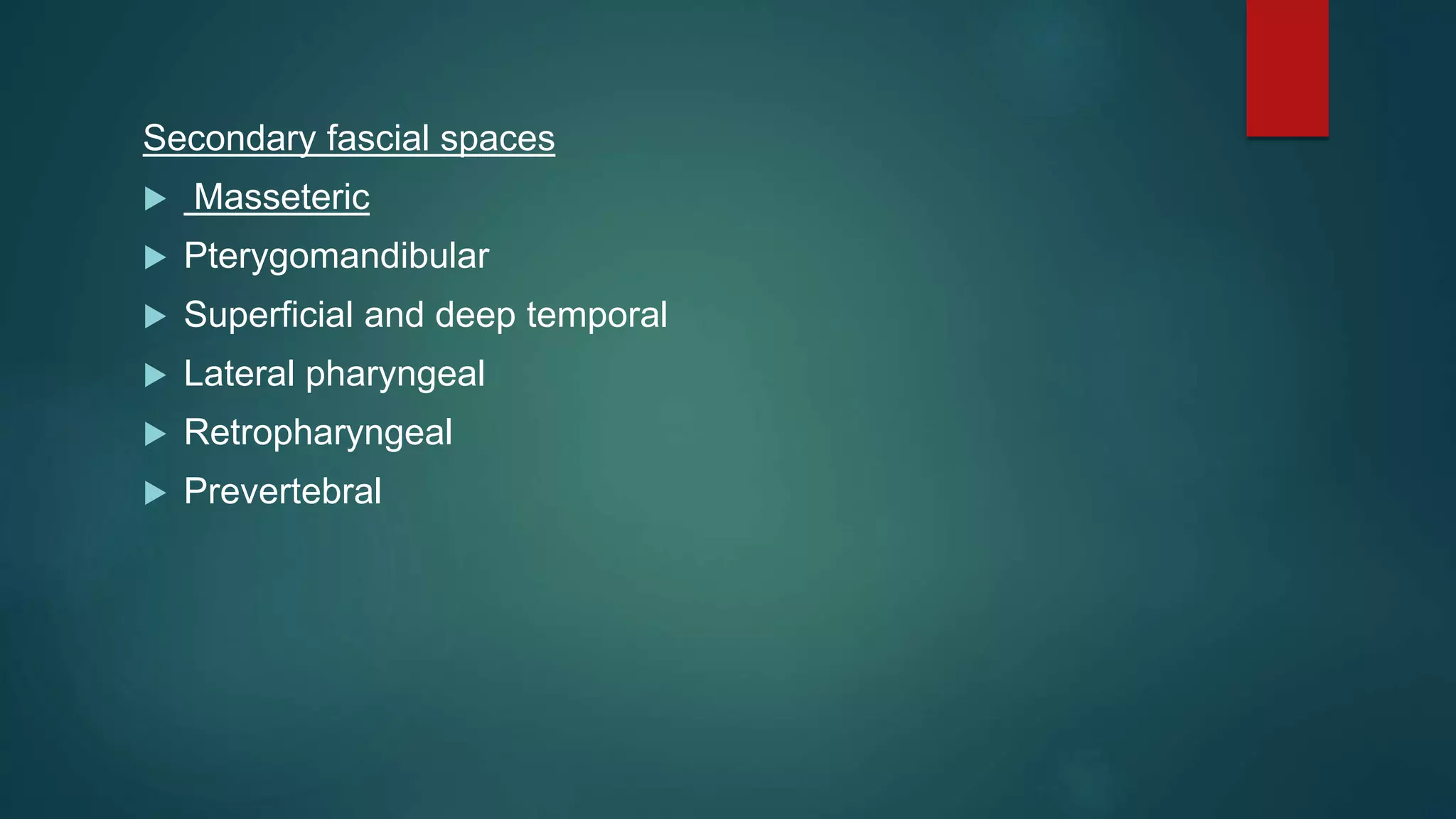
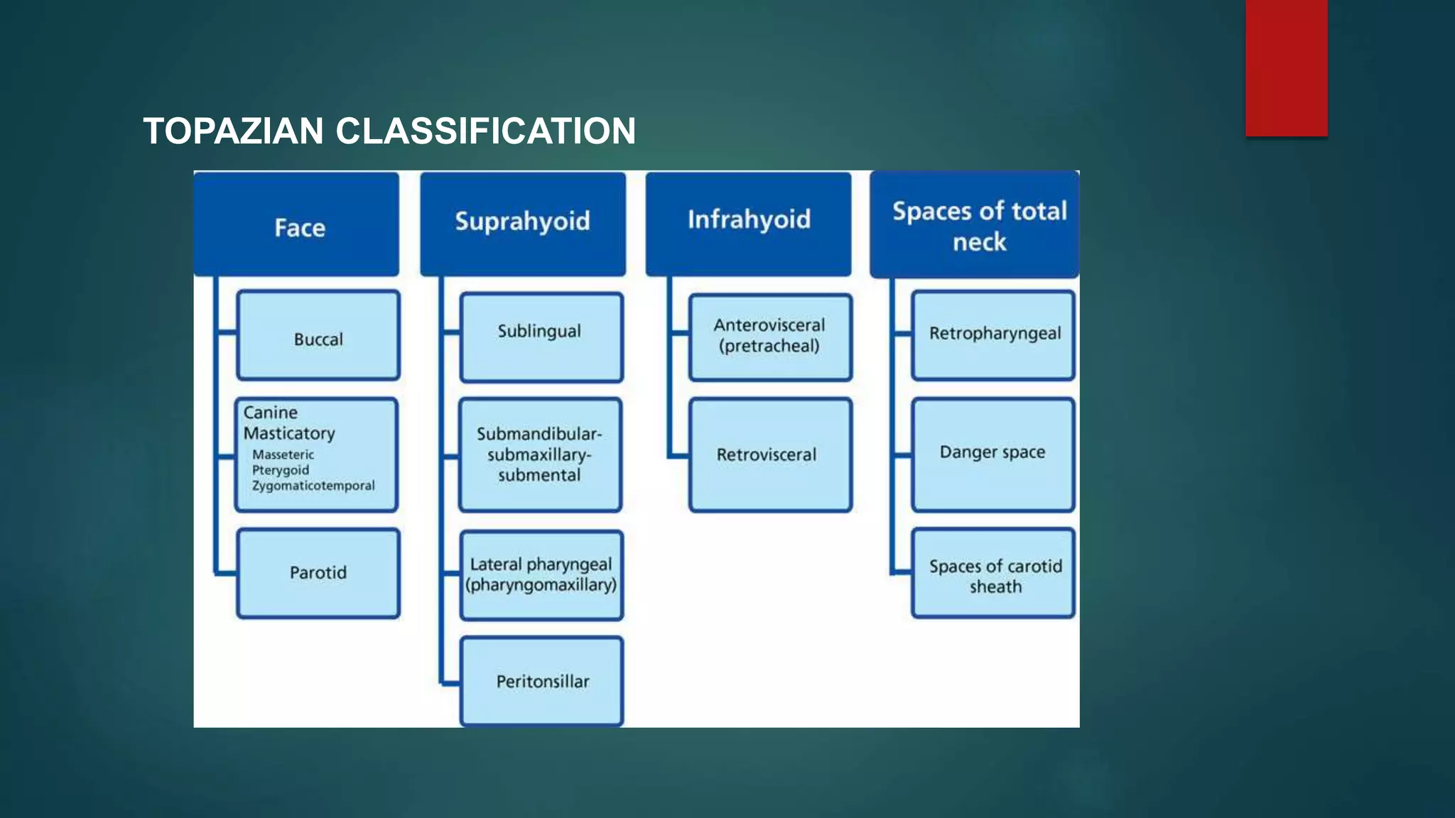
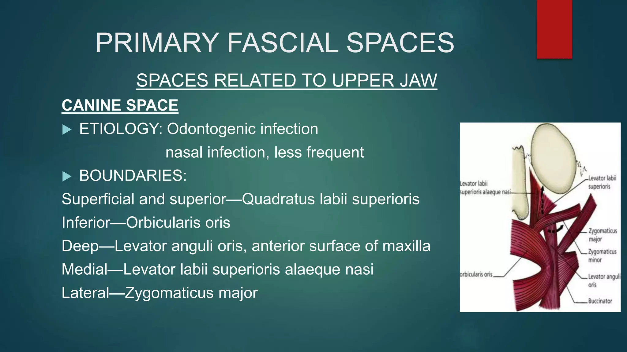
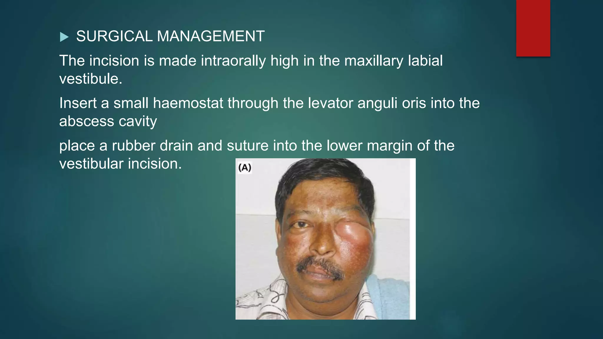
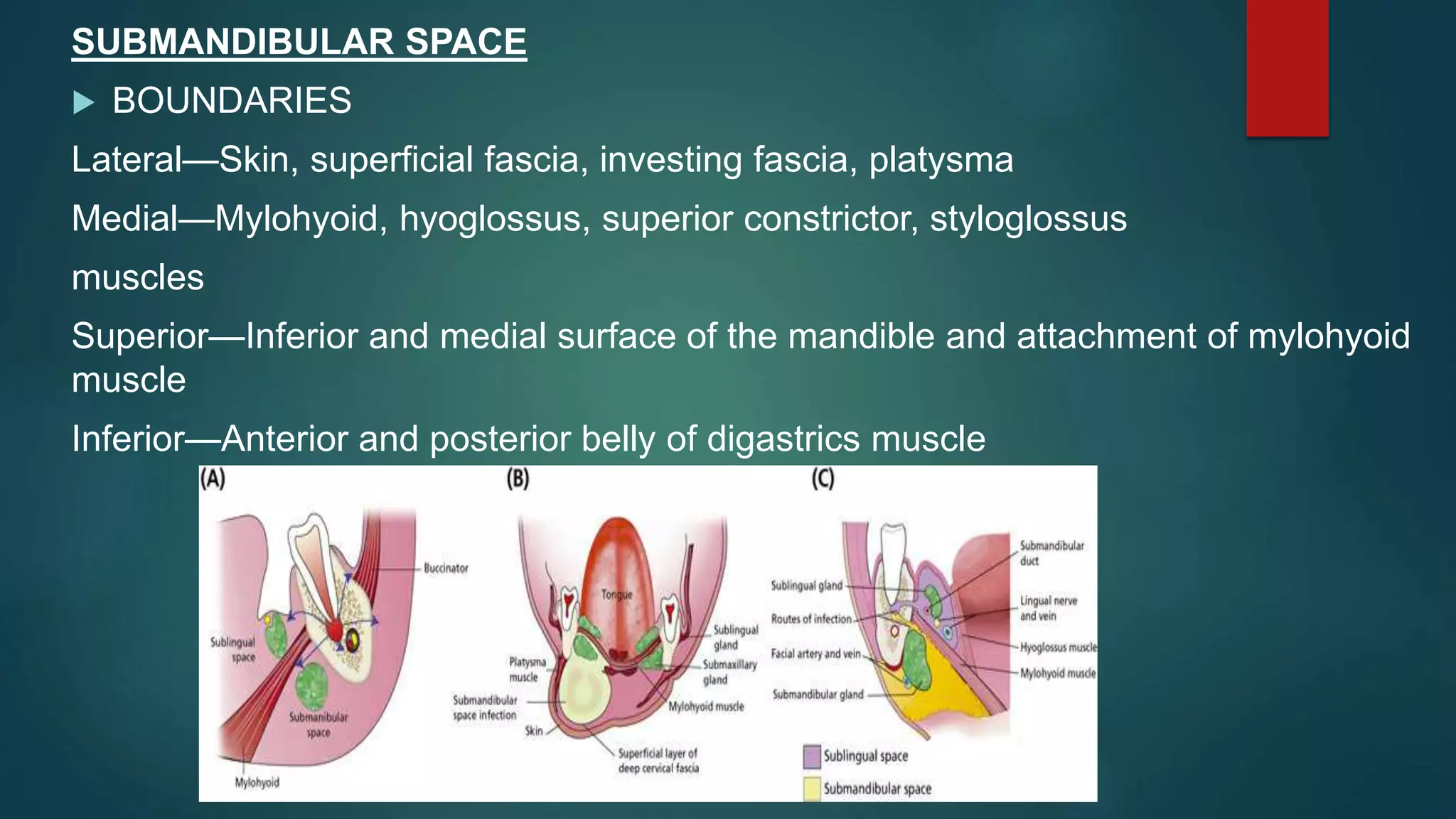
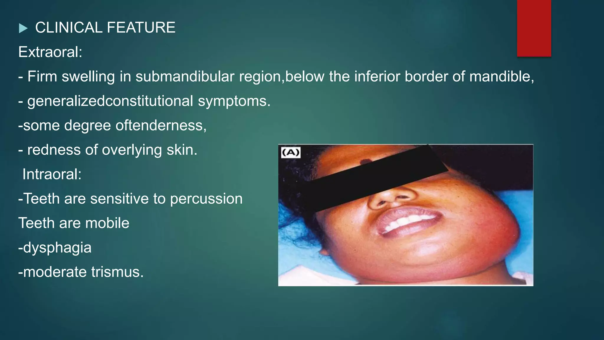
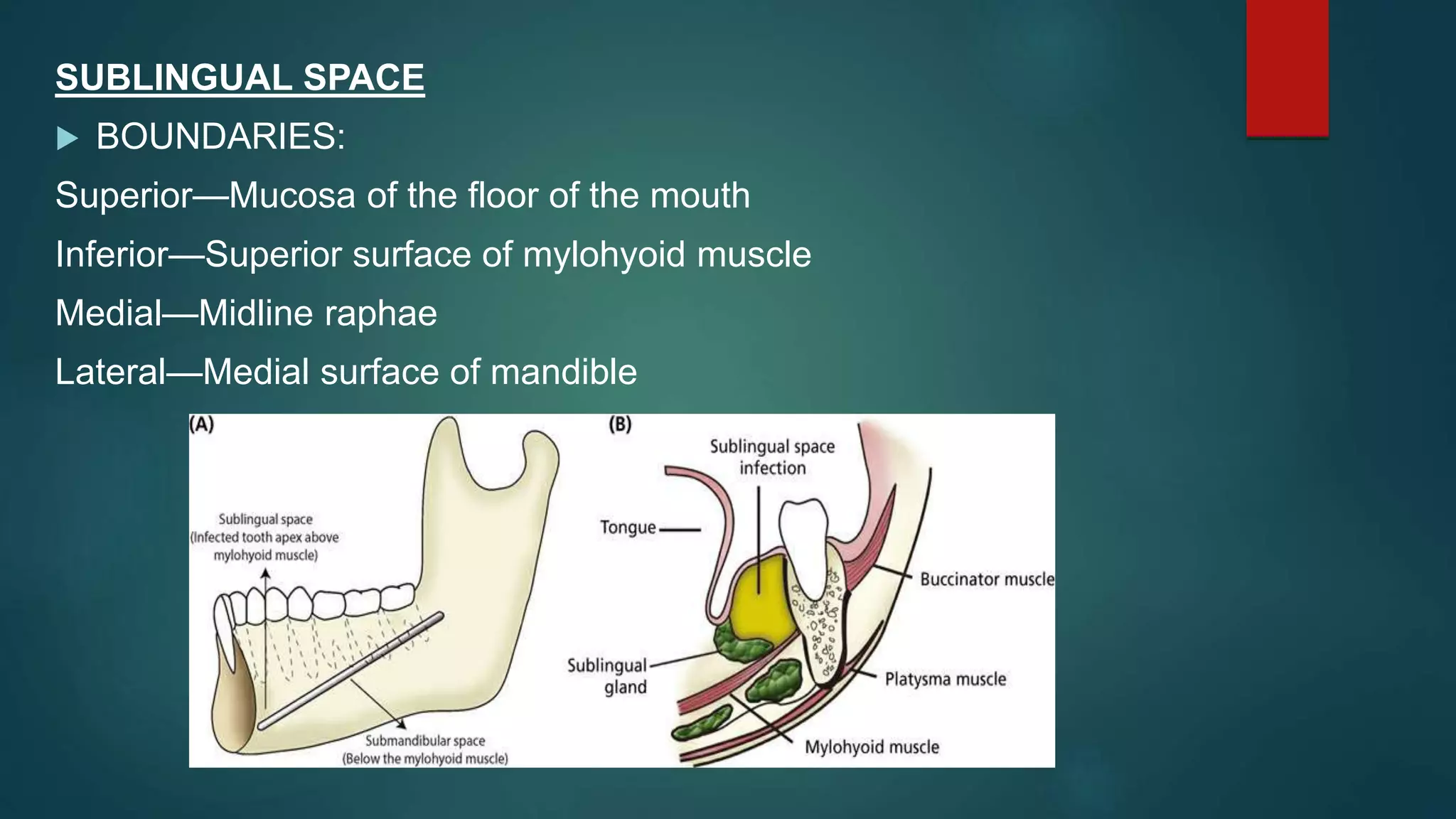
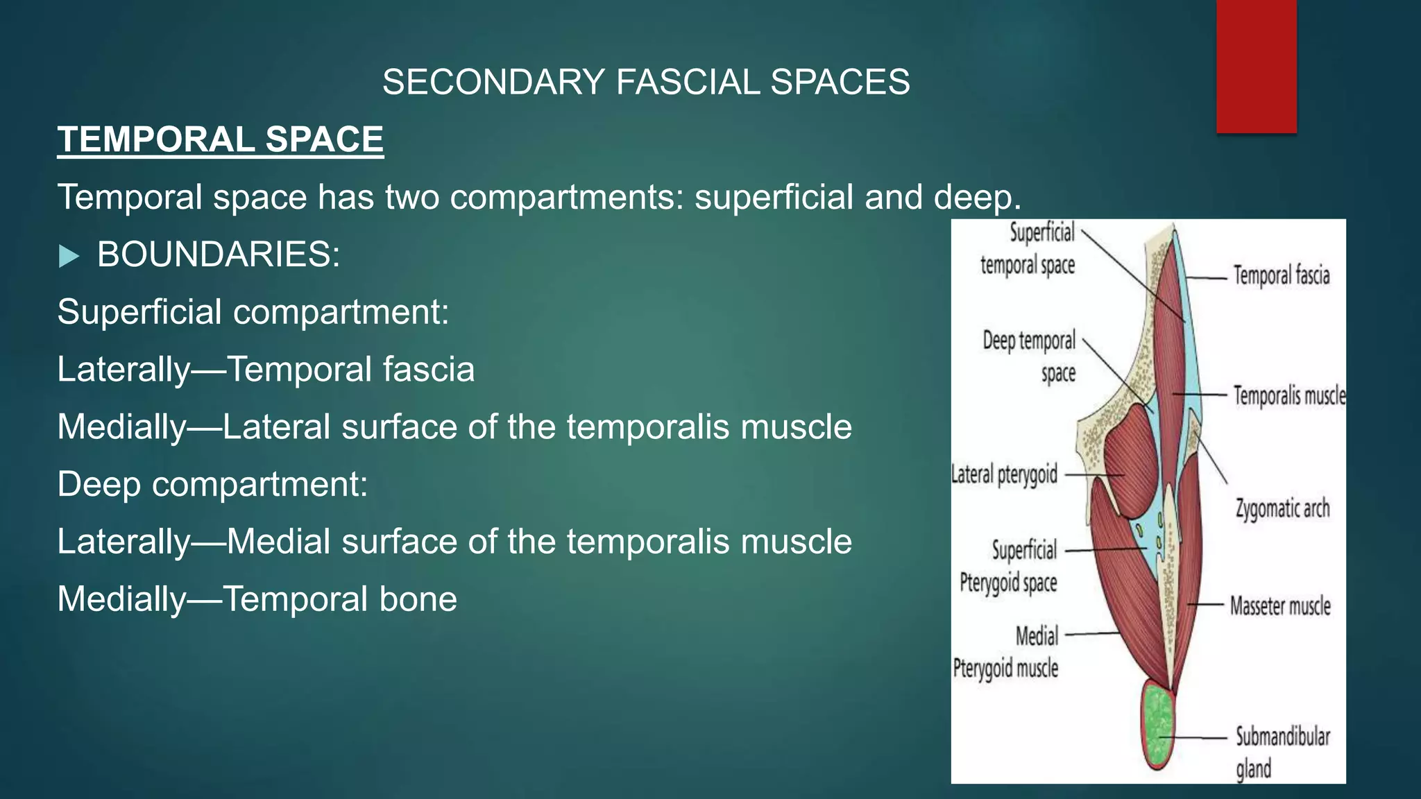
SlideShare a Scribd company logo
Space infection
DR DAVIS NADAKKAVUKARAN .M.D.S
READER
MALABAR DENTAL COLLEGE
CONTENTS
 INTRODUCTION
 CLASSIFIACTION
 SUPRASTERNAL SPACE
 PRIMARY FASCIAL SPACE
- RELATED TO UPPER JAW
- RELATED TO LOWER JAW
 SECONDARY FASCIAL SPACE
 LIFE THREATENING COMPLICATIONS
 CONCLUSION
 REFERENCE
INTRODUCTION
 Soft tissue infections of head and neck are commonly
encountered in routine practice of oral and maxillofacial surgery,
 In case of infection the classic signs and symptoms—pain,
swelling, surface erythema, lymphadenopathy, and systemically-
fever, malaise, toxic appearance, and an elevated white blood
cellcount is present
DEFINITION
 Shapiro defined fascial spaces as potential spaces between the
layer of fascia. These spaces are normally filled with loose
connective tissues and various structures like veins, arteries,
glands, lymph nodes, etc.
CLASSIFICATION OF FASCIAL
SPACE
BASED ON CLINICAL SIGNIFICANCE
Primary maxillary spaces
 Canine (infraorbital)
 Buccal
 Infratemporal
Primary mandibular spaces
 Submental
 Submandibular
 Sublingual
 Buccal
Secondary fascial spaces
 Masseteric
 Pterygomandibular
 Superficial and deep temporal
 Lateral pharyngeal
 Retropharyngeal
 Prevertebral
TOPAZIAN CLASSIFICATION
GRODINSKY & HOLYOKE CLASSIFICATION
 Space 1 : potential space superficial & deep to platysma muscle
 Space 2 : space behind the anterior layer of deep cervical fascia
 Space 3 : pretracheal space lies anterior to trachea
 Space 3 A : viscera vascular space( lincolns highway as coined by
Mosher)
 Space 4 : Danger area b/w alar & prevertebral fascia
BASED ON MODE OF INVOLVEMENT
Direct involvement:
 Primary spaces
1. Maxillary spaces
2. Mandibular spaces
Indirect involvement:
 Secondary spaces
Anatomic spaces of head & neck
SUPRASTERNAL SPACE( OF BURNS)
 The suprasternal space (of Burns) is a space of the inferior
neck. Superficial fascia splits below the level of the hyoid
bone to form two spaces:
1. It forms the lower part of the root of the posterior triangle, the
fascia splits into two layers, both of which are attached to the
clavicle.
2. It forms the lower part of the roof of the anterior triangle and
the fascia splits in form the suprasternal space or the space of
the ‘burns’.
CONTENTS
 Sternal head of sternocleidomastoid muscle
 Anterior Jugular vein anastomoses
 Lymph nodes
 Interclavicular ligament
PRIMARY FASCIAL SPACES
SPACES RELATED TO UPPER JAW
CANINE SPACE
 ETIOLOGY: Odontogenic infection
nasal infection, less frequent
 BOUNDARIES:
Superficial and superior—Quadratus labii superioris
Inferior—Orbicularis oris
Deep—Levator anguli oris, anterior surface of maxilla
Medial—Levator labii superioris alaeque nasi
Lateral—Zygomaticus major
 CONTENT:
Angular artery and vein
Infraorbital nerve
 TEETH INVOLVED
Maxillary canine, 1st premolar infection and sometimes mesiobuccal root of first
molars
 CLINICAL FEATURES
‱ Periapical abscess of canine usually present as labial sulcus swelling
and less commonly as palatal swelling
‱ Swelling of the cheek and upper lip (vestibular abscess)
‱ Obliteration of the nasolabial fold
‱ Oedema of the lower eyelid.
‱ Marked periorbital oedema forcing the eyelid to close.
‱ Marked tenderness and redness in the facial tissue.
 SURGICAL MANAGEMENT
The incision is made intraorally high in the maxillary labial
vestibule.
Insert a small haemostat through the levator anguli oris into the
abscess cavity
place a rubber drain and suture into the lower margin of the
vestibular incision.
BUCCAL SPACE
 BOUNDARIES
Superior—Zygomatic arch
Inferior—Inferior border of mandible
Anterior—Posterior border of the zygomatic bone above and
depressor angulioris below
Posterior—Anterior border of the masseter muscle
Medial—Buccinator muscle and its fascia
Lateral—Skin and subcutaneous tissue.
 CONTENTS
Space filled with buccal pad of fat (adipose tissues)
Parotid duct
Anterior and transverse facial artery and vein.
 TEETH INVOLVED
Maxillary and mandibular premolars and molars.
 CLINICAL FEATURES
-Pus acumination leads to gum boil seen in vestibule
-Prominent extra oral swelling
-swelling extending from lower border of mandible to infraorbital
margin & from anterior margin of masseter to corner of mouth.
-Edema of lower eyelid.
 SURGICAL MANAGEMENT
Horizontal incision through the oral mucosa of the cheek in the
premolar molar region
If the pus is lateral to the muscle, then the muscle is penetrated
with curved mosquito forceps to enter the buccal space
Drain is placed & secured with suture
INFRATEMPORAL SPACE
Also called as retrozygomatic space as it is partly situated behind the
zygomatic bone
 BOUNDARIES
Superior—Skull base-sphenoid crest
Inferior—Lateral pterygoid muscle
Medial—Lateral pterygoid plate
Lateral—Temporalis muscle and tendon
Anterior—Maxillary tuberosity
Posterior—Mandibular condyle
 CONTENTS
-Internal maxillary artery (second part)
- Pterygoid venous plexus
-Mandibular division of trigeminal nerve
-Medial and lateral pterygoid muscles
 INVOLVEMENT
Infratemporal fossa may also become secondarily infected from
infections of the submasseteric, parotid and lateral pharyngeal
spaces.
 CLINICAL FEATURES
EXTRA ORAL- trimus
- bulging of temporalis muscle
- marked swelling of the face
- eye is closed & often proptosed
INTRAORAL – swelling in the tuberosity zone
 SURGICAL MANAGEMENT
Infratemporal space can be reached either intraorally or extraorally.
-Internal approach (Kruger) consists of an incision made in the
buccolabial fold lateral to the maxillary third molar.
-A curved haemostat is introduced carefully behind the tuberosity of
the maxilla and directed medially andsuperiorly within the cavity.
-A drain is then inserted.
-According to Laskin, a vertical incision is made medial to the upper
extentof the anterior border of ramus of the mandible.
- A haemostat is introduced and passed superiorly into the
infratemporal region and a drain is introduced.
SPACES RELATED TO LOWER JAW
SUBMENTAL SPACE
 BOUNDARIES
Superior—Mylohyoid muscle
Inferior—Skin and subcutaneous tissue, platysma and deep cervical
fascia
Medial—Single midline space with no medial wall
Lateral—Anterior belly of digastric (bilateral)
Anterior—Mandible
Posterior—Hyoid bone
 CONTENTS
no vital structures
Lymph nodes and anterior jugular veins
 INVOLVEMENT
Infection from lower incisors, lower lip, chin, tip of the tongue and
anterior part of floor of the mouth
 CLINICAL FEATURE
Extraoral findings- Distinct,
-firm swelling in midline,beneath the chin.
-Skin overlying the swelling is board-like and taut.
- Fluctuation may be present.
Intraoral findings: The anterior teeth are either nonvital, fractured or
carious.
The offending tooth may exhibit tenderness to percussion and may
showmobility.
The patient may experience considerable discomfort on swallowing.
 SURGICAL MANAGEMENT
-The incision for drainage is made bilaterally through the skin,
subcutaneous tissue and platysma muscle at the most inferior aspect
of the swelling.
-Rubber drain is inserted through one incision, exited through the
other and secured with the help of sutures and dressing applied
SUBMANDIBULAR SPACE
 BOUNDARIES
Lateral—Skin, superficial fascia, investing fascia, platysma
Medial—Mylohyoid, hyoglossus, superior constrictor, styloglossus
muscles
Superior—Inferior and medial surface of the mandible and attachment of mylohyoid
muscle
Inferior—Anterior and posterior belly of digastrics muscle
 CONTENTS
-Submandibular salivary gland and lymph nodes
- Facial artery
- Lingual nerve
-Lymph nodes
 INVOLVEMENT
-Infection from the mandibular molars, most commonly second and
third molar
-Infection from submental and sublingual spaces
-Infection from the submandibular salivary gland
-Infection from the middle third of the tongue, posterior part of the floor
of the mouth, maxillary teeth, cheek, maxillary sinus and palate
 CLINICAL FEATURE
Extraoral:
- Firm swelling in submandibular region,below the inferior border of mandible,
- generalizedconstitutional symptoms.
-some degree oftenderness,
- redness of overlying skin.
Intraoral:
-Teeth are sensitive to percussion
Teeth are mobile
-dysphagia
-moderate trismus.
 SURGICAL MANAGEMENT
-Two stab incisions are made at the inferior aspect of the swelling in
the shadow of the mandible.
-The dissection is carried out through one of the incisions with the
curved haemostat in the abscess cavity.
-Blunt dissection avoids the risk of injuring the facial artery, anterior
facial vein and facial nerve.
-The haemostat is passed through one incision and out through the
other.
-A thin rubber drain is passed through the stab incisions with the help
of the haemostat.
-The ends of the drain are sutured to prevent dislodgement.
SUBLINGUAL SPACE
 BOUNDARIES:
Superior—Mucosa of the floor of the mouth
Inferior—Superior surface of mylohyoid muscle
Medial—Midline raphae
Lateral—Medial surface of mandible
 CONTENTS:
-Deep part of submandibular gland, sublingual gland and their
draining ducts (Wharton’s duct and ducts of Rivinus)
-Lingual nerve
 CLINICAL FEATURE:
Extraoral:
- There is little or no swelling.
-The lymnhnodes may be enlarged and tender.
- Pain and discomfoon deglutition.
- Speech may be affected.
Intraoral:
Firm, painful swelling seen in the floor of themouth on the affected side.
-The floor of the mouth is raised.
-The tongue may be pushed superiorly.
-airway obstruction.
-The ability to protrudethe tongue beyond the vermillion border of upper
lip is affected.
 SURGICAL MANAGEMENT:
Drainage of the abscess is obtained through
Extra oral approach—an external transverse skin incision between the
hyoid bone and the inferior border of the mandible.
Intra oral approach—Drainage can be obtained transorally by incising
the mucosa in the anterior part of the floor of the mouth, the incision
should be placed parallel to the submandibular duct.
Blunt dissection is indicated so as to not injure the lingual nerve or the
submandibular gland
SECONDARY FASCIAL SPACES
TEMPORAL SPACE
Temporal space has two compartments: superficial and deep.
 BOUNDARIES:
Superficial compartment:
Laterally—Temporal fascia
Medially—Lateral surface of the temporalis muscle
Deep compartment:
Laterally—Medial surface of the temporalis muscle
Medially—Temporal bone
 CONTENTS:
Superficial temporal vessels, auriculotemporal nerve and temporal fat
pad.
 CLINICAL FEATURE:
- Pain and swelling
- Swelling over the temporal region
 SURGICAL MANAGEMENT:
-extra oral incirion in temporal region, above hairline 45 degree to
zygomatic arch
-hemostat is entered above & below the temporalis muscle
PAROTID SPACE
Parotid space is enclosed by the superficial layer of the deep
cervical fascia surrounding the parotid gland.
 BOUNDARIES
-Space is formed by splitting of superficial layer of deep cervical
fascia surrounding parotid gland
- lies posterior to masticatory space.
Inferiorly : stylomandibular ligament separates parotid from
mandibular space.
 CONTENTS:
-parotid gland &lymph nodes
-facial nerve
-retromandibular vein
-external carotid artery
 CLINICAL FEATURE:
-Swelling.
Swelling everts the lobule of the ear and presents with severe pain
especially while eating.
-Intraorally pus may be milked from the parotid duct.
 MANAGEMENT
-Large incision is made in the retromandibular area from lower aspect
of lobule of the ear to angle of the mandible.
-Blunt dissection with a haemostat is done avoiding injury to the
branches of the facial nerve.
-Multiple drains are used for drainage of the pus.
-A curved incision at the angle of the mandible can also be made;
blunt dissection is done with a haemostat and a drain is placed.
SUBMASSETERIC SPACE
 BOUNDARIES:
Anterior—Buccal space, parotidomasseteric fascia
Posterior—Parotid gland and its fascia
Superior—Zygomatic arch
Inferior—Inferior border of mandible
Superficial or medial—Ascending ramus
Deep or lateral—Masseter muscle
 CONTENTS
Masseteric artery & vein
 CLINICAL FEATURE:
-Extraorally, the swelling is seen mainly over the angle of the
mandible.
-severe trismus and throbbing pain
-Chronic submasseteric space infection can be punctuated by
recurrent exacerbation
-subperiosteal new bone deposition beneath the periosteum, an
important clue to the diagnosis.
 SURGICAL MANAGEMENT
-A vertical incision is made intraorally along the external oblique line of
the mandible.
-A haemostat is inserted through this incision and passed posteriorly
along the lateral aspect of the ramus beneath the masseter muscle and
the beaks are opened for free escape of the pus.
-A rubber drain is inserted and sutured to the incision margin.
-Extraoral approach involves a small incision beneath the angle of the
mandible and blunt dissection is done with the help of the haemostat.
- A rubber catheter is inserted in the wound for drainage
PTERYGOMANDIBULAR SPACE
 BOUNDARY:
Anterior—Buccal space
Posterior—Parotid gland with lateral pharyngeal space
Superior—Lateral pterygoid muscle Inferior—Inferior border of
mandible
Superficial or medial—Lateral surface of medial pterygoid muscle
Deep or lateral—Medial surface of ascending ramus of mandible
 CLINICAL FEATURE
-Extraorally swelling
-Intraorally, there is visible swelling of the soft palate on the same
side
-swelling of the anterior tonsillar pillar
-deviation of the uvula to the opposite side
-severe trismus and dysphagia.
 SURGICAL MANAGEMENT
-The incision for drainage is made between medial aspect of the
ramus of mandible and the pterygomandibular raphe,
-the abscess cavity is opened by blunt dissection using a haemostat.
-Rubber drain is placed and sutured to one of the margins of the
incision to prevent dislodgement.
-This would help in sufficientdrainage.
LATERAL PHARYNGEAL SPACE
 BOUNDARIES:
Anterior—Superior and middle pharyngeal constrictor
Posterior—Carotid sheath, stylohyoid, styloglossus and
stylopharyngeus
Superior—Skull base
Inferior—Hyoid bone
Superficial or medial—Superior pharyngeal constrictors and
retropharyngeal space
Deep or lateral—Medial pterygoid muscle and capsule of parotid
gland
 CLINICAL FEATURE
-Severe pain on the affected side of throat and dysphagia are present
-tonsil, tonsillar pillar and uvula are displaced to the medial side.
-The four cardinal signs of lateral pharyngeal abscess are trismus, induration
and swelling of angle of the jaw, fever and pharyngeal bulging.
-Rotation of the neck away from the side of the swelling causes severe pain
-Complications of lateral pharyngeal abscess include septic jugular thrombophlebitis
and carotid artery erosion.
-Inequality of the pupils due to involvement of cervical sympathetic and
bleeding from nose, mouth or ear can be a warning of such a disastrous sequel.
-infections have a potential to spread upwards causing cavernous sinus
thrombosis, meningitis and brain abscess.
-They can also spread into theretropharyngeal space.
 SURGICAL MANAGEMENT
-intraoral,extraoral and a combination of both.
-Intraoral incision can be either transpharyngeal or lateral. The
transpharyngeal approach is made through the tonsillar fossa,
-Extraoral submandibular incision is the safest approach and should be
used if there is any involvement of posterior compartments.
-In the combined intraoral and extraoral approach, the lateral mucosal
incision is made and a large curved haemostat is passed lateral to the
superior constrictor and medial to the medial pterygoid muscle.
A blunt dissection is carried out posteroinferiorly below the angle of the
mandible.
RETROPHARYNGEAL SPACE
 BOUNDARIES:
Anterior—Superior and middle constrictors
Posterior—Alar fascia
Superior—Skull base
Inferior—Fusion of alar and prevertebral fascia at T4
Superficial or medial—Common space, no wall
Deep or lateral—Carotid sheath and lateral pharyngeal space
 CONTENTS:
Lymph node, no major structures
 CLINICAL FEATURES:
-symptoms include pain, fever, stiffness of the neck, dyspnoea,
drooling and dysphagia.
-Bulging of the posterior pharyngeal wall
- Retropharyngeal abscess is considered the most dangerous deep
neck space abscess, because complications include supraglottic
oedema with airway obstruction, aspiration pneumonia due to
rupture of the abscess and acute mediastinitis.
 MANAGEMENT
-an intraoral approach is made.
-A vertical incision is made on the pharyngeal wall lateral to the
midline.
- Using a haemostat, abscess cavity is opened by blunt dissection
while the patient is in Trendelenburg position to avoid aspiration of
the pus.
-In case of concern about the rupture of the abscess, extraoral
approach is used for drainage.
 PERITONSILLAR ABSCESS ( QUINSY)
-Peritonsillar abscess or quinsy is a deep neck infection usually
secondary to contiguous spread from the local sites or as a
complication of acute tonsillitis
-rarely life threatening in itself
 CLINICAL FEATURE
-The infection is characterised by swelling of the tonsils
-uvular displacement
-trismus and muffled voice.
-Quinsy is usually unilateral
-Most abscesses occur in younger patients who present
with fever, sore throat and dysphagia
 COMPLICATIONS
Spontaneous rupture and aspiration, contiguous spread to
pterygomaxillary space.
 SURGICAL MANAGEMENT
If the patient is not seen until the pus is formed or if the antibiotic
therapy fails, the abscess must be drained.
But since peritonsillar abscess often tends to recur, tonsillectomy
should be performed 6–8 weeks after formation of the abscess.
LIFE THREATEMNING
COMPLICATIONS
 RELATED TO LOWER JAW
-lugwigs angina
-descending deep cellulitis of neck, resulting in mediastinitis
-carotid sheath invasion
 RELATED TO UPPER JAW
-Cavernous sinus thrombosis, brain abscess, dural meningitis &
osteomyelitis of skull
-retrobulbar cellulitis with possibility of blindness
CONCLUSION
 Infection of orofacial region & neck have one of the most
common disease in human being
 Despite great advancement in the healthcare , these infection
remains a major problem
 These infection range from periapical abscess to superficial &
deep abscess of neck
 Early recoginition and prompt appropriate treatment is
absolutely essential
REFRENCE
 Neelima anil malik; text book of oral & maxillofacial
surgery:5th edition
 S M Bhalaji;textbook of oral & maxillofacial surgery; 3rd
edition

More Related Content

What's hot

Management of condylar fractures
Management of condylar fracturesManagement of condylar fractures
Management of condylar fracturesdralimohammedhasan
 
Diseases of pulp and periapical tissues
Diseases of pulp and periapical tissues Diseases of pulp and periapical tissues
Diseases of pulp and periapical tissues
madhusudhan reddy
 
Clinical features and histopathology of dental caries
Clinical features and histopathology of dental cariesClinical features and histopathology of dental caries
Clinical features and histopathology of dental cariesSAGAR HIWALE
 
Primary spaces of space infection
Primary spaces of space infectionPrimary spaces of space infection
Primary spaces of space infection
Avishek Panda
 
space infection
space infectionspace infection
space infection
Dr Ramesh R
 
Dentinogenesis Imperfecta
Dentinogenesis ImperfectaDentinogenesis Imperfecta
Dentinogenesis Imperfecta
Dr. Santhu Sadasivan
 
PULP AND PERIAPICAL LESIONS OF THE TOOTH ppt
PULP AND PERIAPICAL LESIONS OF THE TOOTH pptPULP AND PERIAPICAL LESIONS OF THE TOOTH ppt
PULP AND PERIAPICAL LESIONS OF THE TOOTH ppt
K BHATTACHARJEE
 
Zones of enamel and dentinal caries completed
Zones of enamel and dentinal caries completedZones of enamel and dentinal caries completed
Zones of enamel and dentinal caries completed
Muhmmad Ammar Attari
 
Fascial space infection
Fascial space infectionFascial space infection
Fascial space infection
Abdullah Al-Mamun Tushar
 
Enamel hypoplasia ppt
Enamel hypoplasia pptEnamel hypoplasia ppt
Enamel hypoplasia ppt
Alankrita Sisodia
 
Case of space infection
Case of space infectionCase of space infection
Case of space infection
Dr Bhavik Miyani
 
Major Connectors
Major ConnectorsMajor Connectors
Major Connectors
Apurva Thampi
 
Glass ionomer cement
Glass ionomer cementGlass ionomer cement
Glass ionomer cement
Abhijeet Pallewar
 
Infections of head and neck
Infections of head and neckInfections of head and neck
Infections of head and neck
Mohammed Haneef Farooq
 
Juvenile Periodontitis
Juvenile Periodontitis Juvenile Periodontitis
Juvenile Periodontitis
Nusrat Fahmida
 
Glass Ionomer Cement (GIC) - Science of Dental materials
Glass Ionomer Cement (GIC) - Science of Dental materialsGlass Ionomer Cement (GIC) - Science of Dental materials
Glass Ionomer Cement (GIC) - Science of Dental materials
drabbasnaseem
 
Nerv block - PSA & GREATER PALATINE NERVE BLOCK
Nerv block - PSA & GREATER PALATINE NERVE BLOCKNerv block - PSA & GREATER PALATINE NERVE BLOCK
Nerv block - PSA & GREATER PALATINE NERVE BLOCK
Saadia Ashraf
 
Sequelae of dental caries
Sequelae of dental cariesSequelae of dental caries
Sequelae of dental caries
Babin Karmacharya
 
Mandibular nerve blocks techniques
Mandibular nerve blocks techniques Mandibular nerve blocks techniques
Mandibular nerve blocks techniques
ANNOOR DENTAL COLLEGE,MUVATTUPUZHA
 
maxillary nerve blocks
maxillary nerve blocksmaxillary nerve blocks

What's hot (20)

Management of condylar fractures
Management of condylar fracturesManagement of condylar fractures
Management of condylar fractures
 
Diseases of pulp and periapical tissues
Diseases of pulp and periapical tissues Diseases of pulp and periapical tissues
Diseases of pulp and periapical tissues
 
Clinical features and histopathology of dental caries
Clinical features and histopathology of dental cariesClinical features and histopathology of dental caries
Clinical features and histopathology of dental caries
 
Primary spaces of space infection
Primary spaces of space infectionPrimary spaces of space infection
Primary spaces of space infection
 
space infection
space infectionspace infection
space infection
 
Dentinogenesis Imperfecta
Dentinogenesis ImperfectaDentinogenesis Imperfecta
Dentinogenesis Imperfecta
 
PULP AND PERIAPICAL LESIONS OF THE TOOTH ppt
PULP AND PERIAPICAL LESIONS OF THE TOOTH pptPULP AND PERIAPICAL LESIONS OF THE TOOTH ppt
PULP AND PERIAPICAL LESIONS OF THE TOOTH ppt
 
Zones of enamel and dentinal caries completed
Zones of enamel and dentinal caries completedZones of enamel and dentinal caries completed
Zones of enamel and dentinal caries completed
 
Fascial space infection
Fascial space infectionFascial space infection
Fascial space infection
 
Enamel hypoplasia ppt
Enamel hypoplasia pptEnamel hypoplasia ppt
Enamel hypoplasia ppt
 
Case of space infection
Case of space infectionCase of space infection
Case of space infection
 
Major Connectors
Major ConnectorsMajor Connectors
Major Connectors
 
Glass ionomer cement
Glass ionomer cementGlass ionomer cement
Glass ionomer cement
 
Infections of head and neck
Infections of head and neckInfections of head and neck
Infections of head and neck
 
Juvenile Periodontitis
Juvenile Periodontitis Juvenile Periodontitis
Juvenile Periodontitis
 
Glass Ionomer Cement (GIC) - Science of Dental materials
Glass Ionomer Cement (GIC) - Science of Dental materialsGlass Ionomer Cement (GIC) - Science of Dental materials
Glass Ionomer Cement (GIC) - Science of Dental materials
 
Nerv block - PSA & GREATER PALATINE NERVE BLOCK
Nerv block - PSA & GREATER PALATINE NERVE BLOCKNerv block - PSA & GREATER PALATINE NERVE BLOCK
Nerv block - PSA & GREATER PALATINE NERVE BLOCK
 
Sequelae of dental caries
Sequelae of dental cariesSequelae of dental caries
Sequelae of dental caries
 
Mandibular nerve blocks techniques
Mandibular nerve blocks techniques Mandibular nerve blocks techniques
Mandibular nerve blocks techniques
 
maxillary nerve blocks
maxillary nerve blocksmaxillary nerve blocks
maxillary nerve blocks
 

Similar to space infection.pptx

OMFS mandibular space infection.pptx
OMFS mandibular space infection.pptxOMFS mandibular space infection.pptx
OMFS mandibular space infection.pptx
sooraj40
 
2 mandibular_spaceinfection-.pptx
2 mandibular_spaceinfection-.pptx2 mandibular_spaceinfection-.pptx
2 mandibular_spaceinfection-.pptx
DR DAVIS NADAKKAVUKARAN
 
Fascial spaces of jaws and management
Fascial spaces of jaws and managementFascial spaces of jaws and management
Fascial spaces of jaws and management
soumya12345678910
 
MAXILLARY SPACE INFECTION.pptx
MAXILLARY SPACE INFECTION.pptxMAXILLARY SPACE INFECTION.pptx
MAXILLARY SPACE INFECTION.pptx
DR DAVIS NADAKKAVUKARAN
 
fascial space.pptx
fascial space.pptxfascial space.pptx
fascial space.pptx
mayankgupta672202
 
DAVIS.pptx
DAVIS.pptxDAVIS.pptx
Fascialspaceinfections 140219122023-phpapp01
Fascialspaceinfections 140219122023-phpapp01Fascialspaceinfections 140219122023-phpapp01
Fascialspaceinfections 140219122023-phpapp01Pradyumna Khairnar
 
Floor of the mouth
Floor of the mouthFloor of the mouth
Floor of the mouth
Nida Sumra
 
SURGICAL ANATOMY OF DEEP NECK SPACES
SURGICAL ANATOMY OF DEEP NECK SPACESSURGICAL ANATOMY OF DEEP NECK SPACES
SURGICAL ANATOMY OF DEEP NECK SPACES
Ajay Manickam
 
Space infection Presented by- Dr. himanshu gorawat
Space infection Presented by-  Dr. himanshu gorawat Space infection Presented by-  Dr. himanshu gorawat
Space infection Presented by- Dr. himanshu gorawat
Dr. Himanshu Gorawat
 
Space-infection ih
Space-infection  ihSpace-infection  ih
Space-infection ih
itrat hussain
 
Fascial space & infections
Fascial space & infectionsFascial space & infections
Fascial space & infectionsSurbhi Singh
 
Deep neck space infections
Deep neck space infectionsDeep neck space infections
Deep neck space infections
Ahlam Alzuway
 
Temporal and infratemporal region
Temporal and infratemporal regionTemporal and infratemporal region
Temporal and infratemporal region
Dr. Swathi Yennemadi
 
INFRATEMPORAL FOSSA AND PTERYGOPALATINE FOSSA NEW.pptx
INFRATEMPORAL FOSSA AND PTERYGOPALATINE FOSSA NEW.pptxINFRATEMPORAL FOSSA AND PTERYGOPALATINE FOSSA NEW.pptx
INFRATEMPORAL FOSSA AND PTERYGOPALATINE FOSSA NEW.pptx
Sudin Kayastha
 
Facial spaces and spread of odontogenic infection
Facial spaces and spread of odontogenic infectionFacial spaces and spread of odontogenic infection
Facial spaces and spread of odontogenic infection
Lubna Abu Alrub,DDS
 
Facial spaces and spread of odontogenic infection
Facial spaces and spread of odontogenic infectionFacial spaces and spread of odontogenic infection
Facial spaces and spread of odontogenic infection
Lubna Abu Alrub,DDS
 
ORAL INFECTIONS and facial space infections
ORAL INFECTIONS and facial space infectionsORAL INFECTIONS and facial space infections
ORAL INFECTIONS and facial space infections
WezzySinkala
 
SPACE INFECTIONS
SPACE INFECTIONSSPACE INFECTIONS
SPACE INFECTIONS
MINDS MAHE
 
SPACE INFECTIONS-TYPES, DIAGNSOSIS AND MANAGEMENT
SPACE INFECTIONS-TYPES, DIAGNSOSIS AND MANAGEMENTSPACE INFECTIONS-TYPES, DIAGNSOSIS AND MANAGEMENT
SPACE INFECTIONS-TYPES, DIAGNSOSIS AND MANAGEMENT
drash9955
 

Similar to space infection.pptx (20)

OMFS mandibular space infection.pptx
OMFS mandibular space infection.pptxOMFS mandibular space infection.pptx
OMFS mandibular space infection.pptx
 
2 mandibular_spaceinfection-.pptx
2 mandibular_spaceinfection-.pptx2 mandibular_spaceinfection-.pptx
2 mandibular_spaceinfection-.pptx
 
Fascial spaces of jaws and management
Fascial spaces of jaws and managementFascial spaces of jaws and management
Fascial spaces of jaws and management
 
MAXILLARY SPACE INFECTION.pptx
MAXILLARY SPACE INFECTION.pptxMAXILLARY SPACE INFECTION.pptx
MAXILLARY SPACE INFECTION.pptx
 
fascial space.pptx
fascial space.pptxfascial space.pptx
fascial space.pptx
 
DAVIS.pptx
DAVIS.pptxDAVIS.pptx
DAVIS.pptx
 
Fascialspaceinfections 140219122023-phpapp01
Fascialspaceinfections 140219122023-phpapp01Fascialspaceinfections 140219122023-phpapp01
Fascialspaceinfections 140219122023-phpapp01
 
Floor of the mouth
Floor of the mouthFloor of the mouth
Floor of the mouth
 
SURGICAL ANATOMY OF DEEP NECK SPACES
SURGICAL ANATOMY OF DEEP NECK SPACESSURGICAL ANATOMY OF DEEP NECK SPACES
SURGICAL ANATOMY OF DEEP NECK SPACES
 
Space infection Presented by- Dr. himanshu gorawat
Space infection Presented by-  Dr. himanshu gorawat Space infection Presented by-  Dr. himanshu gorawat
Space infection Presented by- Dr. himanshu gorawat
 
Space-infection ih
Space-infection  ihSpace-infection  ih
Space-infection ih
 
Fascial space & infections
Fascial space & infectionsFascial space & infections
Fascial space & infections
 
Deep neck space infections
Deep neck space infectionsDeep neck space infections
Deep neck space infections
 
Temporal and infratemporal region
Temporal and infratemporal regionTemporal and infratemporal region
Temporal and infratemporal region
 
INFRATEMPORAL FOSSA AND PTERYGOPALATINE FOSSA NEW.pptx
INFRATEMPORAL FOSSA AND PTERYGOPALATINE FOSSA NEW.pptxINFRATEMPORAL FOSSA AND PTERYGOPALATINE FOSSA NEW.pptx
INFRATEMPORAL FOSSA AND PTERYGOPALATINE FOSSA NEW.pptx
 
Facial spaces and spread of odontogenic infection
Facial spaces and spread of odontogenic infectionFacial spaces and spread of odontogenic infection
Facial spaces and spread of odontogenic infection
 
Facial spaces and spread of odontogenic infection
Facial spaces and spread of odontogenic infectionFacial spaces and spread of odontogenic infection
Facial spaces and spread of odontogenic infection
 
ORAL INFECTIONS and facial space infections
ORAL INFECTIONS and facial space infectionsORAL INFECTIONS and facial space infections
ORAL INFECTIONS and facial space infections
 
SPACE INFECTIONS
SPACE INFECTIONSSPACE INFECTIONS
SPACE INFECTIONS
 
SPACE INFECTIONS-TYPES, DIAGNSOSIS AND MANAGEMENT
SPACE INFECTIONS-TYPES, DIAGNSOSIS AND MANAGEMENTSPACE INFECTIONS-TYPES, DIAGNSOSIS AND MANAGEMENT
SPACE INFECTIONS-TYPES, DIAGNSOSIS AND MANAGEMENT
 

More from DR DAVIS NADAKKAVUKARAN

MYOFACIAL PAIN DYSFUNCTION SYNDROME.pptx
MYOFACIAL PAIN DYSFUNCTION SYNDROME.pptxMYOFACIAL PAIN DYSFUNCTION SYNDROME.pptx
MYOFACIAL PAIN DYSFUNCTION SYNDROME.pptx
DR DAVIS NADAKKAVUKARAN
 
ODONTOGENIC TUMORS.pptx
ODONTOGENIC TUMORS.pptxODONTOGENIC TUMORS.pptx
ODONTOGENIC TUMORS.pptx
DR DAVIS NADAKKAVUKARAN
 
6 canine impaction .pptx
6 canine impaction .pptx6 canine impaction .pptx
6 canine impaction .pptx
DR DAVIS NADAKKAVUKARAN
 
MANDIBULAR FRACTURE.pptx
MANDIBULAR FRACTURE.pptxMANDIBULAR FRACTURE.pptx
MANDIBULAR FRACTURE.pptx
DR DAVIS NADAKKAVUKARAN
 
LUDWIG’S ANGINA - DAVISpptx
LUDWIG’S ANGINA - DAVISpptxLUDWIG’S ANGINA - DAVISpptx
LUDWIG’S ANGINA - DAVISpptx
DR DAVIS NADAKKAVUKARAN
 
ZYGOMATIC COMPLEX FRACTURE DAVISpptx
ZYGOMATIC COMPLEX FRACTURE DAVISpptxZYGOMATIC COMPLEX FRACTURE DAVISpptx
ZYGOMATIC COMPLEX FRACTURE DAVISpptx
DR DAVIS NADAKKAVUKARAN
 
5 AUTOIMMUNE DISEASES OF SALIVARY GLAND SNISHA.pptx
5 AUTOIMMUNE DISEASES OF SALIVARY GLAND SNISHA.pptx5 AUTOIMMUNE DISEASES OF SALIVARY GLAND SNISHA.pptx
5 AUTOIMMUNE DISEASES OF SALIVARY GLAND SNISHA.pptx
DR DAVIS NADAKKAVUKARAN
 
MANDIBULAR OSTEOTOMIES.pptx
MANDIBULAR OSTEOTOMIES.pptxMANDIBULAR OSTEOTOMIES.pptx
MANDIBULAR OSTEOTOMIES.pptx
DR DAVIS NADAKKAVUKARAN
 
CONDYLAR FRACTURE.pptx
CONDYLAR FRACTURE.pptxCONDYLAR FRACTURE.pptx
CONDYLAR FRACTURE.pptx
DR DAVIS NADAKKAVUKARAN
 
MAXILLARY FRACTURE.pptx
MAXILLARY FRACTURE.pptxMAXILLARY FRACTURE.pptx
MAXILLARY FRACTURE.pptx
DR DAVIS NADAKKAVUKARAN
 
SURGICAL ANATOMY OF TEMPOROMANDIBULAR JOINT.pptx
SURGICAL ANATOMY OF TEMPOROMANDIBULAR JOINT.pptxSURGICAL ANATOMY OF TEMPOROMANDIBULAR JOINT.pptx
SURGICAL ANATOMY OF TEMPOROMANDIBULAR JOINT.pptx
DR DAVIS NADAKKAVUKARAN
 
SIALOLITHIASIS - OMFS.pptx
SIALOLITHIASIS -  OMFS.pptxSIALOLITHIASIS -  OMFS.pptx
SIALOLITHIASIS - OMFS.pptx
DR DAVIS NADAKKAVUKARAN
 
BENIGN ODONTOGENIC TUMORS.pptx
BENIGN ODONTOGENIC TUMORS.pptxBENIGN ODONTOGENIC TUMORS.pptx
BENIGN ODONTOGENIC TUMORS.pptx
DR DAVIS NADAKKAVUKARAN
 
DIABETES MELLITUS - sb.pptx
DIABETES MELLITUS - sb.pptxDIABETES MELLITUS - sb.pptx
DIABETES MELLITUS - sb.pptx
DR DAVIS NADAKKAVUKARAN
 
BLOOD SUPPLY OF HEAD & NECK.pptx
BLOOD SUPPLY OF HEAD & NECK.pptxBLOOD SUPPLY OF HEAD & NECK.pptx
BLOOD SUPPLY OF HEAD & NECK.pptx
DR DAVIS NADAKKAVUKARAN
 
Biomedical waste management
Biomedical waste management  Biomedical waste management
Biomedical waste management
DR DAVIS NADAKKAVUKARAN
 
Dry socket
Dry socket Dry socket
Chair position and finger rest
Chair position and finger restChair position and finger rest
Chair position and finger rest
DR DAVIS NADAKKAVUKARAN
 
5 tnm staging
5 tnm staging 5 tnm staging
5 tnm staging
DR DAVIS NADAKKAVUKARAN
 

More from DR DAVIS NADAKKAVUKARAN (20)

MYOFACIAL PAIN DYSFUNCTION SYNDROME.pptx
MYOFACIAL PAIN DYSFUNCTION SYNDROME.pptxMYOFACIAL PAIN DYSFUNCTION SYNDROME.pptx
MYOFACIAL PAIN DYSFUNCTION SYNDROME.pptx
 
ODONTOGENIC TUMORS.pptx
ODONTOGENIC TUMORS.pptxODONTOGENIC TUMORS.pptx
ODONTOGENIC TUMORS.pptx
 
6 canine impaction .pptx
6 canine impaction .pptx6 canine impaction .pptx
6 canine impaction .pptx
 
MANDIBULAR FRACTURE.pptx
MANDIBULAR FRACTURE.pptxMANDIBULAR FRACTURE.pptx
MANDIBULAR FRACTURE.pptx
 
LUDWIG’S ANGINA - DAVISpptx
LUDWIG’S ANGINA - DAVISpptxLUDWIG’S ANGINA - DAVISpptx
LUDWIG’S ANGINA - DAVISpptx
 
ZYGOMATIC COMPLEX FRACTURE DAVISpptx
ZYGOMATIC COMPLEX FRACTURE DAVISpptxZYGOMATIC COMPLEX FRACTURE DAVISpptx
ZYGOMATIC COMPLEX FRACTURE DAVISpptx
 
5 AUTOIMMUNE DISEASES OF SALIVARY GLAND SNISHA.pptx
5 AUTOIMMUNE DISEASES OF SALIVARY GLAND SNISHA.pptx5 AUTOIMMUNE DISEASES OF SALIVARY GLAND SNISHA.pptx
5 AUTOIMMUNE DISEASES OF SALIVARY GLAND SNISHA.pptx
 
MANDIBULAR OSTEOTOMIES.pptx
MANDIBULAR OSTEOTOMIES.pptxMANDIBULAR OSTEOTOMIES.pptx
MANDIBULAR OSTEOTOMIES.pptx
 
CONDYLAR FRACTURE.pptx
CONDYLAR FRACTURE.pptxCONDYLAR FRACTURE.pptx
CONDYLAR FRACTURE.pptx
 
MAXILLARY FRACTURE.pptx
MAXILLARY FRACTURE.pptxMAXILLARY FRACTURE.pptx
MAXILLARY FRACTURE.pptx
 
SURGICAL ANATOMY OF TEMPOROMANDIBULAR JOINT.pptx
SURGICAL ANATOMY OF TEMPOROMANDIBULAR JOINT.pptxSURGICAL ANATOMY OF TEMPOROMANDIBULAR JOINT.pptx
SURGICAL ANATOMY OF TEMPOROMANDIBULAR JOINT.pptx
 
SIALOLITHIASIS - OMFS.pptx
SIALOLITHIASIS -  OMFS.pptxSIALOLITHIASIS -  OMFS.pptx
SIALOLITHIASIS - OMFS.pptx
 
5 TNM STAGING .pptx
5 TNM STAGING .pptx5 TNM STAGING .pptx
5 TNM STAGING .pptx
 
BENIGN ODONTOGENIC TUMORS.pptx
BENIGN ODONTOGENIC TUMORS.pptxBENIGN ODONTOGENIC TUMORS.pptx
BENIGN ODONTOGENIC TUMORS.pptx
 
DIABETES MELLITUS - sb.pptx
DIABETES MELLITUS - sb.pptxDIABETES MELLITUS - sb.pptx
DIABETES MELLITUS - sb.pptx
 
BLOOD SUPPLY OF HEAD & NECK.pptx
BLOOD SUPPLY OF HEAD & NECK.pptxBLOOD SUPPLY OF HEAD & NECK.pptx
BLOOD SUPPLY OF HEAD & NECK.pptx
 
Biomedical waste management
Biomedical waste management  Biomedical waste management
Biomedical waste management
 
Dry socket
Dry socket Dry socket
Dry socket
 
Chair position and finger rest
Chair position and finger restChair position and finger rest
Chair position and finger rest
 
5 tnm staging
5 tnm staging 5 tnm staging
5 tnm staging
 

Recently uploaded

💘Ludhiana ℂall Girls 📞]][89011★83002][[ đŸ“± ❀ESCORTS service in Ludhiana💃💩Ludhi...
💘Ludhiana ℂall Girls 📞]][89011★83002][[ đŸ“± ❀ESCORTS service in Ludhiana💃💩Ludhi...💘Ludhiana ℂall Girls 📞]][89011★83002][[ đŸ“± ❀ESCORTS service in Ludhiana💃💩Ludhi...
💘Ludhiana ℂall Girls 📞]][89011★83002][[ đŸ“± ❀ESCORTS service in Ludhiana💃💩Ludhi...
ranishasharma67
 
R3 Stem Cells and Kidney Repair A New Horizon in Nephrology.pptx
R3 Stem Cells and Kidney Repair A New Horizon in Nephrology.pptxR3 Stem Cells and Kidney Repair A New Horizon in Nephrology.pptx
R3 Stem Cells and Kidney Repair A New Horizon in Nephrology.pptx
R3 Stem Cell
 
Antibiotic Stewardship by Anushri Srivastava.pptx
Antibiotic Stewardship by Anushri Srivastava.pptxAntibiotic Stewardship by Anushri Srivastava.pptx
Antibiotic Stewardship by Anushri Srivastava.pptx
AnushriSrivastav
 
Yemen National Tuberculosis Program .ppt
Yemen National Tuberculosis Program .pptYemen National Tuberculosis Program .ppt
Yemen National Tuberculosis Program .ppt
Esam43
 
CHAPTER 1 SEMESTER V PREVENTIVE-PEDIATRICS.pdf
CHAPTER 1 SEMESTER V PREVENTIVE-PEDIATRICS.pdfCHAPTER 1 SEMESTER V PREVENTIVE-PEDIATRICS.pdf
CHAPTER 1 SEMESTER V PREVENTIVE-PEDIATRICS.pdf
Sachin Sharma
 
India Clinical Trials Market: Industry Size and Growth Trends [2030] Analyzed...
India Clinical Trials Market: Industry Size and Growth Trends [2030] Analyzed...India Clinical Trials Market: Industry Size and Growth Trends [2030] Analyzed...
India Clinical Trials Market: Industry Size and Growth Trends [2030] Analyzed...
Kumar Satyam
 
GURGAON Call Girls ❀8901183002❀ #ℂALL# #gIRLS# In GURGAON â‚č,2500 Cash Payment...
GURGAON Call Girls ❀8901183002❀ #ℂALL# #gIRLS# In GURGAON â‚č,2500 Cash Payment...GURGAON Call Girls ❀8901183002❀ #ℂALL# #gIRLS# In GURGAON â‚č,2500 Cash Payment...
GURGAON Call Girls ❀8901183002❀ #ℂALL# #gIRLS# In GURGAON â‚č,2500 Cash Payment...
ranishasharma67
 
A Community health , health for prisoners
A Community health  , health for prisonersA Community health  , health for prisoners
A Community health , health for prisoners
Ahmed Elmi
 
Navigating Healthcare with Telemedicine
Navigating Healthcare with  TelemedicineNavigating Healthcare with  Telemedicine
Navigating Healthcare with Telemedicine
Iris Thiele Isip-Tan
 
Jaipur ❀cALL gIRLS 89O1183002 ❀ℂall Girls IN JaiPuR ESCORT SERVICE
Jaipur ❀cALL gIRLS 89O1183002 ❀ℂall Girls IN JaiPuR ESCORT SERVICEJaipur ❀cALL gIRLS 89O1183002 ❀ℂall Girls IN JaiPuR ESCORT SERVICE
Jaipur ❀cALL gIRLS 89O1183002 ❀ℂall Girls IN JaiPuR ESCORT SERVICE
ranishasharma67
 
The Importance of Community Nursing Care.pdf
The Importance of Community Nursing Care.pdfThe Importance of Community Nursing Care.pdf
The Importance of Community Nursing Care.pdf
AD Healthcare
 
The Docs PPG - 30.05.2024.pptx..........
The Docs PPG - 30.05.2024.pptx..........The Docs PPG - 30.05.2024.pptx..........
The Docs PPG - 30.05.2024.pptx..........
TheDocs
 
Dimensions of Healthcare Quality
Dimensions of Healthcare QualityDimensions of Healthcare Quality
Dimensions of Healthcare Quality
Naeemshahzad51
 
Surgery-Mini-OSCE-All-Past-Years-Questions-Modified.
Surgery-Mini-OSCE-All-Past-Years-Questions-Modified.Surgery-Mini-OSCE-All-Past-Years-Questions-Modified.
Surgery-Mini-OSCE-All-Past-Years-Questions-Modified.
preciousstephanie75
 
Introduction to Forensic Pathology course
Introduction to Forensic Pathology courseIntroduction to Forensic Pathology course
Introduction to Forensic Pathology course
fprxsqvnz5
 
How many patients does case series should have In comparison to case reports.pdf
How many patients does case series should have In comparison to case reports.pdfHow many patients does case series should have In comparison to case reports.pdf
How many patients does case series should have In comparison to case reports.pdf
pubrica101
 
Contact ME {89011**83002} Haridwar ℂall Girls By Full Service Call Girl In Ha...
Contact ME {89011**83002} Haridwar ℂall Girls By Full Service Call Girl In Ha...Contact ME {89011**83002} Haridwar ℂall Girls By Full Service Call Girl In Ha...
Contact ME {89011**83002} Haridwar ℂall Girls By Full Service Call Girl In Ha...
ranishasharma67
 
POLYCYSTIC OVARIAN SYNDROME (PCOS)......
POLYCYSTIC OVARIAN SYNDROME (PCOS)......POLYCYSTIC OVARIAN SYNDROME (PCOS)......
POLYCYSTIC OVARIAN SYNDROME (PCOS)......
Ameena Kadar
 
10 Ideas for Enhancing Your Meeting Experience
10 Ideas for Enhancing Your Meeting Experience10 Ideas for Enhancing Your Meeting Experience
10 Ideas for Enhancing Your Meeting Experience
ranishasharma67
 
Navigating Challenges: Mental Health, Legislation, and the Prison System in B...
Navigating Challenges: Mental Health, Legislation, and the Prison System in B...Navigating Challenges: Mental Health, Legislation, and the Prison System in B...
Navigating Challenges: Mental Health, Legislation, and the Prison System in B...
Guillermo Rivera
 

Recently uploaded (20)

💘Ludhiana ℂall Girls 📞]][89011★83002][[ đŸ“± ❀ESCORTS service in Ludhiana💃💩Ludhi...
💘Ludhiana ℂall Girls 📞]][89011★83002][[ đŸ“± ❀ESCORTS service in Ludhiana💃💩Ludhi...💘Ludhiana ℂall Girls 📞]][89011★83002][[ đŸ“± ❀ESCORTS service in Ludhiana💃💩Ludhi...
💘Ludhiana ℂall Girls 📞]][89011★83002][[ đŸ“± ❀ESCORTS service in Ludhiana💃💩Ludhi...
 
R3 Stem Cells and Kidney Repair A New Horizon in Nephrology.pptx
R3 Stem Cells and Kidney Repair A New Horizon in Nephrology.pptxR3 Stem Cells and Kidney Repair A New Horizon in Nephrology.pptx
R3 Stem Cells and Kidney Repair A New Horizon in Nephrology.pptx
 
Antibiotic Stewardship by Anushri Srivastava.pptx
Antibiotic Stewardship by Anushri Srivastava.pptxAntibiotic Stewardship by Anushri Srivastava.pptx
Antibiotic Stewardship by Anushri Srivastava.pptx
 
Yemen National Tuberculosis Program .ppt
Yemen National Tuberculosis Program .pptYemen National Tuberculosis Program .ppt
Yemen National Tuberculosis Program .ppt
 
CHAPTER 1 SEMESTER V PREVENTIVE-PEDIATRICS.pdf
CHAPTER 1 SEMESTER V PREVENTIVE-PEDIATRICS.pdfCHAPTER 1 SEMESTER V PREVENTIVE-PEDIATRICS.pdf
CHAPTER 1 SEMESTER V PREVENTIVE-PEDIATRICS.pdf
 
India Clinical Trials Market: Industry Size and Growth Trends [2030] Analyzed...
India Clinical Trials Market: Industry Size and Growth Trends [2030] Analyzed...India Clinical Trials Market: Industry Size and Growth Trends [2030] Analyzed...
India Clinical Trials Market: Industry Size and Growth Trends [2030] Analyzed...
 
GURGAON Call Girls ❀8901183002❀ #ℂALL# #gIRLS# In GURGAON â‚č,2500 Cash Payment...
GURGAON Call Girls ❀8901183002❀ #ℂALL# #gIRLS# In GURGAON â‚č,2500 Cash Payment...GURGAON Call Girls ❀8901183002❀ #ℂALL# #gIRLS# In GURGAON â‚č,2500 Cash Payment...
GURGAON Call Girls ❀8901183002❀ #ℂALL# #gIRLS# In GURGAON â‚č,2500 Cash Payment...
 
A Community health , health for prisoners
A Community health  , health for prisonersA Community health  , health for prisoners
A Community health , health for prisoners
 
Navigating Healthcare with Telemedicine
Navigating Healthcare with  TelemedicineNavigating Healthcare with  Telemedicine
Navigating Healthcare with Telemedicine
 
Jaipur ❀cALL gIRLS 89O1183002 ❀ℂall Girls IN JaiPuR ESCORT SERVICE
Jaipur ❀cALL gIRLS 89O1183002 ❀ℂall Girls IN JaiPuR ESCORT SERVICEJaipur ❀cALL gIRLS 89O1183002 ❀ℂall Girls IN JaiPuR ESCORT SERVICE
Jaipur ❀cALL gIRLS 89O1183002 ❀ℂall Girls IN JaiPuR ESCORT SERVICE
 
The Importance of Community Nursing Care.pdf
The Importance of Community Nursing Care.pdfThe Importance of Community Nursing Care.pdf
The Importance of Community Nursing Care.pdf
 
The Docs PPG - 30.05.2024.pptx..........
The Docs PPG - 30.05.2024.pptx..........The Docs PPG - 30.05.2024.pptx..........
The Docs PPG - 30.05.2024.pptx..........
 
Dimensions of Healthcare Quality
Dimensions of Healthcare QualityDimensions of Healthcare Quality
Dimensions of Healthcare Quality
 
Surgery-Mini-OSCE-All-Past-Years-Questions-Modified.
Surgery-Mini-OSCE-All-Past-Years-Questions-Modified.Surgery-Mini-OSCE-All-Past-Years-Questions-Modified.
Surgery-Mini-OSCE-All-Past-Years-Questions-Modified.
 
Introduction to Forensic Pathology course
Introduction to Forensic Pathology courseIntroduction to Forensic Pathology course
Introduction to Forensic Pathology course
 
How many patients does case series should have In comparison to case reports.pdf
How many patients does case series should have In comparison to case reports.pdfHow many patients does case series should have In comparison to case reports.pdf
How many patients does case series should have In comparison to case reports.pdf
 
Contact ME {89011**83002} Haridwar ℂall Girls By Full Service Call Girl In Ha...
Contact ME {89011**83002} Haridwar ℂall Girls By Full Service Call Girl In Ha...Contact ME {89011**83002} Haridwar ℂall Girls By Full Service Call Girl In Ha...
Contact ME {89011**83002} Haridwar ℂall Girls By Full Service Call Girl In Ha...
 
POLYCYSTIC OVARIAN SYNDROME (PCOS)......
POLYCYSTIC OVARIAN SYNDROME (PCOS)......POLYCYSTIC OVARIAN SYNDROME (PCOS)......
POLYCYSTIC OVARIAN SYNDROME (PCOS)......
 
10 Ideas for Enhancing Your Meeting Experience
10 Ideas for Enhancing Your Meeting Experience10 Ideas for Enhancing Your Meeting Experience
10 Ideas for Enhancing Your Meeting Experience
 
Navigating Challenges: Mental Health, Legislation, and the Prison System in B...
Navigating Challenges: Mental Health, Legislation, and the Prison System in B...Navigating Challenges: Mental Health, Legislation, and the Prison System in B...
Navigating Challenges: Mental Health, Legislation, and the Prison System in B...
 

space infection.pptx

  • 1. Space infection DR DAVIS NADAKKAVUKARAN .M.D.S READER MALABAR DENTAL COLLEGE
  • 2. CONTENTS  INTRODUCTION  CLASSIFIACTION  SUPRASTERNAL SPACE  PRIMARY FASCIAL SPACE - RELATED TO UPPER JAW - RELATED TO LOWER JAW  SECONDARY FASCIAL SPACE  LIFE THREATENING COMPLICATIONS  CONCLUSION  REFERENCE
  • 3. INTRODUCTION  Soft tissue infections of head and neck are commonly encountered in routine practice of oral and maxillofacial surgery,  In case of infection the classic signs and symptoms—pain, swelling, surface erythema, lymphadenopathy, and systemically- fever, malaise, toxic appearance, and an elevated white blood cellcount is present DEFINITION  Shapiro defined fascial spaces as potential spaces between the layer of fascia. These spaces are normally filled with loose connective tissues and various structures like veins, arteries, glands, lymph nodes, etc.
  • 4. CLASSIFICATION OF FASCIAL SPACE BASED ON CLINICAL SIGNIFICANCE Primary maxillary spaces  Canine (infraorbital)  Buccal  Infratemporal Primary mandibular spaces  Submental  Submandibular  Sublingual  Buccal
  • 5. Secondary fascial spaces  Masseteric  Pterygomandibular  Superficial and deep temporal  Lateral pharyngeal  Retropharyngeal  Prevertebral
  • 7. GRODINSKY & HOLYOKE CLASSIFICATION  Space 1 : potential space superficial & deep to platysma muscle  Space 2 : space behind the anterior layer of deep cervical fascia  Space 3 : pretracheal space lies anterior to trachea  Space 3 A : viscera vascular space( lincolns highway as coined by Mosher)  Space 4 : Danger area b/w alar & prevertebral fascia
  • 8. BASED ON MODE OF INVOLVEMENT Direct involvement:  Primary spaces 1. Maxillary spaces 2. Mandibular spaces Indirect involvement:  Secondary spaces
  • 9. Anatomic spaces of head & neck
  • 10. SUPRASTERNAL SPACE( OF BURNS)  The suprasternal space (of Burns) is a space of the inferior neck. Superficial fascia splits below the level of the hyoid bone to form two spaces: 1. It forms the lower part of the root of the posterior triangle, the fascia splits into two layers, both of which are attached to the clavicle. 2. It forms the lower part of the roof of the anterior triangle and the fascia splits in form the suprasternal space or the space of the ‘burns’.
  • 11. CONTENTS  Sternal head of sternocleidomastoid muscle  Anterior Jugular vein anastomoses  Lymph nodes  Interclavicular ligament
  • 12. PRIMARY FASCIAL SPACES SPACES RELATED TO UPPER JAW CANINE SPACE  ETIOLOGY: Odontogenic infection nasal infection, less frequent  BOUNDARIES: Superficial and superior—Quadratus labii superioris Inferior—Orbicularis oris Deep—Levator anguli oris, anterior surface of maxilla Medial—Levator labii superioris alaeque nasi Lateral—Zygomaticus major
  • 13.  CONTENT: Angular artery and vein Infraorbital nerve  TEETH INVOLVED Maxillary canine, 1st premolar infection and sometimes mesiobuccal root of first molars  CLINICAL FEATURES ‱ Periapical abscess of canine usually present as labial sulcus swelling and less commonly as palatal swelling ‱ Swelling of the cheek and upper lip (vestibular abscess) ‱ Obliteration of the nasolabial fold ‱ Oedema of the lower eyelid. ‱ Marked periorbital oedema forcing the eyelid to close. ‱ Marked tenderness and redness in the facial tissue.
  • 14.  SURGICAL MANAGEMENT The incision is made intraorally high in the maxillary labial vestibule. Insert a small haemostat through the levator anguli oris into the abscess cavity place a rubber drain and suture into the lower margin of the vestibular incision.
  • 15. BUCCAL SPACE  BOUNDARIES Superior—Zygomatic arch Inferior—Inferior border of mandible Anterior—Posterior border of the zygomatic bone above and depressor angulioris below Posterior—Anterior border of the masseter muscle Medial—Buccinator muscle and its fascia Lateral—Skin and subcutaneous tissue.
  • 16.  CONTENTS Space filled with buccal pad of fat (adipose tissues) Parotid duct Anterior and transverse facial artery and vein.  TEETH INVOLVED Maxillary and mandibular premolars and molars.  CLINICAL FEATURES -Pus acumination leads to gum boil seen in vestibule -Prominent extra oral swelling -swelling extending from lower border of mandible to infraorbital margin & from anterior margin of masseter to corner of mouth. -Edema of lower eyelid.
  • 17.  SURGICAL MANAGEMENT Horizontal incision through the oral mucosa of the cheek in the premolar molar region If the pus is lateral to the muscle, then the muscle is penetrated with curved mosquito forceps to enter the buccal space Drain is placed & secured with suture
  • 18. INFRATEMPORAL SPACE Also called as retrozygomatic space as it is partly situated behind the zygomatic bone  BOUNDARIES Superior—Skull base-sphenoid crest Inferior—Lateral pterygoid muscle Medial—Lateral pterygoid plate Lateral—Temporalis muscle and tendon Anterior—Maxillary tuberosity Posterior—Mandibular condyle
  • 19.  CONTENTS -Internal maxillary artery (second part) - Pterygoid venous plexus -Mandibular division of trigeminal nerve -Medial and lateral pterygoid muscles  INVOLVEMENT Infratemporal fossa may also become secondarily infected from infections of the submasseteric, parotid and lateral pharyngeal spaces.
  • 20.  CLINICAL FEATURES EXTRA ORAL- trimus - bulging of temporalis muscle - marked swelling of the face - eye is closed & often proptosed INTRAORAL – swelling in the tuberosity zone
  • 21.  SURGICAL MANAGEMENT Infratemporal space can be reached either intraorally or extraorally. -Internal approach (Kruger) consists of an incision made in the buccolabial fold lateral to the maxillary third molar. -A curved haemostat is introduced carefully behind the tuberosity of the maxilla and directed medially andsuperiorly within the cavity. -A drain is then inserted. -According to Laskin, a vertical incision is made medial to the upper extentof the anterior border of ramus of the mandible. - A haemostat is introduced and passed superiorly into the infratemporal region and a drain is introduced.
  • 22. SPACES RELATED TO LOWER JAW SUBMENTAL SPACE  BOUNDARIES Superior—Mylohyoid muscle Inferior—Skin and subcutaneous tissue, platysma and deep cervical fascia Medial—Single midline space with no medial wall Lateral—Anterior belly of digastric (bilateral) Anterior—Mandible Posterior—Hyoid bone
  • 23.  CONTENTS no vital structures Lymph nodes and anterior jugular veins  INVOLVEMENT Infection from lower incisors, lower lip, chin, tip of the tongue and anterior part of floor of the mouth  CLINICAL FEATURE Extraoral findings- Distinct, -firm swelling in midline,beneath the chin. -Skin overlying the swelling is board-like and taut. - Fluctuation may be present.
  • 24. Intraoral findings: The anterior teeth are either nonvital, fractured or carious. The offending tooth may exhibit tenderness to percussion and may showmobility. The patient may experience considerable discomfort on swallowing.  SURGICAL MANAGEMENT -The incision for drainage is made bilaterally through the skin, subcutaneous tissue and platysma muscle at the most inferior aspect of the swelling. -Rubber drain is inserted through one incision, exited through the other and secured with the help of sutures and dressing applied
  • 25. SUBMANDIBULAR SPACE  BOUNDARIES Lateral—Skin, superficial fascia, investing fascia, platysma Medial—Mylohyoid, hyoglossus, superior constrictor, styloglossus muscles Superior—Inferior and medial surface of the mandible and attachment of mylohyoid muscle Inferior—Anterior and posterior belly of digastrics muscle
  • 26.  CONTENTS -Submandibular salivary gland and lymph nodes - Facial artery - Lingual nerve -Lymph nodes  INVOLVEMENT -Infection from the mandibular molars, most commonly second and third molar -Infection from submental and sublingual spaces -Infection from the submandibular salivary gland -Infection from the middle third of the tongue, posterior part of the floor of the mouth, maxillary teeth, cheek, maxillary sinus and palate
  • 27.  CLINICAL FEATURE Extraoral: - Firm swelling in submandibular region,below the inferior border of mandible, - generalizedconstitutional symptoms. -some degree oftenderness, - redness of overlying skin. Intraoral: -Teeth are sensitive to percussion Teeth are mobile -dysphagia -moderate trismus.
  • 28.  SURGICAL MANAGEMENT -Two stab incisions are made at the inferior aspect of the swelling in the shadow of the mandible. -The dissection is carried out through one of the incisions with the curved haemostat in the abscess cavity. -Blunt dissection avoids the risk of injuring the facial artery, anterior facial vein and facial nerve. -The haemostat is passed through one incision and out through the other. -A thin rubber drain is passed through the stab incisions with the help of the haemostat. -The ends of the drain are sutured to prevent dislodgement.
  • 29. SUBLINGUAL SPACE  BOUNDARIES: Superior—Mucosa of the floor of the mouth Inferior—Superior surface of mylohyoid muscle Medial—Midline raphae Lateral—Medial surface of mandible
  • 30.  CONTENTS: -Deep part of submandibular gland, sublingual gland and their draining ducts (Wharton’s duct and ducts of Rivinus) -Lingual nerve  CLINICAL FEATURE: Extraoral: - There is little or no swelling. -The lymnhnodes may be enlarged and tender. - Pain and discomfoon deglutition. - Speech may be affected.
  • 31. Intraoral: Firm, painful swelling seen in the floor of themouth on the affected side. -The floor of the mouth is raised. -The tongue may be pushed superiorly. -airway obstruction. -The ability to protrudethe tongue beyond the vermillion border of upper lip is affected.  SURGICAL MANAGEMENT: Drainage of the abscess is obtained through Extra oral approach—an external transverse skin incision between the hyoid bone and the inferior border of the mandible.
  • 32. Intra oral approach—Drainage can be obtained transorally by incising the mucosa in the anterior part of the floor of the mouth, the incision should be placed parallel to the submandibular duct. Blunt dissection is indicated so as to not injure the lingual nerve or the submandibular gland
  • 33. SECONDARY FASCIAL SPACES TEMPORAL SPACE Temporal space has two compartments: superficial and deep.  BOUNDARIES: Superficial compartment: Laterally—Temporal fascia Medially—Lateral surface of the temporalis muscle Deep compartment: Laterally—Medial surface of the temporalis muscle Medially—Temporal bone
  • 34.  CONTENTS: Superficial temporal vessels, auriculotemporal nerve and temporal fat pad.  CLINICAL FEATURE: - Pain and swelling - Swelling over the temporal region  SURGICAL MANAGEMENT: -extra oral incirion in temporal region, above hairline 45 degree to zygomatic arch -hemostat is entered above & below the temporalis muscle
  • 35. PAROTID SPACE Parotid space is enclosed by the superficial layer of the deep cervical fascia surrounding the parotid gland.  BOUNDARIES -Space is formed by splitting of superficial layer of deep cervical fascia surrounding parotid gland - lies posterior to masticatory space. Inferiorly : stylomandibular ligament separates parotid from mandibular space.  CONTENTS: -parotid gland &lymph nodes -facial nerve -retromandibular vein -external carotid artery
  • 36.  CLINICAL FEATURE: -Swelling. Swelling everts the lobule of the ear and presents with severe pain especially while eating. -Intraorally pus may be milked from the parotid duct.  MANAGEMENT -Large incision is made in the retromandibular area from lower aspect of lobule of the ear to angle of the mandible. -Blunt dissection with a haemostat is done avoiding injury to the branches of the facial nerve. -Multiple drains are used for drainage of the pus. -A curved incision at the angle of the mandible can also be made; blunt dissection is done with a haemostat and a drain is placed.
  • 37. SUBMASSETERIC SPACE  BOUNDARIES: Anterior—Buccal space, parotidomasseteric fascia Posterior—Parotid gland and its fascia Superior—Zygomatic arch Inferior—Inferior border of mandible Superficial or medial—Ascending ramus Deep or lateral—Masseter muscle  CONTENTS Masseteric artery & vein
  • 38.  CLINICAL FEATURE: -Extraorally, the swelling is seen mainly over the angle of the mandible. -severe trismus and throbbing pain -Chronic submasseteric space infection can be punctuated by recurrent exacerbation -subperiosteal new bone deposition beneath the periosteum, an important clue to the diagnosis.
  • 39.  SURGICAL MANAGEMENT -A vertical incision is made intraorally along the external oblique line of the mandible. -A haemostat is inserted through this incision and passed posteriorly along the lateral aspect of the ramus beneath the masseter muscle and the beaks are opened for free escape of the pus. -A rubber drain is inserted and sutured to the incision margin. -Extraoral approach involves a small incision beneath the angle of the mandible and blunt dissection is done with the help of the haemostat. - A rubber catheter is inserted in the wound for drainage
  • 40. PTERYGOMANDIBULAR SPACE  BOUNDARY: Anterior—Buccal space Posterior—Parotid gland with lateral pharyngeal space Superior—Lateral pterygoid muscle Inferior—Inferior border of mandible Superficial or medial—Lateral surface of medial pterygoid muscle Deep or lateral—Medial surface of ascending ramus of mandible
  • 41.  CLINICAL FEATURE -Extraorally swelling -Intraorally, there is visible swelling of the soft palate on the same side -swelling of the anterior tonsillar pillar -deviation of the uvula to the opposite side -severe trismus and dysphagia.
  • 42.  SURGICAL MANAGEMENT -The incision for drainage is made between medial aspect of the ramus of mandible and the pterygomandibular raphe, -the abscess cavity is opened by blunt dissection using a haemostat. -Rubber drain is placed and sutured to one of the margins of the incision to prevent dislodgement. -This would help in sufficientdrainage.
  • 43. LATERAL PHARYNGEAL SPACE  BOUNDARIES: Anterior—Superior and middle pharyngeal constrictor Posterior—Carotid sheath, stylohyoid, styloglossus and stylopharyngeus Superior—Skull base Inferior—Hyoid bone Superficial or medial—Superior pharyngeal constrictors and retropharyngeal space Deep or lateral—Medial pterygoid muscle and capsule of parotid gland
  • 44.  CLINICAL FEATURE -Severe pain on the affected side of throat and dysphagia are present -tonsil, tonsillar pillar and uvula are displaced to the medial side. -The four cardinal signs of lateral pharyngeal abscess are trismus, induration and swelling of angle of the jaw, fever and pharyngeal bulging. -Rotation of the neck away from the side of the swelling causes severe pain -Complications of lateral pharyngeal abscess include septic jugular thrombophlebitis and carotid artery erosion. -Inequality of the pupils due to involvement of cervical sympathetic and bleeding from nose, mouth or ear can be a warning of such a disastrous sequel. -infections have a potential to spread upwards causing cavernous sinus thrombosis, meningitis and brain abscess. -They can also spread into theretropharyngeal space.
  • 45.  SURGICAL MANAGEMENT -intraoral,extraoral and a combination of both. -Intraoral incision can be either transpharyngeal or lateral. The transpharyngeal approach is made through the tonsillar fossa, -Extraoral submandibular incision is the safest approach and should be used if there is any involvement of posterior compartments. -In the combined intraoral and extraoral approach, the lateral mucosal incision is made and a large curved haemostat is passed lateral to the superior constrictor and medial to the medial pterygoid muscle. A blunt dissection is carried out posteroinferiorly below the angle of the mandible.
  • 46. RETROPHARYNGEAL SPACE  BOUNDARIES: Anterior—Superior and middle constrictors Posterior—Alar fascia Superior—Skull base Inferior—Fusion of alar and prevertebral fascia at T4 Superficial or medial—Common space, no wall Deep or lateral—Carotid sheath and lateral pharyngeal space  CONTENTS: Lymph node, no major structures
  • 47.  CLINICAL FEATURES: -symptoms include pain, fever, stiffness of the neck, dyspnoea, drooling and dysphagia. -Bulging of the posterior pharyngeal wall - Retropharyngeal abscess is considered the most dangerous deep neck space abscess, because complications include supraglottic oedema with airway obstruction, aspiration pneumonia due to rupture of the abscess and acute mediastinitis.
  • 48.  MANAGEMENT -an intraoral approach is made. -A vertical incision is made on the pharyngeal wall lateral to the midline. - Using a haemostat, abscess cavity is opened by blunt dissection while the patient is in Trendelenburg position to avoid aspiration of the pus. -In case of concern about the rupture of the abscess, extraoral approach is used for drainage.
  • 49.  PERITONSILLAR ABSCESS ( QUINSY) -Peritonsillar abscess or quinsy is a deep neck infection usually secondary to contiguous spread from the local sites or as a complication of acute tonsillitis -rarely life threatening in itself  CLINICAL FEATURE -The infection is characterised by swelling of the tonsils -uvular displacement -trismus and muffled voice. -Quinsy is usually unilateral -Most abscesses occur in younger patients who present with fever, sore throat and dysphagia
  • 50.  COMPLICATIONS Spontaneous rupture and aspiration, contiguous spread to pterygomaxillary space.  SURGICAL MANAGEMENT If the patient is not seen until the pus is formed or if the antibiotic therapy fails, the abscess must be drained. But since peritonsillar abscess often tends to recur, tonsillectomy should be performed 6–8 weeks after formation of the abscess.
  • 51. LIFE THREATEMNING COMPLICATIONS  RELATED TO LOWER JAW -lugwigs angina -descending deep cellulitis of neck, resulting in mediastinitis -carotid sheath invasion  RELATED TO UPPER JAW -Cavernous sinus thrombosis, brain abscess, dural meningitis & osteomyelitis of skull -retrobulbar cellulitis with possibility of blindness
  • 52. CONCLUSION  Infection of orofacial region & neck have one of the most common disease in human being  Despite great advancement in the healthcare , these infection remains a major problem  These infection range from periapical abscess to superficial & deep abscess of neck  Early recoginition and prompt appropriate treatment is absolutely essential
  • 53. REFRENCE  Neelima anil malik; text book of oral & maxillofacial surgery:5th edition  S M Bhalaji;textbook of oral & maxillofacial surgery; 3rd edition