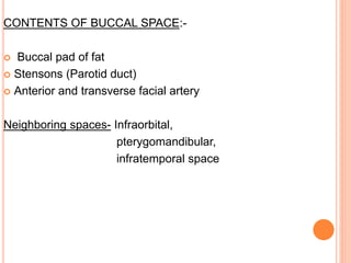
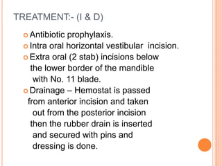
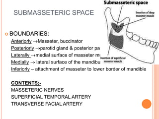
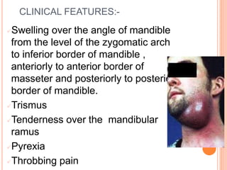
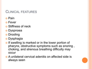
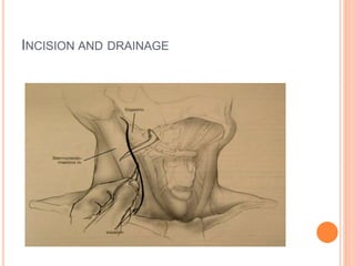
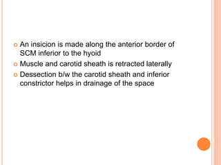
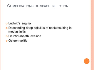
MANDIBULAR SPACE
INFECTIONS
DR DAVIS NADAKKAVUKARAN M.D.S
READER
MALABAR DENTAL COLLEGE EDAPPAL
INTRODUCTION
 Tissue spaces or fascial spaces are potential spaces situated b/w
planes
of fascia that form natural pathway along which infection may
spread producing a cellulitis or within which infection may localise with
actual
abscess formation
 Infection is defined as invasion and colanization of pathogenic micro-
or
ganisms in body tissues , resulting in local intracellular replication or
antigen-antibody response
 The infection generally spreads by following the path of least
resistance through
connective tissue and along fascial planes
 The infection spread to such an extend, distant from the site of site of
origin,
causing considerable morbidity and occasional death
CLASSIFICATION OF FASCIAL SPACES
BASED ON MODE OF INVOLVEMENT
1. Direct Involvement. (Primary Spaces)
 Maxillary Spaces – Canine, buccal
infratemporal
 Mandibular Spaces – Submental,
Submandibular, Sublingual, Buccal
2. Indirect involvement (Secondary Spaces)
 Masseteric
 Pterygomandibular
 Superficial and deep temporal
 Lateral and retro pharyngeal
 Prevertebral
 Parotid
BASED ON CLINICAL SIGNIFICANCE
a) FACE- buccal, cannine , masticatory, parotid
b) SUPRAHYOID-sublingual, submandibular,
pharyngomaxillary, peritonsillar
c) INFRAHYOID- anterovisceral(paratracheal)
d) SPACES OF TOTAL NECK- retropharyngeal,
spaces of carotid sheath
SUBMANDIBULAR SPACE
BOUNDARIES
• SUPERIORLY- inferior and
medial surface of mandible
and attachment of
mylohyoid muscle
• INFERIORLY- anterior and
posterior belly of digastric
• LATERALY- skin,
superficial fascia,
platysma, investing fascia
• MEDIALY- mylohyoid ,
hyoglossus , superoir
constrictor, styloglossus
CONTENTS:
o Submandibular gland
o Facial artery
o Lymph nodes
NEIGHBOURING SPACES:
o Sublingual
o Submental
o Lateral pharyngeal
o buccal
CAUSE –
 Infection from Mandibular molars.
 From sublingual space
 Infections from middle third of the tongue, posterior
part of floor of the mouth.
 From submental space / submental lymph nodes
 Infection from the submandibular gland
CLINICAL FEATURES
•Swelling begins at lower border
of mandible extends to the level
of hyoid bone in a shape of
inverted cone.
•No trismus
•Redness of overlying skin
•Dysphagia
•Teeth sensitive to percussion &
mobility present.
 Incision and drainage through Extra-oral incision.
 Incision – 2 stab incisions of about 1.5 to 2cm in length are
given over the dependent part 2cm below the lower border
of mandible in the neck of the mandible
TREATMENT
 Sinus forceps inserted superiorly &posteriorly on
lingual side of mandible
below mylohyoid & Blunt dissection through
subcutaneous fat
not to damage facial A, anterior facial vein and the
facial nerve.
 Drainage – Corrugated rubber drain is placed &
dressing is given
SPREAD
 Submental
 Sublingual
 Contralateral submandibular
SUBLINGUAL SPACE
BOUNDARIES:-
o ANTERIORLY- lingual surface of mandible
 POSTERIORLY - hyoid bone
 SUPERIORLY- oral mucosa
 INFERIORLY - Mylohyoid muscle
 MEDIALLY- geniohyoid, genioglossus &
hyoglossus (muscles of tongue)
 LATERALLY - lingual aspect of mandible,
above mylohyoid
CONTENTS
 Lingual nerve
 Hypoglossal nerve
 Submandibular gland(deep part) and its duct
 Sublingual gland
 Hyoglossus
 Geniohyoid
 Genioglossus
CAUSE
 Mandibular premolars and molars, trauma
CLINICAL FEATURES
 Swelling in anterior part of floor of the mouth on the
affected side displacing tongue medially and
superiorly.
 Interferes with swallowing and is extremely painful.
 Elevation of tongue to palate causes airway
compromise.
TREATMENT:-
 Antibiotic prophylaxis
 Incision is made Intraorally over lingual cortical plate
lateral to sublingual plica.
 Sinus forceps is passed beneath sublingual gland in
an
antero posterior direction and drain is placed.
 Extraorally incision is placed skin overlying
submndibular space.
 When infection crosses midline, same incision is
made bilaterally,
sinus forceps is passed through floor of mouth from
one side to other & drain is placed
SPREAD
 Contralateral sublingual space
 Submandibular space
 Pterygomandibular space
 Parapharyngeal space
 Submental space
SUBMENTAL SPACE
BOUNDARIES:-
 ANTERIORLY – Inferior
border of mandible
 POSTEROIRLY- Hyoid
 LATERALLY – Anterior
belly of the digastric
muscle,
 MEDIALLY- Common
space ,no medial wall
 SUPERIORILY –
Mylohyoid muscle
 INFERIORILY –
suprahyoid portion of
investing layer of deep
cervical fascia ,
platysma, superficial
fascia, skin.
CONTENTS
• subental lymph nodes
• anterior jugular vein.
CAUSE
 From lower anteriors.
 Secondarily due to infection from submental lymph
nodes which drain lower lip, skin overlying chin,
anterior part of floor of the mouth, tip of the tongue
& sublingual tissues.
 Symphysis fracture.
CLINICAL FEATURES
Swelling is limited to the point of
the
chin & to the region immediately
below it
TREATMENT:-
Transverse incisions on skin below
symphysis.
Sinus forceps inserted upwrd & backward
Corrugated rubber drain & dressings are
place.
BUCCAL SPACE
BOUNDARIES:-
 ANTERIOMEDIALLY - buccinator muscle
 POSTERIOMEDIALLY- Masseter muscle
overlying anterior border
of mandible
 SUPERIORILY -zygomatic process of
maxilla
zygomaticus major and
minor
 INFERIORLY - deep fascia attached to
mandible
& depressor anguli
oris
LATERALLY - platysma musle and
extension of
deep fascia from
subcutaneous space between the fascial skin and
buccinator muscle.
CLINICAL FEATURES:
•Dome shaped swelling on the
anterior aspect of cheek
• anteroposteriorly from corner of
mouth to angle of mandible
• superoinferiorly from level of
zygomatic arch to inferior border of
mandible.
CONTENTS OF BUCCAL SPACE:-
 Buccal pad of fat
 Stensons (Parotid duct)
 Anterior and transverse facial artery
Neighboring spaces- Infraorbital,
pterygomandibular,
infratemporal space
TREATMENT:- (I & D)
 Antibiotic prophylaxis.
 Intra oral horizontal vestibular incision.
 Extra oral (2 stab) incisions below
the lower border of the mandible
with No. 11 blade.
 Drainage – Hemostat is passed
from anterior incision and taken
out from the posterior incision
then the rubber drain is inserted
and secured with pins and
dressing is done.
SUBMASSETERIC SPACE
 BOUNDARIES:
Anteriorly Masseter, buccinator
Posteriorly parotid gland & posterior part of masseter
Laterally medial surface of masseter muscle
Medially  lateral surface of the mandibular ramus
Inferiorly – attachment of masseter to lower border of mandible
CONTENTS;-
MASSETERIC NERVES
SUPERFICIAL TEMPORAL ARTERY
TRANSVERSE FACIAL ARTERY
CLINICAL FEATURES:-
Swelling over the angle of mandible
from the level of the zygomatic arch
to inferior border of mandible ,
anteriorly to anterior border of
masseter and posteriorly to posterior
border of mandible.
Trismus
Tenderness over the mandibular
ramus
Pyrexia
Throbbing pain
SURGICAL MANAGMENT
 Intraorally – a vertical incision is made over the
lower part of anterior border of ramus of mandible
 A sinus foreceps is passed along lateral surface of
ramus downwards and backwards and pus is
drained
 Drain is inserted and secured with a suture
 Extraorally – an incision is placed in the skin behind
the angle of mandible to open the abcess by
hilton’s method
 A rubber drain is inserted and secured with a suture
PTERYGOMANDIBULAR SPACE
 BOUNDARIES:
 Anteriorly 
pterygomandibular
raphe
 Posteriorly  deep
portion of parotid gland
 Superiorly  lateral
pterygoid muscle
 inferiorly inferior
border of the mandible
 Laterally  medial
surface of ramus of
mandible
 Medialy  Lateral
CONTENTS
1. Lingual nerve
2. Mandibular nerve
3. IAN
4. Mylohyoid nerves and vessels
5. Loose areolar CT
NEIGHBOURING SPACES
 Buccal space
 Lateral pharyngeal space
 Submassetric space
 Deep temporal space
 Parotid space
 Peritonsillar space
CLINICAL FEATURES:
 No external evidence of swelling extra orally
 Intraorally visible swelling over soft palate
 Redness and tendrness over 3rd molar region
 Anterior bulging of half the soft palate and the anterior
tonsillar pillar with deviation of uvula to the unaffected
side.
 Severe trismus and dysphagia
TREATMENT:
If trismus is severe.
-Extraoral mandibular nerve block or G.A. is
given
 intra oral incision in the mucosal area between
medial aspect
of ramus and the pterygomandibular
raphae.Blunt dissection using hemostat.
Drainage.
 Extra oral incision is made below the angle of
mandible.
 A sinus forceps is inserted towards medial side of
mandible in upward and backward direction
 Pus evacuated . Drain is inserted
LATERAL PHARYNGEAL SPACE
INFECTIONS
It lies immediately posterior and
lateral to the pharynx
Anatomically the lateral pharyngeal
space may be thought of
as an inverted pyramid shape-the
base of the pyramid being the
skull base and the apex the hyoid
bone.
 BOUNDARIES:-
 Superiorly  Base
of skull
 Inferiorly  Hyoid
bone
 Medially 
superior
pharyngeal
constrictor
 Laterally 
medial pterygoid
m., capsule of
parotid gland
 Posteriorly 
carotid sheath,
CONTENTS:
 Anterior compartment:
Ascending pharyngeal A.
Loose areolar connective tissue.
 Posterior compartments:-
Cervical sympathetic trunk
Carotid sheath with its contents
Neighboring spaces -
1) Pterygomandibular,
2) submandibular,
3) sublingual,
4) peritonsillar,
5) retropharyngeal space.
CLINICAL FEATURES
 Brawny induration of face
above the angle of mandible,
extents downward to
submandibular region and
upward to parotid region
 Difficuty in flexing and turning
of neck.
 Trismus secondary pterygoid
muscle involvement
 Dysphagia.
 Dyspnoea.
 Displacement of tonsil,
tonsillar pillar, uvula to the
MANAGMENT
 Incision and drainage
 Extraoral approach- a 3-5cm incision is placed along the
anterior border of SCM , extending from below the angle
of mandible to the middle 3rd of submandibular salivary
gland
 The fascia behind the gland is incised and a curved
hemostat is inserted and carefully directed medialy behind
the mandible
 A ruber drain is inserted and sutured
 Intraoral approach- a vertical insicion is made over the
pterygomandibular raphae
 A sinus foreceps is inserted through pterygomandibular
raphae along the medial side of mandible medial to medial
pterygoid and just lateral to superior constrictor is then
divided posteriorly to pus pocket
COMPLICATIONS
Suppurative jugular venous
thrombosis.
Patient will have shaking chills,
high fever.
Tenderness at the mandibular
angle and along
sternocleidomastoid muscle.
Retropharyngeal space
Two compartments:
Suprahyoid
1. Lymph nodes and fat.
Infrahyoid
1. Only fat
Retropharyngeal space is the potential
space sandwiched between alar and
prevertebral layers of deep layer of
the deep investing fascia
posteriorly&anteriorly.
Most dangerous of all types of deep neck infections
(Danger space)
BOUNDARIES
 ANTERIORLY- superior and middle pharygeal
constrictor muscle
 POSTERIORLY- carotid sheath, stylohyoid,
styloglossus, and stylopharyngenous
 SUPERIORLY- base of skull
 INFERORLY- hyoid bone
 MEDIALY- superior pharyngeal constrictor
 LATERALY- medial pterygoid muscle and capsule
of parotid gland
CONTENTS
 Carotid artery
 IJV
 Vagus nerve
 Cervical sympathetic chain
CLINICAL FEATURES
 Pain
 Fever
 Stiffness of neck
 Dyspnoea
 Drooling
 Dysphagia
 If swelling is marked or in the lower portion of
pharynx, obstructive symptoms such as snoring ,
choking, and strenous breathing difficulty may
occur
 A unilateral cervical adenitis on affected side is
always seen
INCISION AND DRAINAGE
 An insicion is made along the anterior border of
SCM inferior to the hyoid
 Muscle and carotid sheath is retracted laterally
 Dessection b/w the carotid sheath and inferior
constrictor helps in drainage of the space
COMPLICATIONS OF SPACE INFECTION
 Ludwig’s angina
 Descending deep cellulitis of neck’resulting in
mediastinitis
 Carotid sheeth invasion
 Osteomyelitis
REFERENCES
 Text book of oral and maxillofacial surgery
neelima anil malik
 Text book of oral and maxillofacial surgey –
S M Balaji
2 mandibular_spaceinfection-.pptx

2 mandibular_spaceinfection-.pptx

  • 1.
    MANDIBULAR SPACE INFECTIONS DR DAVISNADAKKAVUKARAN M.D.S READER MALABAR DENTAL COLLEGE EDAPPAL
  • 2.
    INTRODUCTION  Tissue spacesor fascial spaces are potential spaces situated b/w planes of fascia that form natural pathway along which infection may spread producing a cellulitis or within which infection may localise with actual abscess formation  Infection is defined as invasion and colanization of pathogenic micro- or ganisms in body tissues , resulting in local intracellular replication or antigen-antibody response  The infection generally spreads by following the path of least resistance through connective tissue and along fascial planes  The infection spread to such an extend, distant from the site of site of origin, causing considerable morbidity and occasional death
  • 3.
  • 4.
    BASED ON MODEOF INVOLVEMENT 1. Direct Involvement. (Primary Spaces)  Maxillary Spaces – Canine, buccal infratemporal  Mandibular Spaces – Submental, Submandibular, Sublingual, Buccal 2. Indirect involvement (Secondary Spaces)  Masseteric  Pterygomandibular  Superficial and deep temporal  Lateral and retro pharyngeal  Prevertebral  Parotid
  • 5.
    BASED ON CLINICALSIGNIFICANCE a) FACE- buccal, cannine , masticatory, parotid b) SUPRAHYOID-sublingual, submandibular, pharyngomaxillary, peritonsillar c) INFRAHYOID- anterovisceral(paratracheal) d) SPACES OF TOTAL NECK- retropharyngeal, spaces of carotid sheath
  • 6.
    SUBMANDIBULAR SPACE BOUNDARIES • SUPERIORLY-inferior and medial surface of mandible and attachment of mylohyoid muscle • INFERIORLY- anterior and posterior belly of digastric • LATERALY- skin, superficial fascia, platysma, investing fascia • MEDIALY- mylohyoid , hyoglossus , superoir constrictor, styloglossus
  • 7.
    CONTENTS: o Submandibular gland oFacial artery o Lymph nodes NEIGHBOURING SPACES: o Sublingual o Submental o Lateral pharyngeal o buccal
  • 8.
    CAUSE –  Infectionfrom Mandibular molars.  From sublingual space  Infections from middle third of the tongue, posterior part of floor of the mouth.  From submental space / submental lymph nodes  Infection from the submandibular gland
  • 9.
    CLINICAL FEATURES •Swelling beginsat lower border of mandible extends to the level of hyoid bone in a shape of inverted cone. •No trismus •Redness of overlying skin •Dysphagia •Teeth sensitive to percussion & mobility present.
  • 10.
     Incision anddrainage through Extra-oral incision.  Incision – 2 stab incisions of about 1.5 to 2cm in length are given over the dependent part 2cm below the lower border of mandible in the neck of the mandible TREATMENT
  • 11.
     Sinus forcepsinserted superiorly &posteriorly on lingual side of mandible below mylohyoid & Blunt dissection through subcutaneous fat not to damage facial A, anterior facial vein and the facial nerve.  Drainage – Corrugated rubber drain is placed & dressing is given
  • 13.
    SPREAD  Submental  Sublingual Contralateral submandibular
  • 14.
    SUBLINGUAL SPACE BOUNDARIES:- o ANTERIORLY-lingual surface of mandible  POSTERIORLY - hyoid bone  SUPERIORLY- oral mucosa  INFERIORLY - Mylohyoid muscle  MEDIALLY- geniohyoid, genioglossus & hyoglossus (muscles of tongue)  LATERALLY - lingual aspect of mandible, above mylohyoid
  • 16.
    CONTENTS  Lingual nerve Hypoglossal nerve  Submandibular gland(deep part) and its duct  Sublingual gland  Hyoglossus  Geniohyoid  Genioglossus
  • 17.
  • 18.
    CLINICAL FEATURES  Swellingin anterior part of floor of the mouth on the affected side displacing tongue medially and superiorly.  Interferes with swallowing and is extremely painful.  Elevation of tongue to palate causes airway compromise.
  • 19.
    TREATMENT:-  Antibiotic prophylaxis Incision is made Intraorally over lingual cortical plate lateral to sublingual plica.  Sinus forceps is passed beneath sublingual gland in an antero posterior direction and drain is placed.
  • 20.
     Extraorally incisionis placed skin overlying submndibular space.  When infection crosses midline, same incision is made bilaterally, sinus forceps is passed through floor of mouth from one side to other & drain is placed
  • 21.
    SPREAD  Contralateral sublingualspace  Submandibular space  Pterygomandibular space  Parapharyngeal space  Submental space
  • 22.
    SUBMENTAL SPACE BOUNDARIES:-  ANTERIORLY– Inferior border of mandible  POSTEROIRLY- Hyoid  LATERALLY – Anterior belly of the digastric muscle,  MEDIALLY- Common space ,no medial wall  SUPERIORILY – Mylohyoid muscle  INFERIORILY – suprahyoid portion of investing layer of deep cervical fascia , platysma, superficial fascia, skin.
  • 23.
    CONTENTS • subental lymphnodes • anterior jugular vein.
  • 24.
    CAUSE  From loweranteriors.  Secondarily due to infection from submental lymph nodes which drain lower lip, skin overlying chin, anterior part of floor of the mouth, tip of the tongue & sublingual tissues.  Symphysis fracture.
  • 25.
    CLINICAL FEATURES Swelling islimited to the point of the chin & to the region immediately below it
  • 26.
    TREATMENT:- Transverse incisions onskin below symphysis. Sinus forceps inserted upwrd & backward Corrugated rubber drain & dressings are place.
  • 27.
    BUCCAL SPACE BOUNDARIES:-  ANTERIOMEDIALLY- buccinator muscle  POSTERIOMEDIALLY- Masseter muscle overlying anterior border of mandible  SUPERIORILY -zygomatic process of maxilla zygomaticus major and minor  INFERIORLY - deep fascia attached to mandible & depressor anguli oris LATERALLY - platysma musle and extension of deep fascia from subcutaneous space between the fascial skin and buccinator muscle.
  • 29.
    CLINICAL FEATURES: •Dome shapedswelling on the anterior aspect of cheek • anteroposteriorly from corner of mouth to angle of mandible • superoinferiorly from level of zygomatic arch to inferior border of mandible.
  • 30.
    CONTENTS OF BUCCALSPACE:-  Buccal pad of fat  Stensons (Parotid duct)  Anterior and transverse facial artery Neighboring spaces- Infraorbital, pterygomandibular, infratemporal space
  • 31.
    TREATMENT:- (I &D)  Antibiotic prophylaxis.  Intra oral horizontal vestibular incision.  Extra oral (2 stab) incisions below the lower border of the mandible with No. 11 blade.  Drainage – Hemostat is passed from anterior incision and taken out from the posterior incision then the rubber drain is inserted and secured with pins and dressing is done.
  • 32.
    SUBMASSETERIC SPACE  BOUNDARIES: AnteriorlyMasseter, buccinator Posteriorly parotid gland & posterior part of masseter Laterally medial surface of masseter muscle Medially  lateral surface of the mandibular ramus Inferiorly – attachment of masseter to lower border of mandible CONTENTS;- MASSETERIC NERVES SUPERFICIAL TEMPORAL ARTERY TRANSVERSE FACIAL ARTERY
  • 33.
    CLINICAL FEATURES:- Swelling overthe angle of mandible from the level of the zygomatic arch to inferior border of mandible , anteriorly to anterior border of masseter and posteriorly to posterior border of mandible. Trismus Tenderness over the mandibular ramus Pyrexia Throbbing pain
  • 34.
    SURGICAL MANAGMENT  Intraorally– a vertical incision is made over the lower part of anterior border of ramus of mandible  A sinus foreceps is passed along lateral surface of ramus downwards and backwards and pus is drained  Drain is inserted and secured with a suture  Extraorally – an incision is placed in the skin behind the angle of mandible to open the abcess by hilton’s method  A rubber drain is inserted and secured with a suture
  • 35.
    PTERYGOMANDIBULAR SPACE  BOUNDARIES: Anteriorly  pterygomandibular raphe  Posteriorly  deep portion of parotid gland  Superiorly  lateral pterygoid muscle  inferiorly inferior border of the mandible  Laterally  medial surface of ramus of mandible  Medialy  Lateral
  • 36.
    CONTENTS 1. Lingual nerve 2.Mandibular nerve 3. IAN 4. Mylohyoid nerves and vessels 5. Loose areolar CT
  • 37.
    NEIGHBOURING SPACES  Buccalspace  Lateral pharyngeal space  Submassetric space  Deep temporal space  Parotid space  Peritonsillar space
  • 38.
    CLINICAL FEATURES:  Noexternal evidence of swelling extra orally  Intraorally visible swelling over soft palate  Redness and tendrness over 3rd molar region  Anterior bulging of half the soft palate and the anterior tonsillar pillar with deviation of uvula to the unaffected side.  Severe trismus and dysphagia
  • 39.
    TREATMENT: If trismus issevere. -Extraoral mandibular nerve block or G.A. is given  intra oral incision in the mucosal area between medial aspect of ramus and the pterygomandibular raphae.Blunt dissection using hemostat. Drainage.
  • 40.
     Extra oralincision is made below the angle of mandible.  A sinus forceps is inserted towards medial side of mandible in upward and backward direction  Pus evacuated . Drain is inserted
  • 41.
    LATERAL PHARYNGEAL SPACE INFECTIONS Itlies immediately posterior and lateral to the pharynx Anatomically the lateral pharyngeal space may be thought of as an inverted pyramid shape-the base of the pyramid being the skull base and the apex the hyoid bone.
  • 42.
     BOUNDARIES:-  Superiorly Base of skull  Inferiorly  Hyoid bone  Medially  superior pharyngeal constrictor  Laterally  medial pterygoid m., capsule of parotid gland  Posteriorly  carotid sheath,
  • 43.
    CONTENTS:  Anterior compartment: Ascendingpharyngeal A. Loose areolar connective tissue.  Posterior compartments:- Cervical sympathetic trunk Carotid sheath with its contents Neighboring spaces - 1) Pterygomandibular, 2) submandibular, 3) sublingual, 4) peritonsillar, 5) retropharyngeal space.
  • 44.
    CLINICAL FEATURES  Brawnyinduration of face above the angle of mandible, extents downward to submandibular region and upward to parotid region  Difficuty in flexing and turning of neck.  Trismus secondary pterygoid muscle involvement  Dysphagia.  Dyspnoea.  Displacement of tonsil, tonsillar pillar, uvula to the
  • 46.
    MANAGMENT  Incision anddrainage  Extraoral approach- a 3-5cm incision is placed along the anterior border of SCM , extending from below the angle of mandible to the middle 3rd of submandibular salivary gland  The fascia behind the gland is incised and a curved hemostat is inserted and carefully directed medialy behind the mandible  A ruber drain is inserted and sutured  Intraoral approach- a vertical insicion is made over the pterygomandibular raphae  A sinus foreceps is inserted through pterygomandibular raphae along the medial side of mandible medial to medial pterygoid and just lateral to superior constrictor is then divided posteriorly to pus pocket
  • 47.
    COMPLICATIONS Suppurative jugular venous thrombosis. Patientwill have shaking chills, high fever. Tenderness at the mandibular angle and along sternocleidomastoid muscle.
  • 48.
    Retropharyngeal space Two compartments: Suprahyoid 1.Lymph nodes and fat. Infrahyoid 1. Only fat Retropharyngeal space is the potential space sandwiched between alar and prevertebral layers of deep layer of the deep investing fascia posteriorly&anteriorly. Most dangerous of all types of deep neck infections (Danger space)
  • 49.
    BOUNDARIES  ANTERIORLY- superiorand middle pharygeal constrictor muscle  POSTERIORLY- carotid sheath, stylohyoid, styloglossus, and stylopharyngenous  SUPERIORLY- base of skull  INFERORLY- hyoid bone  MEDIALY- superior pharyngeal constrictor  LATERALY- medial pterygoid muscle and capsule of parotid gland
  • 50.
    CONTENTS  Carotid artery IJV  Vagus nerve  Cervical sympathetic chain
  • 51.
    CLINICAL FEATURES  Pain Fever  Stiffness of neck  Dyspnoea  Drooling  Dysphagia  If swelling is marked or in the lower portion of pharynx, obstructive symptoms such as snoring , choking, and strenous breathing difficulty may occur  A unilateral cervical adenitis on affected side is always seen
  • 52.
  • 53.
     An insicionis made along the anterior border of SCM inferior to the hyoid  Muscle and carotid sheath is retracted laterally  Dessection b/w the carotid sheath and inferior constrictor helps in drainage of the space
  • 54.
    COMPLICATIONS OF SPACEINFECTION  Ludwig’s angina  Descending deep cellulitis of neck’resulting in mediastinitis  Carotid sheeth invasion  Osteomyelitis
  • 55.
    REFERENCES  Text bookof oral and maxillofacial surgery neelima anil malik  Text book of oral and maxillofacial surgey – S M Balaji

Editor's Notes

  • #11 Blunt dissection to prevent damage to facial artery, vein and nerve
  • #19  Infection may cross genial muscles to involve space of other side. Hot potato voice.
  • #20 Care is taken not to injure sublingual galnd, lingual nerve , submand duct
  • #28 Anteriorly- orbicularis oris , zygomaticus major Deep – buccopharygeal fascial Superiorly -Zygomatic arch
  • #30 Repated buccal space infection suspects crohn’s disease
  • #34 COMPLICATIONS:- Osteomyelitis with sequestrum in the ramus of mandible. Necrosis of muscle
  • #43 Space divided into 2 compartments anterior and posterior by the styloid process. Its connections with carotid sheath alarms a great danger when this space is involved. 
  • #44 Post comprtmnt --9th ,11th ,12th cranial Nerves
  • #45 Diagnostic evaluation Chest CT scan, Chest radiographs