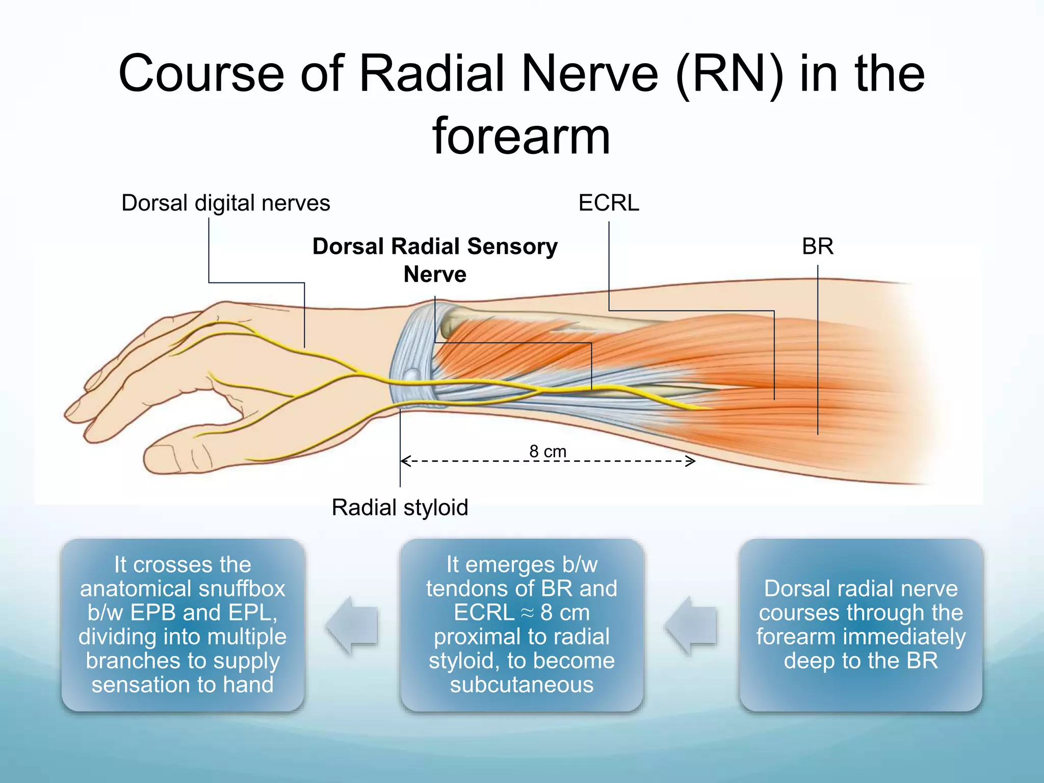
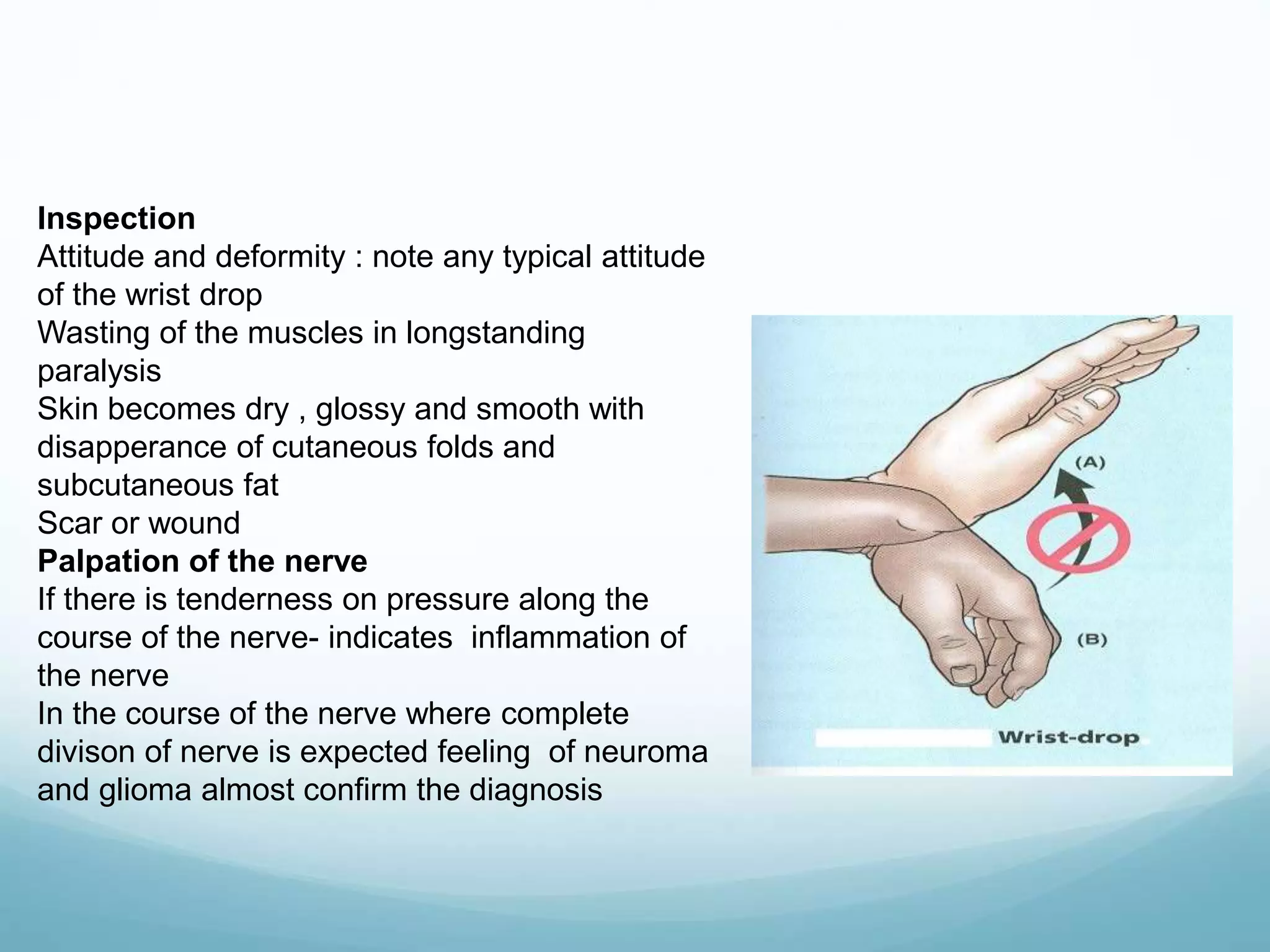
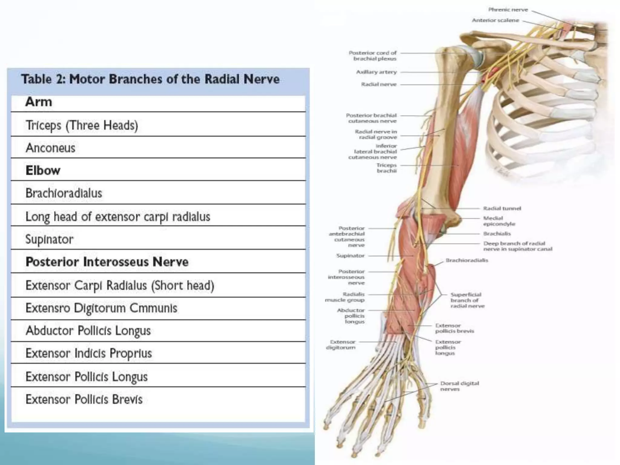
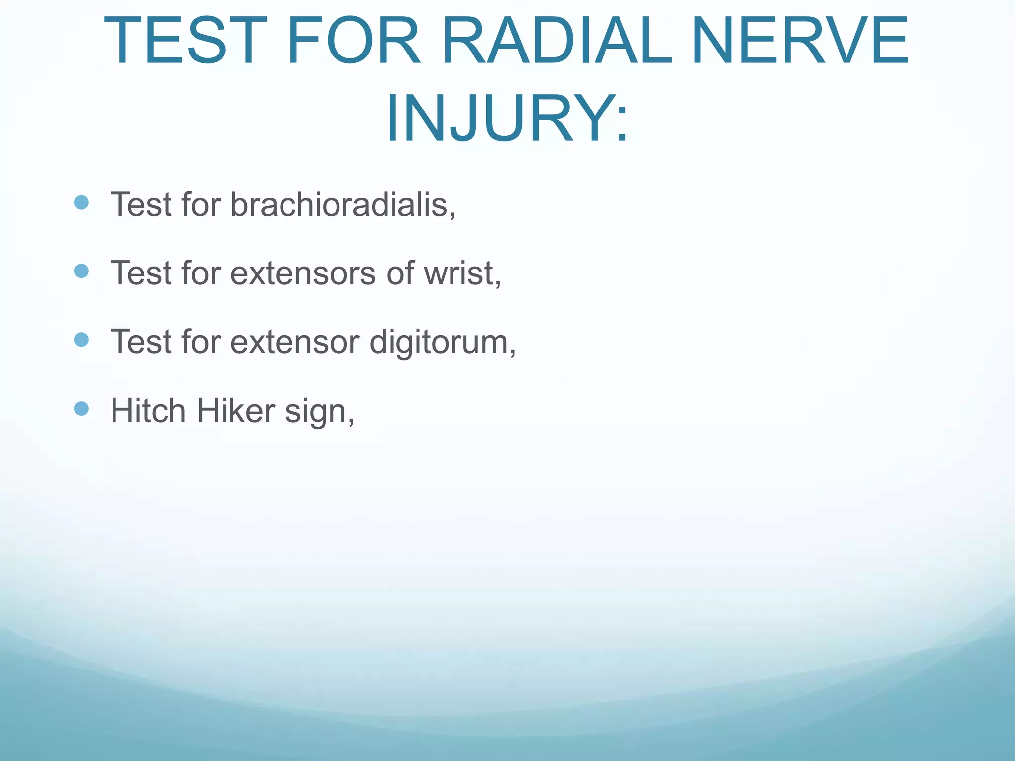
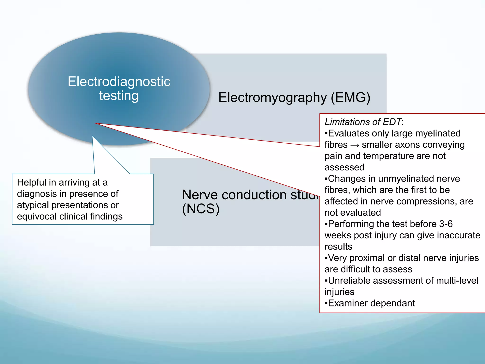
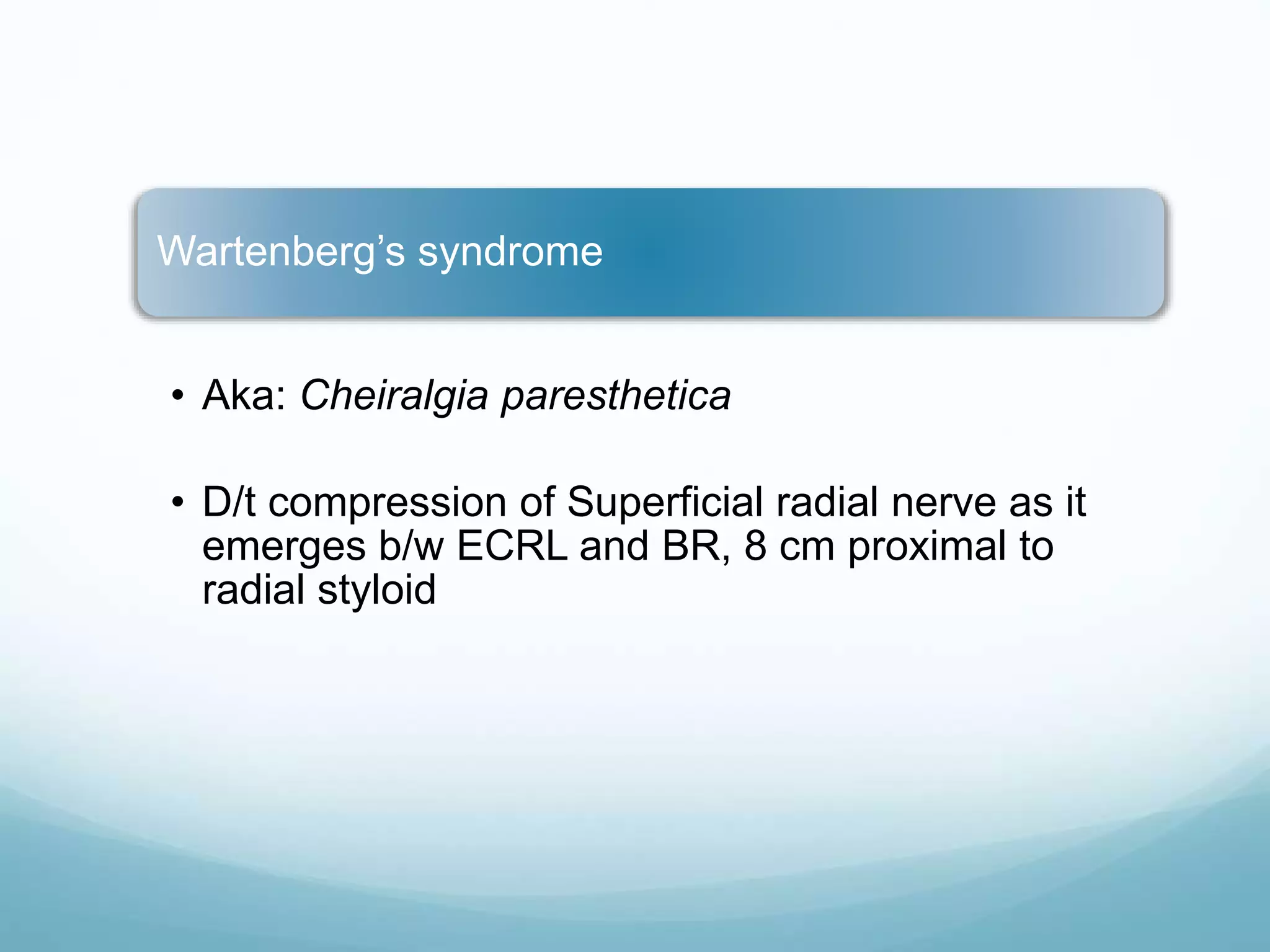
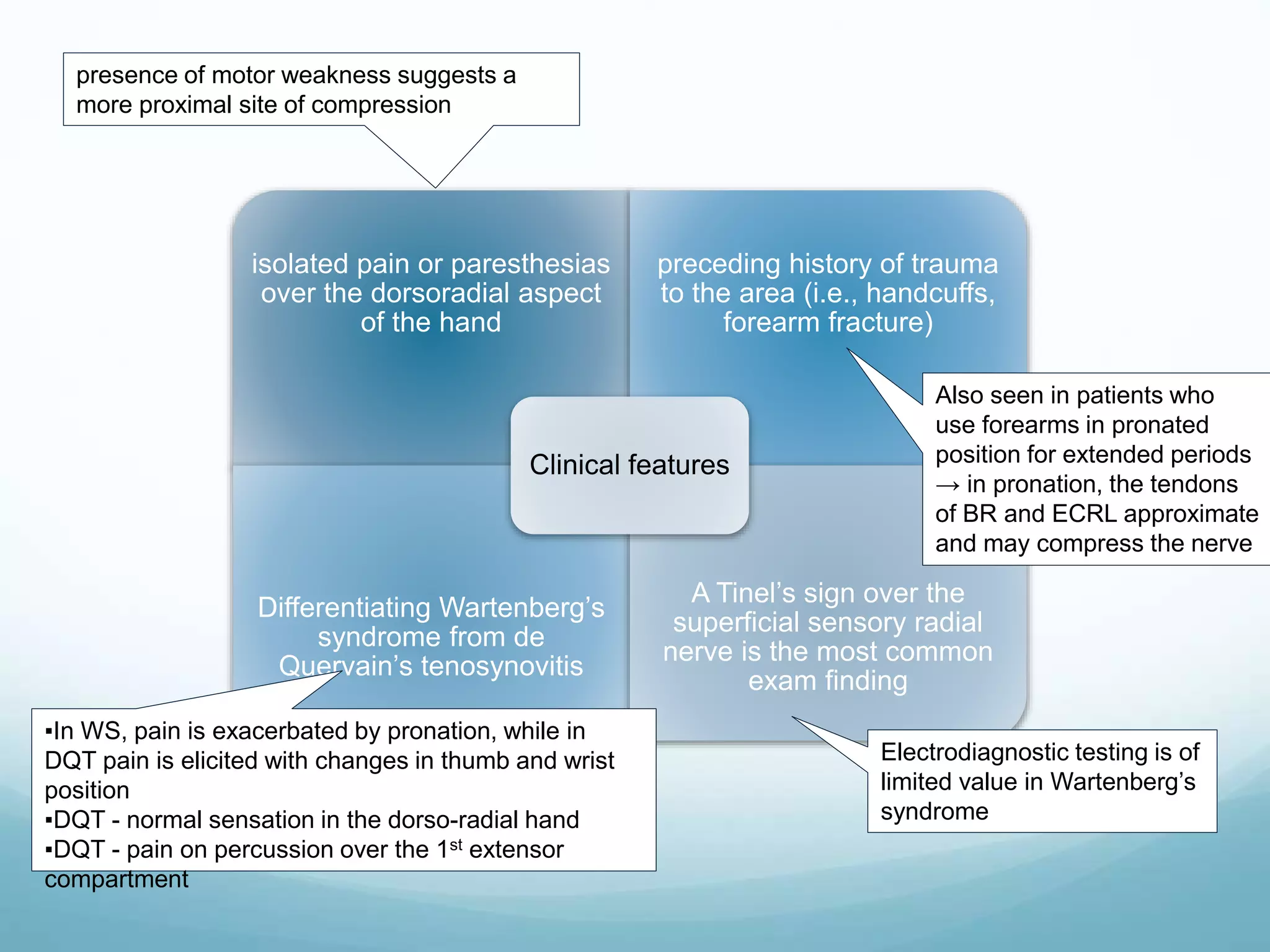
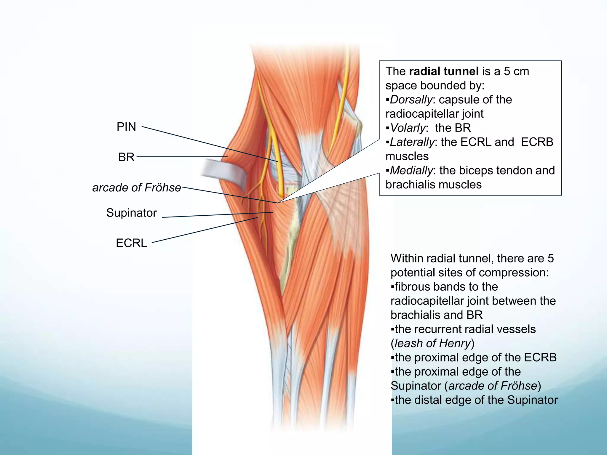
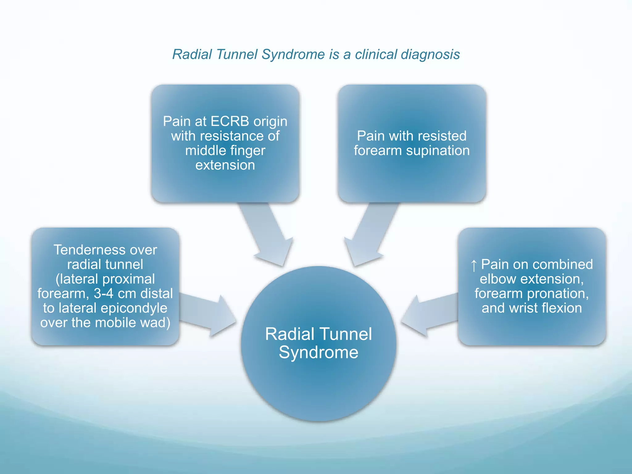
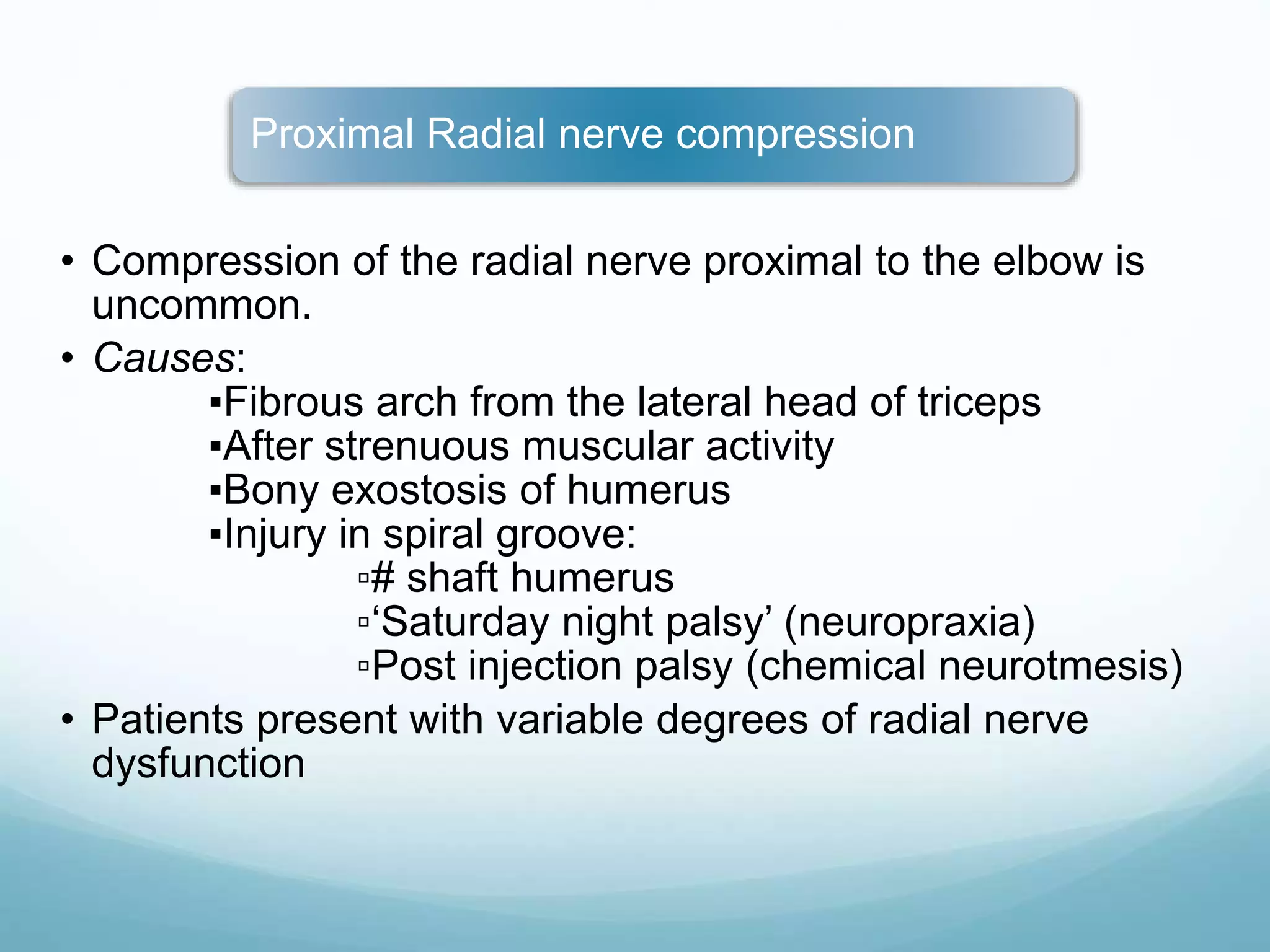
SlideShare a Scribd company logo
RADIAL NERVE
INJURY AND
MANAGEMENT
SUSHANT S. SONARKAR
ANATOMY:
 Posterior cord,(C5-6,7,8,T1),
 Largest branch,
 Predominantly motor,
Course of Radial Nerve (RN) in the
arm
In the axilla, RN lies
anterior to
subscapularis, teres
major and LD
• Sensory supply:
Posterior cutaneous
nerve of arm
RN leaves the axilla
via the triangular
space
• Motor supply:
long head of
Triceps
It then comes to lie
along spiral groove
on posterior aspect of
humeral shaft along
with arteria profunda
brachii
• Motor: medial and lateral
heads of triceps,
Anconeus
• Sensory: posterior
cutaneous nerve of
forearm, lower lateral
cutaneous nerve of arm
RN then leaves the spiral
groove by piercing the lateral
intermuscular septum to enter
the anterior compartment of
the arm, 10-12 cm above the
lateral epicondyle
• Motor supply: Brachialis
(lateral part), BR, ECRL
Anterior to lateral
epicondyle, RN divides into its
terminal branches
• Terminal branches:
Posterior
Interosseous Nerve
(PIN) and Dorsal or
Superficial radial
sensory nerve
Here it lies b/w brachialis
and BR
Radial nerve (C5, 6, 7 , 8,
T1) exiting axilla via the
triangular space
Brachialis (lateral part)
Anconeus
Triceps brachii
Posterior cutaneous nerve
of forearm
Posterior cutaneous nerve
of arm
Lower lateral cutaneous
nerve of arm
Radial nerve in the spiral
groove (posterior aspect
of humeral shaft)
Brachioradialis (BR)
ECRL (last branch of
radial nerve proper)
Lateral intermuscular
septum
BR
ECRL
Dorsal Radial Sensory
Nerve
Dorsal digital nerves
Radial styloid
8 cm
Dorsal radial nerve
courses through the
forearm immediately
deep to the BR
It emerges b/w
tendons of BR and
ECRL ≈ 8 cm
proximal to radial
styloid, to become
subcutaneous
It crosses the
anatomical snuffbox
b/w EPB and EPL,
dividing into multiple
branches to supply
sensation to hand
Course of Radial Nerve (RN) in the
forearm
Deep terminal branch →
Posterior interosseous
nerve (PIN)
Supinator
EIP
EDC and EDM
ECU
ECRB
Superficial terminal branch
Radial Nerve Proper
EPL
EPB
APL
PIN reaches the back of
forearm by passing
around the lateral aspect
of the radius b/w the
superficial and deep
heads of the Supinator
to supply all extensor
compartment.
Finally, PIN ends by
supplying carpal joint
sensation
Lower lateral cutaneous
nerve of arm
Posterior cutaneous
nerve of arm
Posterior cutaneous
nerve of forearm
Dorsal radial sensory nerve
Gives sensibility to the dorsum of
the hand over the radial two-thirds,
the dorsum of the thumb, and the
index, long, and half of the ring
finger proximal to the distal
interphalangeal joint.
Cutaneous innervation from radial
nerve
Inspection
Attitude and deformity : note any typical attitude
of the wrist drop
Wasting of the muscles in longstanding
paralysis
Skin becomes dry , glossy and smooth with
disapperance of cutaneous folds and
subcutaneous fat
Scar or wound
Palpation of the nerve
If there is tenderness on pressure along the
course of the nerve- indicates inflammation of
the nerve
In the course of the nerve where complete
divison of nerve is expected feeling of neuroma
and glioma almost confirm the diagnosis
ETIOLOGY:
 In the Axilla,
 In the shoulder,
 In the Radial groove,
 Between spiral groove and the lateral epicondyle,
 At the elbow,
 In the forearm,
 Other causes,
TYPES OF INJURY:
 Primary injury,
 Secondary Injury,
Clinical
Features
Functional motor deficit
Inability to extend the wrist (in
case of injury at level of PIN,
wrist extension is weak with
radial deviation since ECRL
innervation is intact)
Inability to extend the fingers at
the MCP joints
Inability to extend and radially
abduct the thumb
Weakness of grip strength d/t
loss of mechanical advantage
that wrist extension provides
for grasp and power grip
Area of sensory loss in
radial nerve injury in the
axilla
Unlike the median and ulnar
nerves, sensory loss
following radial nerve injury is
not functionally disabling
unless the patient develops a
painful neuroma
Sensory Loss
Autonomous sensory zone
for radial nerve → dorsum of
1st webspace
The Lateral cutaneous nerve
of forearm has a significant
overlap pattern with the
Superficial radial sensory
nerve
CLINICAL FEATURES:
 VERY HIGH RADIAL NERVE PALSY: ( in axilla)
 Motor paralysis:
1. Triceps, anconeus,
2. Brachioradialis,
3. Supinator,
4. Long Extensors of wrist.
 Sensory Paralysis:
1. Loss of skin sensations in posterior surface of lower arm and back of
forearm,
2. Lateral three and one half fingers(dorsum)
HIGH RADIAL NERVE PALSY:
(Radial Groove)
 Motor paralysis:
1. Brachioradialis,
2. Supinator,
3. Long extensors of wrist.
 Sensory paralysis:
Back of hand, radial side of thumb, and adjoining part of thenar
eminence
The medial side of thumb , index , middle and lateral part of
ring fingers.
LOW RADIAL NERVE
PALSY:
 Motor paralysis:
1. EDC, EDM,EDI,ECU
2. APL,EPL,EPLB
 Sensory loss:
1. No sensory loss
TEST FOR RADIAL NERVE
INJURY:
 Test for brachioradialis,
 Test for extensors of wrist,
 Test for extensor digitorum,
 Hitch Hiker sign,
Diagnostic Tests:
 Modality tests,
 Functional tests,
 Objective test,
1. Sweat test,
2. Skin resistance test,
3. Wrinkle test,
4. Tinel’s sign,
5. Positive axon reflex test.
ELECTROPHYSIOLOGICAL
STUDY:
 Electromyography,
 Motor nerve conduction studies,
 Strength duration Curve,
Electromyography (EMG)
Nerve conduction studies
(NCS)
Electrodiagnostic
testing
Helpful in arriving at a
diagnosis in presence of
atypical presentations or
equivocal clinical findings
Limitations of EDT:
▪Evaluates only large myelinated
fibres → smaller axons conveying
pain and temperature are not
assessed
▪Changes in unmyelinated nerve
fibres, which are the first to be
affected in nerve compressions, are
not evaluated
▪Performing the test before 3-6
weeks post injury can give inaccurate
results
▪Very proximal or distal nerve injuries
are difficult to assess
▪Unreliable assessment of multi-level
injuries
▪Examiner dependant
Nerve conduction studies (NCS)
2 electrodes are placed along the course of the
nerve. The first electrode stimulates the nerve
to fire, and the second electrode records the
generated action potential
Amplitude
• represents the size of the
response
• proportional to the number
of depolarizing axons in
the nerve
Latency
• the delay in response
following stimulation
Conduction velocity
Sensory nerve action
potential (SNAP)
• Response obtained when
the recording electrodes is
placed proximally along
the sensory nerve, toward
the spinal cord
Compound motor action
potential (CMAP)
• Response obtained when
the recording electrodes is
placed distally at the
target muscle
Electromyography (EMG)
• Activity observed when a needle
electrode is inserted into the muscle
Insertional activity
• Seen when the muscle is at rest
• Absent in normal muscles
▪Fibrillation potentials
▪Fasciculations
• Generated by the muscle during a
voluntary contraction
• Evaluates the integrity of neuro-
muscular junction
Motor unit potentials
(MUPs)
Sequence of events in nerve
compression
Focal demyelination
Axonal damage at the
compression site
Further axonal loss
Axonal sprouting producing
collateral re-innervation
Remyelination following
decompression
▪↑Latency
▪↓Nerve conduction
velocity
Associated Electrodiagnostic
findings
▪↓SNAP
▪↓CMAP
▪↑Insertional activity
▪Fibrillation potentials and
fasciculations
▪’Giant’ MUPs
▪Normalization of NCV
▪Loss of ‘giant’ MUPs
Wartenberg’s syndrome
• Aka: Cheiralgia paresthetica
• D/t compression of Superficial radial nerve as it
emerges b/w ECRL and BR, 8 cm proximal to
radial styloid
isolated pain or paresthesias
over the dorsoradial aspect
of the hand
preceding history of trauma
to the area (i.e., handcuffs,
forearm fracture)
Differentiating Wartenberg’s
syndrome from de
Quervain’s tenosynovitis
A Tinel’s sign over the
superficial sensory radial
nerve is the most common
exam finding
Clinical features
presence of motor weakness suggests a
more proximal site of compression
Also seen in patients who
use forearms in pronated
position for extended periods
→ in pronation, the tendons
of BR and ECRL approximate
and may compress the nerve
▪In WS, pain is exacerbated by pronation, while in
DQT pain is elicited with changes in thumb and wrist
position
▪DQT - normal sensation in the dorso-radial hand
▪DQT - pain on percussion over the 1st extensor
compartment
Electrodiagnostic testing is of
limited value in Wartenberg’s
syndrome
Posterior interosseous nerve (PIN) syndrome
• D/t compression of PIN in the radial tunnel
• Most common causes include:
▪Tumors such as lipomas, ganglia
▪Rheumatoid synovitis
▪Septic arthritis
▪Vasculitis
The radial tunnel is a 5 cm
space bounded by:
▪Dorsally: capsule of the
radiocapitellar joint
▪Volarly: the BR
▪Laterally: the ECRL and ECRB
muscles
▪Medially: the biceps tendon and
brachialis muscles
Within radial tunnel, there are 5
potential sites of compression:
▪fibrous bands to the
radiocapitellar joint between the
brachialis and BR
▪the recurrent radial vessels
(leash of Henry)
▪the proximal edge of the ECRB
▪the proximal edge of the
Supinator (arcade of Fröhse)
▪the distal edge of the Supinator
BR
Supinator
arcade of Fröhse
ECRL
PIN
Radial Tunnel syndrome
• Similar to PIN syndrome, it is also d/t
compression of PIN in the radial tunnel
• Not considered a true compression neuropathy.
Radial Tunnel Syndrome is a clinical diagnosis
Radial Tunnel
Syndrome
Tenderness over
radial tunnel
(lateral proximal
forearm, 3-4 cm distal
to lateral epicondyle
over the mobile wad)
Pain at ECRB origin
with resistance of
middle finger
extension
Pain with resisted
forearm supination
↑ Pain on combined
elbow extension,
forearm pronation,
and wrist flexion
Proximal Radial nerve compression
• Compression of the radial nerve proximal to the elbow is
uncommon.
• Causes:
▪Fibrous arch from the lateral head of triceps
▪After strenuous muscular activity
▪Bony exostosis of humerus
▪Injury in spiral groove:
▫# shaft humerus
▫‘Saturday night palsy’ (neuropraxia)
▫Post injection palsy (chemical neurotmesis)
• Patients present with variable degrees of radial nerve
dysfunction
Management:
 In nerve root avulsion injuries: non operative, early
neurotization,
 Open injuries,
 Closed injuries,
SURGICAL TECHNIQUES
 Techniques of neurorrhaphy :
-partial neurorrhaphy
-epineural neurorrhaphy
-perineural neurorrhaphy
-epiperineural neurorrhaphy
-interfasicular nerve grafting
MANAGEMENT
 Nerve grafting:
Types of graft
-Trunk graft
-Cable graft
-Pedicle nerve graft
-Inter fascicular nerve graft
-Pre vascularized nerve graft
Source of graft
-Sural nerve is probably the best source of graft in majority of cases
-Medial and lateral cutaneous nerve at the wrist is an ideal donor graft for
fascicular nerve after for digital nerve
-Superifical radial nerve is an excellent source of graft generally used in case of radial
nerve injuries
-Dorsal branch of ulnar nerve can also be used as a graft.
RECONSTRUCTIVE PROCEDURES
 Tendon transfers
 Arthodesis
 Tendon transfers work to correct:
 instability
 imbalance
 lack of co-ordination
 restore function by redistributing remaining muscular
forces
TENDON TRANSFER
 Robert jones described 2 sets of tendon transfers
1916: PT - ECRL and ECRB
FCU - EDC III,IV,V
FCR - EDCII,EIP and EPL
1921: PT - ECRL and ECRB
FCU - EDC III,IV,V
FCR - EDCII,EIP , EPL,APL ,EPB
Current standard tendon transfer protocol BRANDT
PT - ECRB
FCR- EDC
PL - EPL
INTERNAL SPLINT
 Burkhalter proposed early transfer of PT-ECRB to restore
wrist extension as an adjunct to nerve repair.
 It restores the power grip quickly and effectively since
wrist extension is restored
Advantages are:
 It works as a substitute during nerve regrowth and largely
eliminates an external splint
 Subsequently the transfer aids the newly innervated and
week wrist extensor
 It continues to act as a substitute in case nerve
regeneration is poor or absent
TENDON TRANSFER
 BOYE’S tendon transfer
PT -ECRL and ECRB
FCR - EPB and APL
FDS middle –EDC
FDS ring - EPL and EI
POST OP CARE:
immobilization for 6 weeks
usually maintained for 4 weeks followed by a spring loaded extension
splint for the wrist and finger.
during the cast immobilization the MCP joints held in 40degee of
reflection wrist should be fully extended with thumb in abduction and
extension
ip joints of fingers in comfortable flexion
NON-OPERATIVE TREATMENT
 SPLINTS
 wrist drop can be treated successfully by splints
 Bark halter has observed that grip strength may be increased
by 3 to 5 times by simply stabilizing the wrist with splints
 Many types of splints have been described
 Each patient individual need should be dictate
the type of splinting used
 splints are used for patients who are
debilitated and who reject surgery.
Cock up splint)
OPPENHEIMER SPLINT
It is a dynamic splint used for radial
nerve palsy
it consists of one palmar bar over the
prox.phalanx,one distal forearm cuff
dorsally and proimal cuff volarly.
Provides global extension of wrist and
fingers
Limitation of this splint:restriction of
grasp due to palmar bar.
Proximal migration of splint cause friction
blisters around the wrist
RECOVERY
 Factors influencing the prognosis for recovery
-age
-level of injury
-delay between the time of injury and repair
-gap between nerve ends
-Condition of the nerve
-type of nerve
-
 Level of injury:
 The more proximal the injury, the more incomplete the overall return of
motor and sensory function, especially in the more distal structures.
• Delay between the time of injury and repair
-Delay of neurorrhaphy affects motor recovery more profoundly than
sensory recovery
-satisfactory reinnervation of muscle can occur after denervation upto 12
months.
-irreversible changes develop in the muscles after 24 months
-with respect to sensory recovery ,nerve can be repaired even after 2 yrs
for satisfactory recovery. However results are best if done earlier.
 Gap between nerve ends
 The methods for closing the gap are
 nerve mobilization
 nerve transposition,
 positioning of the extremity ,
 nerve grafts,
 bone shortening.
Condition of the nerve: A clear cut sharp nerve injury has got better prognosis
following primary repair than crushed or avulsed nerve injuries which needs
secondary repair
Type of nerves: pure motor ,pure sensory nerves recover better than mixed nerves
because the consequence of mismatching are not so great in pure motor or
sensory nerves.

More Related Content

What's hot

De quervain's
De quervain'sDe quervain's
De quervain's
Lee Yew
 
Hand examination
Hand examination Hand examination
Hand examination
Ahmed Atef
 
Rotator cuff injuries
Rotator cuff injuriesRotator cuff injuries
Rotator cuff injuries
rajusvmc
 
Median nerve palsy final
Median nerve palsy finalMedian nerve palsy final
Median nerve palsy final
animesh kunwar
 
Leg length measurements
Leg length measurementsLeg length measurements
Leg length measurements
Dr. Akshita Duha Juneja (PT)
 
Sacroiliac Joint
Sacroiliac JointSacroiliac Joint
Sacroiliac Joint
Saeid Safari
 
Peripheral nerve injuries
Peripheral nerve injuriesPeripheral nerve injuries
Peripheral nerve injuries
Binod Chaudhary
 
Periarthritis shoulder
Periarthritis shoulderPeriarthritis shoulder
De quervain’s
De quervain’sDe quervain’s
Piriformis syndrome
Piriformis syndromePiriformis syndrome
Piriformis syndromeAndy Coleman
 
Modified ashworth scale application
Modified ashworth scale applicationModified ashworth scale application
Modified ashworth scale application
Dr. Vijay Kumar Pathak
 
Wrist drop
Wrist dropWrist drop
Wrist drop
DrDivya Chandil
 
Hand deformity in rheumatoid arthritis
Hand deformity in rheumatoid arthritisHand deformity in rheumatoid arthritis
Hand deformity in rheumatoid arthritis
sushilonlines
 
Trigger finger,causes,symptom,management
Trigger finger,causes,symptom,managementTrigger finger,causes,symptom,management
Trigger finger,causes,symptom,management
Dr.Md.Monsur Rahman
 
Radial nerve injury
Radial nerve injuryRadial nerve injury
Radial nerve injury
EuniceSusan
 
Wrist drop
Wrist dropWrist drop
Wrist drop
PratikDhabalia
 
Frozen Shoulder Physiotherapy Management
Frozen Shoulder Physiotherapy ManagementFrozen Shoulder Physiotherapy Management
Frozen Shoulder Physiotherapy Management
Vishal Deep
 
Cervical Radiculopathy Power Point.ppt
Cervical Radiculopathy Power Point.pptCervical Radiculopathy Power Point.ppt
Cervical Radiculopathy Power Point.pptGenevieve Hall
 
Assessment of shoulder
Assessment of shoulderAssessment of shoulder
Assessment of shoulder
Dr. Nithin Nair (PT)
 

What's hot (20)

De quervain's
De quervain'sDe quervain's
De quervain's
 
Common peroneal nerve lesions
Common peroneal nerve lesionsCommon peroneal nerve lesions
Common peroneal nerve lesions
 
Hand examination
Hand examination Hand examination
Hand examination
 
Rotator cuff injuries
Rotator cuff injuriesRotator cuff injuries
Rotator cuff injuries
 
Median nerve palsy final
Median nerve palsy finalMedian nerve palsy final
Median nerve palsy final
 
Leg length measurements
Leg length measurementsLeg length measurements
Leg length measurements
 
Sacroiliac Joint
Sacroiliac JointSacroiliac Joint
Sacroiliac Joint
 
Peripheral nerve injuries
Peripheral nerve injuriesPeripheral nerve injuries
Peripheral nerve injuries
 
Periarthritis shoulder
Periarthritis shoulderPeriarthritis shoulder
Periarthritis shoulder
 
De quervain’s
De quervain’sDe quervain’s
De quervain’s
 
Piriformis syndrome
Piriformis syndromePiriformis syndrome
Piriformis syndrome
 
Modified ashworth scale application
Modified ashworth scale applicationModified ashworth scale application
Modified ashworth scale application
 
Wrist drop
Wrist dropWrist drop
Wrist drop
 
Hand deformity in rheumatoid arthritis
Hand deformity in rheumatoid arthritisHand deformity in rheumatoid arthritis
Hand deformity in rheumatoid arthritis
 
Trigger finger,causes,symptom,management
Trigger finger,causes,symptom,managementTrigger finger,causes,symptom,management
Trigger finger,causes,symptom,management
 
Radial nerve injury
Radial nerve injuryRadial nerve injury
Radial nerve injury
 
Wrist drop
Wrist dropWrist drop
Wrist drop
 
Frozen Shoulder Physiotherapy Management
Frozen Shoulder Physiotherapy ManagementFrozen Shoulder Physiotherapy Management
Frozen Shoulder Physiotherapy Management
 
Cervical Radiculopathy Power Point.ppt
Cervical Radiculopathy Power Point.pptCervical Radiculopathy Power Point.ppt
Cervical Radiculopathy Power Point.ppt
 
Assessment of shoulder
Assessment of shoulderAssessment of shoulder
Assessment of shoulder
 

Similar to Radial nerve injuries

Radial nerve palsy clinical features and diagnosis
Radial nerve palsy  clinical features and diagnosisRadial nerve palsy  clinical features and diagnosis
Radial nerve palsy clinical features and diagnosis
Subhakanta Mohapatra
 
peripheral nerve injuries
peripheral nerve injuriesperipheral nerve injuries
peripheral nerve injuries
Vivek Mathew Philip
 
Ulnarnerveseminar BY KARNA VENKATESWARA REDDY
Ulnarnerveseminar BY KARNA VENKATESWARA REDDYUlnarnerveseminar BY KARNA VENKATESWARA REDDY
Ulnarnerveseminar BY KARNA VENKATESWARA REDDY
KARNA VENKATESWARA REDDY
 
Peripheral nerve injuries Dr Aditya shrimal
Peripheral nerve injuries Dr Aditya shrimalPeripheral nerve injuries Dr Aditya shrimal
Peripheral nerve injuries Dr Aditya shrimal
Saurabh Chahar
 
Entrapment syndrome
Entrapment syndromeEntrapment syndrome
Entrapment syndrome
Hazel Panabe
 
4c5ff848-18ff-493d-be9e-b2c0e33c3e12.pdf
4c5ff848-18ff-493d-be9e-b2c0e33c3e12.pdf4c5ff848-18ff-493d-be9e-b2c0e33c3e12.pdf
4c5ff848-18ff-493d-be9e-b2c0e33c3e12.pdf
gulamrasoolsalmani63
 
USMLE MSK L018 Upper 07 Nerves upper limb anatomy injury.pdf
USMLE   MSK L018 Upper 07 Nerves upper limb anatomy injury.pdfUSMLE   MSK L018 Upper 07 Nerves upper limb anatomy injury.pdf
USMLE MSK L018 Upper 07 Nerves upper limb anatomy injury.pdf
AHMED ASHOUR
 
VEENA (1).pptx
VEENA (1).pptxVEENA (1).pptx
VEENA (1).pptx
DhanushHarikumar
 
Median nerve palsy
Median nerve palsy Median nerve palsy
Median nerve palsy
Bijay Mehta
 
PERIPHERAL NERVE INJURY.pptx
PERIPHERAL NERVE INJURY.pptxPERIPHERAL NERVE INJURY.pptx
PERIPHERAL NERVE INJURY.pptx
nabilahabdulmalek2
 
Nerve injury and its treatment
Nerve injury and its treatmentNerve injury and its treatment
Nerve injury and its treatment
Akash kumar maddheshiya
 
pni-180616152851.pdfgkhgkgkgkhkhvjvhjvhjvhj
pni-180616152851.pdfgkhgkgkgkhkhvjvhjvhjvhjpni-180616152851.pdfgkhgkgkgkhkhvjvhjvhjvhj
pni-180616152851.pdfgkhgkgkgkhkhvjvhjvhjvhj
SriRam071
 
Radial nerve palsy
Radial nerve palsyRadial nerve palsy
Radial nerve palsy
Aliyas Yeldo
 
Compressive neuropathies of upper limb
Compressive neuropathies of upper limbCompressive neuropathies of upper limb
Compressive neuropathies of upper limb
Prasanthmuddada
 
mediannerve-.ppt
mediannerve-.pptmediannerve-.ppt
mediannerve-.ppt
drahmedpt1
 
pni-180616152851.pptx
pni-180616152851.pptxpni-180616152851.pptx
pni-180616152851.pptx
jomns
 
Radial nerve Course, Clinical Implications Assessment and Physiotherapy Mana...
Radial nerve Course, Clinical Implications Assessment and Physiotherapy  Mana...Radial nerve Course, Clinical Implications Assessment and Physiotherapy  Mana...
Radial nerve Course, Clinical Implications Assessment and Physiotherapy Mana...
KARISHMA SACHDEV
 
Peripheral+Anatomy.pptx
Peripheral+Anatomy.pptxPeripheral+Anatomy.pptx
Peripheral+Anatomy.pptx
ssuser781459
 
Peripheral nerve injuries of upper limb
Peripheral nerve injuries of upper limbPeripheral nerve injuries of upper limb
Peripheral nerve injuries of upper limb
darshann77
 

Similar to Radial nerve injuries (20)

Radial nerve palsy clinical features and diagnosis
Radial nerve palsy  clinical features and diagnosisRadial nerve palsy  clinical features and diagnosis
Radial nerve palsy clinical features and diagnosis
 
peripheral nerve injuries
peripheral nerve injuriesperipheral nerve injuries
peripheral nerve injuries
 
Ulnarnerveseminar BY KARNA VENKATESWARA REDDY
Ulnarnerveseminar BY KARNA VENKATESWARA REDDYUlnarnerveseminar BY KARNA VENKATESWARA REDDY
Ulnarnerveseminar BY KARNA VENKATESWARA REDDY
 
Peripheral nerve injuries Dr Aditya shrimal
Peripheral nerve injuries Dr Aditya shrimalPeripheral nerve injuries Dr Aditya shrimal
Peripheral nerve injuries Dr Aditya shrimal
 
Entrapment syndrome
Entrapment syndromeEntrapment syndrome
Entrapment syndrome
 
4c5ff848-18ff-493d-be9e-b2c0e33c3e12.pdf
4c5ff848-18ff-493d-be9e-b2c0e33c3e12.pdf4c5ff848-18ff-493d-be9e-b2c0e33c3e12.pdf
4c5ff848-18ff-493d-be9e-b2c0e33c3e12.pdf
 
USMLE MSK L018 Upper 07 Nerves upper limb anatomy injury.pdf
USMLE   MSK L018 Upper 07 Nerves upper limb anatomy injury.pdfUSMLE   MSK L018 Upper 07 Nerves upper limb anatomy injury.pdf
USMLE MSK L018 Upper 07 Nerves upper limb anatomy injury.pdf
 
VEENA (1).pptx
VEENA (1).pptxVEENA (1).pptx
VEENA (1).pptx
 
Median nerve palsy
Median nerve palsy Median nerve palsy
Median nerve palsy
 
PERIPHERAL NERVE INJURY.pptx
PERIPHERAL NERVE INJURY.pptxPERIPHERAL NERVE INJURY.pptx
PERIPHERAL NERVE INJURY.pptx
 
Nerve injury and its treatment
Nerve injury and its treatmentNerve injury and its treatment
Nerve injury and its treatment
 
pni-180616152851.pdfgkhgkgkgkhkhvjvhjvhjvhj
pni-180616152851.pdfgkhgkgkgkhkhvjvhjvhjvhjpni-180616152851.pdfgkhgkgkgkhkhvjvhjvhjvhj
pni-180616152851.pdfgkhgkgkgkhkhvjvhjvhjvhj
 
Radial nerve palsy
Radial nerve palsyRadial nerve palsy
Radial nerve palsy
 
Compressive neuropathies of upper limb
Compressive neuropathies of upper limbCompressive neuropathies of upper limb
Compressive neuropathies of upper limb
 
Median nerve
Median nerveMedian nerve
Median nerve
 
mediannerve-.ppt
mediannerve-.pptmediannerve-.ppt
mediannerve-.ppt
 
pni-180616152851.pptx
pni-180616152851.pptxpni-180616152851.pptx
pni-180616152851.pptx
 
Radial nerve Course, Clinical Implications Assessment and Physiotherapy Mana...
Radial nerve Course, Clinical Implications Assessment and Physiotherapy  Mana...Radial nerve Course, Clinical Implications Assessment and Physiotherapy  Mana...
Radial nerve Course, Clinical Implications Assessment and Physiotherapy Mana...
 
Peripheral+Anatomy.pptx
Peripheral+Anatomy.pptxPeripheral+Anatomy.pptx
Peripheral+Anatomy.pptx
 
Peripheral nerve injuries of upper limb
Peripheral nerve injuries of upper limbPeripheral nerve injuries of upper limb
Peripheral nerve injuries of upper limb
 

More from Dr Sushant S. Sonarkar

Frozen shoulder ADHESIVE CAPSULITIS OF SHOULDER
Frozen shoulder ADHESIVE CAPSULITIS OF SHOULDERFrozen shoulder ADHESIVE CAPSULITIS OF SHOULDER
Frozen shoulder ADHESIVE CAPSULITIS OF SHOULDER
Dr Sushant S. Sonarkar
 
Complex TOTAL KNEE ARTHROPLASTY
Complex TOTAL KNEE ARTHROPLASTYComplex TOTAL KNEE ARTHROPLASTY
Complex TOTAL KNEE ARTHROPLASTY
Dr Sushant S. Sonarkar
 
Bursae around knee joint
Bursae around knee jointBursae around knee joint
Bursae around knee joint
Dr Sushant S. Sonarkar
 
Cast application
Cast applicationCast application
Cast application
Dr Sushant S. Sonarkar
 
Ankle anatomy
Ankle anatomyAnkle anatomy
Archilles tendonitis and rupture
Archilles tendonitis and rupture Archilles tendonitis and rupture
Archilles tendonitis and rupture
Dr Sushant S. Sonarkar
 
Halluxvalgus Deformity Correction
Halluxvalgus Deformity CorrectionHalluxvalgus Deformity Correction
Halluxvalgus Deformity Correction
Dr Sushant S. Sonarkar
 
Distal Radius.Fractures
Distal Radius.FracturesDistal Radius.Fractures
Distal Radius.Fractures
Dr Sushant S. Sonarkar
 
Vacuum assisted closure
Vacuum assisted closureVacuum assisted closure
Vacuum assisted closure
Dr Sushant S. Sonarkar
 
Ac joint poster
Ac joint posterAc joint poster
Ac joint poster
Dr Sushant S. Sonarkar
 
Paediatric femur fractures
Paediatric femur fracturesPaediatric femur fractures
Paediatric femur fractures
Dr Sushant S. Sonarkar
 

More from Dr Sushant S. Sonarkar (11)

Frozen shoulder ADHESIVE CAPSULITIS OF SHOULDER
Frozen shoulder ADHESIVE CAPSULITIS OF SHOULDERFrozen shoulder ADHESIVE CAPSULITIS OF SHOULDER
Frozen shoulder ADHESIVE CAPSULITIS OF SHOULDER
 
Complex TOTAL KNEE ARTHROPLASTY
Complex TOTAL KNEE ARTHROPLASTYComplex TOTAL KNEE ARTHROPLASTY
Complex TOTAL KNEE ARTHROPLASTY
 
Bursae around knee joint
Bursae around knee jointBursae around knee joint
Bursae around knee joint
 
Cast application
Cast applicationCast application
Cast application
 
Ankle anatomy
Ankle anatomyAnkle anatomy
Ankle anatomy
 
Archilles tendonitis and rupture
Archilles tendonitis and rupture Archilles tendonitis and rupture
Archilles tendonitis and rupture
 
Halluxvalgus Deformity Correction
Halluxvalgus Deformity CorrectionHalluxvalgus Deformity Correction
Halluxvalgus Deformity Correction
 
Distal Radius.Fractures
Distal Radius.FracturesDistal Radius.Fractures
Distal Radius.Fractures
 
Vacuum assisted closure
Vacuum assisted closureVacuum assisted closure
Vacuum assisted closure
 
Ac joint poster
Ac joint posterAc joint poster
Ac joint poster
 
Paediatric femur fractures
Paediatric femur fracturesPaediatric femur fractures
Paediatric femur fractures
 

Recently uploaded

New Drug Discovery and Development .....
New Drug Discovery and Development .....New Drug Discovery and Development .....
New Drug Discovery and Development .....
NEHA GUPTA
 
heat stroke and heat exhaustion in children
heat stroke and heat exhaustion in childrenheat stroke and heat exhaustion in children
heat stroke and heat exhaustion in children
SumeraAhmad5
 
ACUTE SCROTUM.....pdf. ACUTE SCROTAL CONDITIOND
ACUTE SCROTUM.....pdf. ACUTE SCROTAL CONDITIONDACUTE SCROTUM.....pdf. ACUTE SCROTAL CONDITIOND
ACUTE SCROTUM.....pdf. ACUTE SCROTAL CONDITIOND
DR SETH JOTHAM
 
HOT NEW PRODUCT! BIG SALES FAST SHIPPING NOW FROM CHINA!! EU KU DB BK substit...
HOT NEW PRODUCT! BIG SALES FAST SHIPPING NOW FROM CHINA!! EU KU DB BK substit...HOT NEW PRODUCT! BIG SALES FAST SHIPPING NOW FROM CHINA!! EU KU DB BK substit...
HOT NEW PRODUCT! BIG SALES FAST SHIPPING NOW FROM CHINA!! EU KU DB BK substit...
GL Anaacs
 
micro teaching on communication m.sc nursing.pdf
micro teaching on communication m.sc nursing.pdfmicro teaching on communication m.sc nursing.pdf
micro teaching on communication m.sc nursing.pdf
Anurag Sharma
 
Cervical & Brachial Plexus By Dr. RIG.pptx
Cervical & Brachial Plexus By Dr. RIG.pptxCervical & Brachial Plexus By Dr. RIG.pptx
Cervical & Brachial Plexus By Dr. RIG.pptx
Dr. Rabia Inam Gandapore
 
Flu Vaccine Alert in Bangalore Karnataka
Flu Vaccine Alert in Bangalore KarnatakaFlu Vaccine Alert in Bangalore Karnataka
Flu Vaccine Alert in Bangalore Karnataka
addon Scans
 
Physiology of Special Chemical Sensation of Taste
Physiology of Special Chemical Sensation of TastePhysiology of Special Chemical Sensation of Taste
Physiology of Special Chemical Sensation of Taste
MedicoseAcademics
 
Couples presenting to the infertility clinic- Do they really have infertility...
Couples presenting to the infertility clinic- Do they really have infertility...Couples presenting to the infertility clinic- Do they really have infertility...
Couples presenting to the infertility clinic- Do they really have infertility...
Sujoy Dasgupta
 
Triangles of Neck and Clinical Correlation by Dr. RIG.pptx
Triangles of Neck and Clinical Correlation by Dr. RIG.pptxTriangles of Neck and Clinical Correlation by Dr. RIG.pptx
Triangles of Neck and Clinical Correlation by Dr. RIG.pptx
Dr. Rabia Inam Gandapore
 
Report Back from SGO 2024: What’s the Latest in Cervical Cancer?
Report Back from SGO 2024: What’s the Latest in Cervical Cancer?Report Back from SGO 2024: What’s the Latest in Cervical Cancer?
Report Back from SGO 2024: What’s the Latest in Cervical Cancer?
bkling
 
Are There Any Natural Remedies To Treat Syphilis.pdf
Are There Any Natural Remedies To Treat Syphilis.pdfAre There Any Natural Remedies To Treat Syphilis.pdf
Are There Any Natural Remedies To Treat Syphilis.pdf
Little Cross Family Clinic
 
Hemodialysis: Chapter 3, Dialysis Water Unit - Dr.Gawad
Hemodialysis: Chapter 3, Dialysis Water Unit - Dr.GawadHemodialysis: Chapter 3, Dialysis Water Unit - Dr.Gawad
Hemodialysis: Chapter 3, Dialysis Water Unit - Dr.Gawad
NephroTube - Dr.Gawad
 
Novas diretrizes da OMS para os cuidados perinatais de mais qualidade
Novas diretrizes da OMS para os cuidados perinatais de mais qualidadeNovas diretrizes da OMS para os cuidados perinatais de mais qualidade
Novas diretrizes da OMS para os cuidados perinatais de mais qualidade
Prof. Marcus Renato de Carvalho
 
BENIGN PROSTATIC HYPERPLASIA.BPH. BPHpdf
BENIGN PROSTATIC HYPERPLASIA.BPH. BPHpdfBENIGN PROSTATIC HYPERPLASIA.BPH. BPHpdf
BENIGN PROSTATIC HYPERPLASIA.BPH. BPHpdf
DR SETH JOTHAM
 
basicmodesofventilation2022-220313203758.pdf
basicmodesofventilation2022-220313203758.pdfbasicmodesofventilation2022-220313203758.pdf
basicmodesofventilation2022-220313203758.pdf
aljamhori teaching hospital
 
Physiology of Chemical Sensation of smell.pdf
Physiology of Chemical Sensation of smell.pdfPhysiology of Chemical Sensation of smell.pdf
Physiology of Chemical Sensation of smell.pdf
MedicoseAcademics
 
BRACHYTHERAPY OVERVIEW AND APPLICATORS
BRACHYTHERAPY OVERVIEW  AND  APPLICATORSBRACHYTHERAPY OVERVIEW  AND  APPLICATORS
BRACHYTHERAPY OVERVIEW AND APPLICATORS
Krishan Murari
 
263778731218 Abortion Clinic /Pills In Harare ,
263778731218 Abortion Clinic /Pills In Harare ,263778731218 Abortion Clinic /Pills In Harare ,
263778731218 Abortion Clinic /Pills In Harare ,
sisternakatoto
 
ARTHROLOGY PPT NCISM SYLLABUS AYURVEDA STUDENTS
ARTHROLOGY PPT NCISM SYLLABUS AYURVEDA STUDENTSARTHROLOGY PPT NCISM SYLLABUS AYURVEDA STUDENTS
ARTHROLOGY PPT NCISM SYLLABUS AYURVEDA STUDENTS
Dr. Vinay Pareek
 

Recently uploaded (20)

New Drug Discovery and Development .....
New Drug Discovery and Development .....New Drug Discovery and Development .....
New Drug Discovery and Development .....
 
heat stroke and heat exhaustion in children
heat stroke and heat exhaustion in childrenheat stroke and heat exhaustion in children
heat stroke and heat exhaustion in children
 
ACUTE SCROTUM.....pdf. ACUTE SCROTAL CONDITIOND
ACUTE SCROTUM.....pdf. ACUTE SCROTAL CONDITIONDACUTE SCROTUM.....pdf. ACUTE SCROTAL CONDITIOND
ACUTE SCROTUM.....pdf. ACUTE SCROTAL CONDITIOND
 
HOT NEW PRODUCT! BIG SALES FAST SHIPPING NOW FROM CHINA!! EU KU DB BK substit...
HOT NEW PRODUCT! BIG SALES FAST SHIPPING NOW FROM CHINA!! EU KU DB BK substit...HOT NEW PRODUCT! BIG SALES FAST SHIPPING NOW FROM CHINA!! EU KU DB BK substit...
HOT NEW PRODUCT! BIG SALES FAST SHIPPING NOW FROM CHINA!! EU KU DB BK substit...
 
micro teaching on communication m.sc nursing.pdf
micro teaching on communication m.sc nursing.pdfmicro teaching on communication m.sc nursing.pdf
micro teaching on communication m.sc nursing.pdf
 
Cervical & Brachial Plexus By Dr. RIG.pptx
Cervical & Brachial Plexus By Dr. RIG.pptxCervical & Brachial Plexus By Dr. RIG.pptx
Cervical & Brachial Plexus By Dr. RIG.pptx
 
Flu Vaccine Alert in Bangalore Karnataka
Flu Vaccine Alert in Bangalore KarnatakaFlu Vaccine Alert in Bangalore Karnataka
Flu Vaccine Alert in Bangalore Karnataka
 
Physiology of Special Chemical Sensation of Taste
Physiology of Special Chemical Sensation of TastePhysiology of Special Chemical Sensation of Taste
Physiology of Special Chemical Sensation of Taste
 
Couples presenting to the infertility clinic- Do they really have infertility...
Couples presenting to the infertility clinic- Do they really have infertility...Couples presenting to the infertility clinic- Do they really have infertility...
Couples presenting to the infertility clinic- Do they really have infertility...
 
Triangles of Neck and Clinical Correlation by Dr. RIG.pptx
Triangles of Neck and Clinical Correlation by Dr. RIG.pptxTriangles of Neck and Clinical Correlation by Dr. RIG.pptx
Triangles of Neck and Clinical Correlation by Dr. RIG.pptx
 
Report Back from SGO 2024: What’s the Latest in Cervical Cancer?
Report Back from SGO 2024: What’s the Latest in Cervical Cancer?Report Back from SGO 2024: What’s the Latest in Cervical Cancer?
Report Back from SGO 2024: What’s the Latest in Cervical Cancer?
 
Are There Any Natural Remedies To Treat Syphilis.pdf
Are There Any Natural Remedies To Treat Syphilis.pdfAre There Any Natural Remedies To Treat Syphilis.pdf
Are There Any Natural Remedies To Treat Syphilis.pdf
 
Hemodialysis: Chapter 3, Dialysis Water Unit - Dr.Gawad
Hemodialysis: Chapter 3, Dialysis Water Unit - Dr.GawadHemodialysis: Chapter 3, Dialysis Water Unit - Dr.Gawad
Hemodialysis: Chapter 3, Dialysis Water Unit - Dr.Gawad
 
Novas diretrizes da OMS para os cuidados perinatais de mais qualidade
Novas diretrizes da OMS para os cuidados perinatais de mais qualidadeNovas diretrizes da OMS para os cuidados perinatais de mais qualidade
Novas diretrizes da OMS para os cuidados perinatais de mais qualidade
 
BENIGN PROSTATIC HYPERPLASIA.BPH. BPHpdf
BENIGN PROSTATIC HYPERPLASIA.BPH. BPHpdfBENIGN PROSTATIC HYPERPLASIA.BPH. BPHpdf
BENIGN PROSTATIC HYPERPLASIA.BPH. BPHpdf
 
basicmodesofventilation2022-220313203758.pdf
basicmodesofventilation2022-220313203758.pdfbasicmodesofventilation2022-220313203758.pdf
basicmodesofventilation2022-220313203758.pdf
 
Physiology of Chemical Sensation of smell.pdf
Physiology of Chemical Sensation of smell.pdfPhysiology of Chemical Sensation of smell.pdf
Physiology of Chemical Sensation of smell.pdf
 
BRACHYTHERAPY OVERVIEW AND APPLICATORS
BRACHYTHERAPY OVERVIEW  AND  APPLICATORSBRACHYTHERAPY OVERVIEW  AND  APPLICATORS
BRACHYTHERAPY OVERVIEW AND APPLICATORS
 
263778731218 Abortion Clinic /Pills In Harare ,
263778731218 Abortion Clinic /Pills In Harare ,263778731218 Abortion Clinic /Pills In Harare ,
263778731218 Abortion Clinic /Pills In Harare ,
 
ARTHROLOGY PPT NCISM SYLLABUS AYURVEDA STUDENTS
ARTHROLOGY PPT NCISM SYLLABUS AYURVEDA STUDENTSARTHROLOGY PPT NCISM SYLLABUS AYURVEDA STUDENTS
ARTHROLOGY PPT NCISM SYLLABUS AYURVEDA STUDENTS
 

Radial nerve injuries

  • 2. ANATOMY:  Posterior cord,(C5-6,7,8,T1),  Largest branch,  Predominantly motor,
  • 3. Course of Radial Nerve (RN) in the arm In the axilla, RN lies anterior to subscapularis, teres major and LD • Sensory supply: Posterior cutaneous nerve of arm RN leaves the axilla via the triangular space • Motor supply: long head of Triceps It then comes to lie along spiral groove on posterior aspect of humeral shaft along with arteria profunda brachii • Motor: medial and lateral heads of triceps, Anconeus • Sensory: posterior cutaneous nerve of forearm, lower lateral cutaneous nerve of arm
  • 4. RN then leaves the spiral groove by piercing the lateral intermuscular septum to enter the anterior compartment of the arm, 10-12 cm above the lateral epicondyle • Motor supply: Brachialis (lateral part), BR, ECRL Anterior to lateral epicondyle, RN divides into its terminal branches • Terminal branches: Posterior Interosseous Nerve (PIN) and Dorsal or Superficial radial sensory nerve Here it lies b/w brachialis and BR
  • 5. Radial nerve (C5, 6, 7 , 8, T1) exiting axilla via the triangular space Brachialis (lateral part) Anconeus Triceps brachii Posterior cutaneous nerve of forearm Posterior cutaneous nerve of arm Lower lateral cutaneous nerve of arm Radial nerve in the spiral groove (posterior aspect of humeral shaft) Brachioradialis (BR) ECRL (last branch of radial nerve proper) Lateral intermuscular septum
  • 6. BR ECRL Dorsal Radial Sensory Nerve Dorsal digital nerves Radial styloid 8 cm Dorsal radial nerve courses through the forearm immediately deep to the BR It emerges b/w tendons of BR and ECRL ≈ 8 cm proximal to radial styloid, to become subcutaneous It crosses the anatomical snuffbox b/w EPB and EPL, dividing into multiple branches to supply sensation to hand Course of Radial Nerve (RN) in the forearm
  • 7. Deep terminal branch → Posterior interosseous nerve (PIN) Supinator EIP EDC and EDM ECU ECRB Superficial terminal branch Radial Nerve Proper EPL EPB APL PIN reaches the back of forearm by passing around the lateral aspect of the radius b/w the superficial and deep heads of the Supinator to supply all extensor compartment. Finally, PIN ends by supplying carpal joint sensation
  • 8. Lower lateral cutaneous nerve of arm Posterior cutaneous nerve of arm Posterior cutaneous nerve of forearm Dorsal radial sensory nerve Gives sensibility to the dorsum of the hand over the radial two-thirds, the dorsum of the thumb, and the index, long, and half of the ring finger proximal to the distal interphalangeal joint. Cutaneous innervation from radial nerve
  • 9. Inspection Attitude and deformity : note any typical attitude of the wrist drop Wasting of the muscles in longstanding paralysis Skin becomes dry , glossy and smooth with disapperance of cutaneous folds and subcutaneous fat Scar or wound Palpation of the nerve If there is tenderness on pressure along the course of the nerve- indicates inflammation of the nerve In the course of the nerve where complete divison of nerve is expected feeling of neuroma and glioma almost confirm the diagnosis
  • 10.
  • 11.
  • 12. ETIOLOGY:  In the Axilla,  In the shoulder,  In the Radial groove,  Between spiral groove and the lateral epicondyle,  At the elbow,  In the forearm,  Other causes,
  • 13. TYPES OF INJURY:  Primary injury,  Secondary Injury,
  • 15. Functional motor deficit Inability to extend the wrist (in case of injury at level of PIN, wrist extension is weak with radial deviation since ECRL innervation is intact) Inability to extend the fingers at the MCP joints Inability to extend and radially abduct the thumb Weakness of grip strength d/t loss of mechanical advantage that wrist extension provides for grasp and power grip
  • 16. Area of sensory loss in radial nerve injury in the axilla Unlike the median and ulnar nerves, sensory loss following radial nerve injury is not functionally disabling unless the patient develops a painful neuroma Sensory Loss Autonomous sensory zone for radial nerve → dorsum of 1st webspace The Lateral cutaneous nerve of forearm has a significant overlap pattern with the Superficial radial sensory nerve
  • 17. CLINICAL FEATURES:  VERY HIGH RADIAL NERVE PALSY: ( in axilla)  Motor paralysis: 1. Triceps, anconeus, 2. Brachioradialis, 3. Supinator, 4. Long Extensors of wrist.  Sensory Paralysis: 1. Loss of skin sensations in posterior surface of lower arm and back of forearm, 2. Lateral three and one half fingers(dorsum)
  • 18. HIGH RADIAL NERVE PALSY: (Radial Groove)  Motor paralysis: 1. Brachioradialis, 2. Supinator, 3. Long extensors of wrist.  Sensory paralysis: Back of hand, radial side of thumb, and adjoining part of thenar eminence The medial side of thumb , index , middle and lateral part of ring fingers.
  • 19. LOW RADIAL NERVE PALSY:  Motor paralysis: 1. EDC, EDM,EDI,ECU 2. APL,EPL,EPLB  Sensory loss: 1. No sensory loss
  • 20. TEST FOR RADIAL NERVE INJURY:  Test for brachioradialis,  Test for extensors of wrist,  Test for extensor digitorum,  Hitch Hiker sign,
  • 21. Diagnostic Tests:  Modality tests,  Functional tests,  Objective test, 1. Sweat test, 2. Skin resistance test, 3. Wrinkle test, 4. Tinel’s sign, 5. Positive axon reflex test.
  • 22. ELECTROPHYSIOLOGICAL STUDY:  Electromyography,  Motor nerve conduction studies,  Strength duration Curve,
  • 23. Electromyography (EMG) Nerve conduction studies (NCS) Electrodiagnostic testing Helpful in arriving at a diagnosis in presence of atypical presentations or equivocal clinical findings Limitations of EDT: ▪Evaluates only large myelinated fibres → smaller axons conveying pain and temperature are not assessed ▪Changes in unmyelinated nerve fibres, which are the first to be affected in nerve compressions, are not evaluated ▪Performing the test before 3-6 weeks post injury can give inaccurate results ▪Very proximal or distal nerve injuries are difficult to assess ▪Unreliable assessment of multi-level injuries ▪Examiner dependant
  • 24. Nerve conduction studies (NCS) 2 electrodes are placed along the course of the nerve. The first electrode stimulates the nerve to fire, and the second electrode records the generated action potential Amplitude • represents the size of the response • proportional to the number of depolarizing axons in the nerve Latency • the delay in response following stimulation Conduction velocity Sensory nerve action potential (SNAP) • Response obtained when the recording electrodes is placed proximally along the sensory nerve, toward the spinal cord Compound motor action potential (CMAP) • Response obtained when the recording electrodes is placed distally at the target muscle
  • 25. Electromyography (EMG) • Activity observed when a needle electrode is inserted into the muscle Insertional activity • Seen when the muscle is at rest • Absent in normal muscles ▪Fibrillation potentials ▪Fasciculations • Generated by the muscle during a voluntary contraction • Evaluates the integrity of neuro- muscular junction Motor unit potentials (MUPs)
  • 26. Sequence of events in nerve compression Focal demyelination Axonal damage at the compression site Further axonal loss Axonal sprouting producing collateral re-innervation Remyelination following decompression ▪↑Latency ▪↓Nerve conduction velocity Associated Electrodiagnostic findings ▪↓SNAP ▪↓CMAP ▪↑Insertional activity ▪Fibrillation potentials and fasciculations ▪’Giant’ MUPs ▪Normalization of NCV ▪Loss of ‘giant’ MUPs
  • 27. Wartenberg’s syndrome • Aka: Cheiralgia paresthetica • D/t compression of Superficial radial nerve as it emerges b/w ECRL and BR, 8 cm proximal to radial styloid
  • 28. isolated pain or paresthesias over the dorsoradial aspect of the hand preceding history of trauma to the area (i.e., handcuffs, forearm fracture) Differentiating Wartenberg’s syndrome from de Quervain’s tenosynovitis A Tinel’s sign over the superficial sensory radial nerve is the most common exam finding Clinical features presence of motor weakness suggests a more proximal site of compression Also seen in patients who use forearms in pronated position for extended periods → in pronation, the tendons of BR and ECRL approximate and may compress the nerve ▪In WS, pain is exacerbated by pronation, while in DQT pain is elicited with changes in thumb and wrist position ▪DQT - normal sensation in the dorso-radial hand ▪DQT - pain on percussion over the 1st extensor compartment Electrodiagnostic testing is of limited value in Wartenberg’s syndrome
  • 29. Posterior interosseous nerve (PIN) syndrome • D/t compression of PIN in the radial tunnel • Most common causes include: ▪Tumors such as lipomas, ganglia ▪Rheumatoid synovitis ▪Septic arthritis ▪Vasculitis
  • 30. The radial tunnel is a 5 cm space bounded by: ▪Dorsally: capsule of the radiocapitellar joint ▪Volarly: the BR ▪Laterally: the ECRL and ECRB muscles ▪Medially: the biceps tendon and brachialis muscles Within radial tunnel, there are 5 potential sites of compression: ▪fibrous bands to the radiocapitellar joint between the brachialis and BR ▪the recurrent radial vessels (leash of Henry) ▪the proximal edge of the ECRB ▪the proximal edge of the Supinator (arcade of Fröhse) ▪the distal edge of the Supinator BR Supinator arcade of Fröhse ECRL PIN
  • 31. Radial Tunnel syndrome • Similar to PIN syndrome, it is also d/t compression of PIN in the radial tunnel • Not considered a true compression neuropathy.
  • 32. Radial Tunnel Syndrome is a clinical diagnosis Radial Tunnel Syndrome Tenderness over radial tunnel (lateral proximal forearm, 3-4 cm distal to lateral epicondyle over the mobile wad) Pain at ECRB origin with resistance of middle finger extension Pain with resisted forearm supination ↑ Pain on combined elbow extension, forearm pronation, and wrist flexion
  • 33. Proximal Radial nerve compression • Compression of the radial nerve proximal to the elbow is uncommon. • Causes: ▪Fibrous arch from the lateral head of triceps ▪After strenuous muscular activity ▪Bony exostosis of humerus ▪Injury in spiral groove: ▫# shaft humerus ▫‘Saturday night palsy’ (neuropraxia) ▫Post injection palsy (chemical neurotmesis) • Patients present with variable degrees of radial nerve dysfunction
  • 34. Management:  In nerve root avulsion injuries: non operative, early neurotization,  Open injuries,  Closed injuries,
  • 35. SURGICAL TECHNIQUES  Techniques of neurorrhaphy : -partial neurorrhaphy -epineural neurorrhaphy -perineural neurorrhaphy -epiperineural neurorrhaphy -interfasicular nerve grafting
  • 36. MANAGEMENT  Nerve grafting: Types of graft -Trunk graft -Cable graft -Pedicle nerve graft -Inter fascicular nerve graft -Pre vascularized nerve graft Source of graft -Sural nerve is probably the best source of graft in majority of cases -Medial and lateral cutaneous nerve at the wrist is an ideal donor graft for fascicular nerve after for digital nerve -Superifical radial nerve is an excellent source of graft generally used in case of radial nerve injuries -Dorsal branch of ulnar nerve can also be used as a graft.
  • 37. RECONSTRUCTIVE PROCEDURES  Tendon transfers  Arthodesis  Tendon transfers work to correct:  instability  imbalance  lack of co-ordination  restore function by redistributing remaining muscular forces
  • 38. TENDON TRANSFER  Robert jones described 2 sets of tendon transfers 1916: PT - ECRL and ECRB FCU - EDC III,IV,V FCR - EDCII,EIP and EPL 1921: PT - ECRL and ECRB FCU - EDC III,IV,V FCR - EDCII,EIP , EPL,APL ,EPB Current standard tendon transfer protocol BRANDT PT - ECRB FCR- EDC PL - EPL
  • 39.
  • 40. INTERNAL SPLINT  Burkhalter proposed early transfer of PT-ECRB to restore wrist extension as an adjunct to nerve repair.  It restores the power grip quickly and effectively since wrist extension is restored Advantages are:  It works as a substitute during nerve regrowth and largely eliminates an external splint  Subsequently the transfer aids the newly innervated and week wrist extensor  It continues to act as a substitute in case nerve regeneration is poor or absent
  • 41. TENDON TRANSFER  BOYE’S tendon transfer PT -ECRL and ECRB FCR - EPB and APL FDS middle –EDC FDS ring - EPL and EI POST OP CARE: immobilization for 6 weeks usually maintained for 4 weeks followed by a spring loaded extension splint for the wrist and finger. during the cast immobilization the MCP joints held in 40degee of reflection wrist should be fully extended with thumb in abduction and extension ip joints of fingers in comfortable flexion
  • 42. NON-OPERATIVE TREATMENT  SPLINTS  wrist drop can be treated successfully by splints  Bark halter has observed that grip strength may be increased by 3 to 5 times by simply stabilizing the wrist with splints  Many types of splints have been described  Each patient individual need should be dictate the type of splinting used  splints are used for patients who are debilitated and who reject surgery. Cock up splint)
  • 43. OPPENHEIMER SPLINT It is a dynamic splint used for radial nerve palsy it consists of one palmar bar over the prox.phalanx,one distal forearm cuff dorsally and proimal cuff volarly. Provides global extension of wrist and fingers Limitation of this splint:restriction of grasp due to palmar bar. Proximal migration of splint cause friction blisters around the wrist
  • 44. RECOVERY  Factors influencing the prognosis for recovery -age -level of injury -delay between the time of injury and repair -gap between nerve ends -Condition of the nerve -type of nerve -
  • 45.  Level of injury:  The more proximal the injury, the more incomplete the overall return of motor and sensory function, especially in the more distal structures. • Delay between the time of injury and repair -Delay of neurorrhaphy affects motor recovery more profoundly than sensory recovery -satisfactory reinnervation of muscle can occur after denervation upto 12 months. -irreversible changes develop in the muscles after 24 months -with respect to sensory recovery ,nerve can be repaired even after 2 yrs for satisfactory recovery. However results are best if done earlier.
  • 46.  Gap between nerve ends  The methods for closing the gap are  nerve mobilization  nerve transposition,  positioning of the extremity ,  nerve grafts,  bone shortening. Condition of the nerve: A clear cut sharp nerve injury has got better prognosis following primary repair than crushed or avulsed nerve injuries which needs secondary repair Type of nerves: pure motor ,pure sensory nerves recover better than mixed nerves because the consequence of mismatching are not so great in pure motor or sensory nerves.