Downloaded 788 times

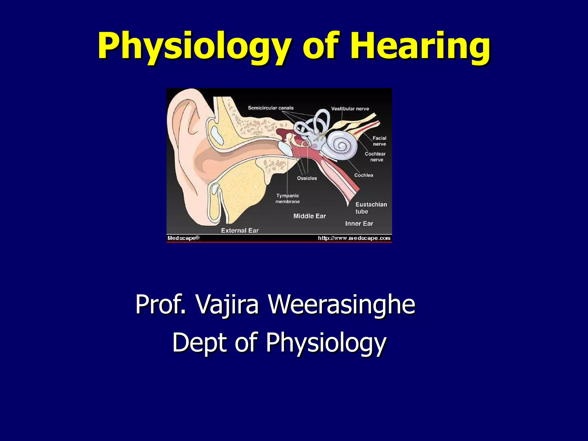

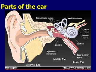
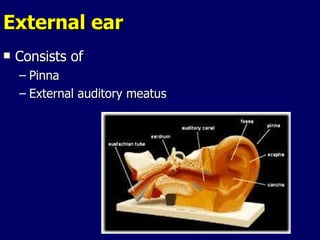
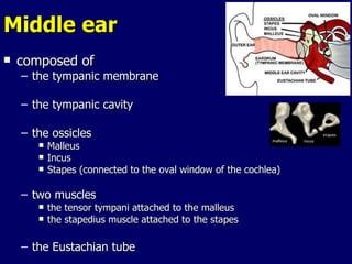
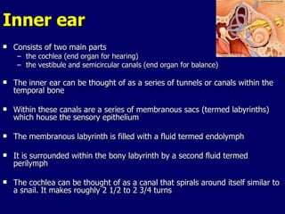
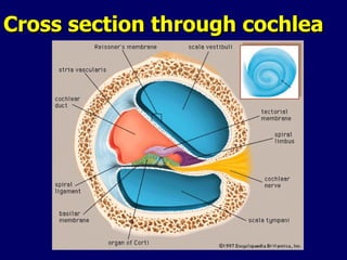
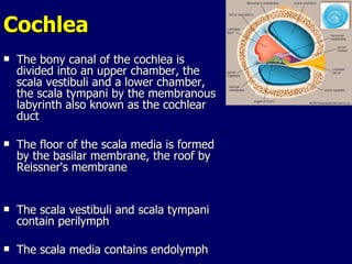

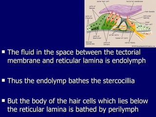
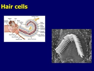
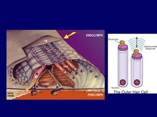

The document summarizes the physiology of hearing. It describes how sound is transmitted through the ear as a pressure wave and perceived as sound information. It details the anatomy of the outer, middle and inner ear. It explains how sound waves cause vibrations that are amplified and transmitted through the ossicles to the cochlea fluid. Within the cochlea, vibrations travel in a wave along the basilar membrane to stimulate hair cells, which transmit signals to the auditory nerve.


