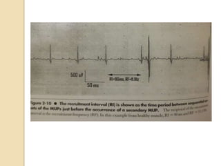
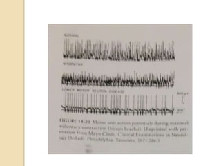
The Motor Unit &
Electromyography
Stanley J. Myers, Bhagwan T. Shahani, and
Frans L. Bruyninckx
Reading Downey and Darling’s Chapter 14
Interference pattern &
recruitment
 Needle emg examination of a normal
skeletal muscle at rest reveals an
isoelectric baseline
 Recruitment :
◦ Successive activation of the same &
additional motor units with increasing
strength of voluntary muscle contraction
Interference pattern &
recruitment
 Minimal voluntary contraction  1 or
several motor units are activated  low
amplitude
 As tension increase  firing frequency
of the individual potential increase
 Motor unit potentials tended to
increase in amplitude with heightened
tension during isometric contraction
(Studies of the human rectus femoris &
gastrocnemius)
Interference pattern &
recruitment
Order of recruitment :
 S type (slow-fatigue , type I) :
◦ Low strength of contraction  few motor units are active
slow rate of discharge & low amplitude
◦ Low-treshold, early activated unit produce relatively small
forces used for fine motor control & postural adjustment.
 Increasing tension  increases the frequency of
discharge by the active motor unit & recruitment of
previously inactive units
 FF (fast-fatigue) or FR (fatigue-resistant) type :
◦ Higher threshold units, larger force contribution
 Motor units are recruited in order of size, from small to
large.
Interference Pattern & Recruitment
 Normal, smooth, clinically sustained
contraction of a muscle, motor units fire
at a rate and in relation to each other 
smooth, continous, nonjerky contraction.
 Recruitment interval :
◦ Interdischarge interval between two
consecutive discharge of a MUAP at the point
at which a second motor unit potential is
recruited with gradually increasing strength of
voluntary muscle contraction
Interference Pattern &
Recruitment
 Recruitment frequency (firing rate)
◦ Firing rate of a MUAP when a different
MUAP first appears, with gradually
increasing strength of voluntary muscle
contraction
◦ The reciprocal of the recruitment interval
◦ Quantitative measures
◦ Most accurately evaluated when a fixed,
low-level percentage of max voluntary
contraction (MVC) of a particular muscle
(25-30%) is maintained
Interference Pattern &
Recruitment
 In neuropathic disease:
◦ First unit usually firing more rapidly at the
moment when the second is recruited.
◦ the recruitment interval is shortened
◦ Recruitment frequency is increased
 In myopathic disease :
◦ first unit is firing more slowly at the moment
when the second unit is recruited
 Ratio of the average firing rate to the
number of active units (N < 5:1)
Interference Pattern &
Recruitment
 It appears that recruitmet has a more
important role in grading of activity than
does changing the frequency, except at
very low and very high contraction
strenghts
 Under most condition (25-75% MVC),
the fastest units respond at frequencies
between 25-35 Hz.
 Over the tension range of 5-60% MVC,
the firing starts and stops abruptly and
frequency increases, but not in
proportion to strength
30/09/2022 11
Effect of age
 The normal mean duration of the
muscle action potential increases with
age.
 Decrease in the propagation velocity
of the impulse over the muscle fiber.
 Peterson & Kugelberg :
◦ in young persons normal values are
obtained from the first-recruited low-
treshold muscle action potentials
◦ Aging may selectively destroy muscle
fibers with the largest calibers and lowest
tresholds.
Effect of age
Stalberg & Thiele :
 Study on EDC muscle in subject aged
10-89 years
 Fiber density increases slowly
throughout life and progress faster
after age 70 years
 Impairment of nerve and
neuromuscular impulse transmission
increases at the same time 
degenerative loss of motor neurons
with aging was compensated for by
Effect of age
 Motor unit density in the m tibialis
anterior decreased with age 
indicate loss of motor units with
increasing age
 Distal muscles show more extensive
changes than proximal ones, & not all
muscles are uniformly affected
 There was no evidenc for loss of
motor neurons p to age 60, but
beyond that age, there was evidence
for a diminishing motor neuron
population
Effect of temperature
 For every 1 ° reduction :
◦ Mean duration of the muscle action potential increases
10-30%
◦ Mean amplitude decreases by 2-5%
 The number of polyphasic potentials can increase
as much as 10-fold with a 10% decrease in
temperature
 Temperature coefficient of the propagation
velocity  prolongation of duration
 The slower propagation velocity of the impulse
over the muscle fiber and the terminal nerve fibers
can cause temporal dispersion of fibers within the
motor unit
Fatigue
• Neuromuscular fatique :
 Any reduction in the force-generating
capacity of the total neuromuscular system,
regardless of the force required in any given
situation
 Increased of force caused by:
◦ Activation of more motor units
◦ Greater frequency of discharge
 Fatiguing muscle :
◦ Relative force developed
◦ Sum of electrical activity remain linearly
related until the level of mechanical activity
cannot be maintained
Fatigue
 EMG results recorded with surface
electrode:
◦ Increased amplitude of the summated
potentials
◦ Decreased frequency
 Jerky firing pattern
 Needle EMG :
◦ Discharges of fibers from different motor
units tend to group during fatiguing muscle
work
◦ Increase in total number of active motor
units
◦ Individual MUAPs are decreased in
amplitude, duration is little changed.
◦ Number of polyphasic potentials is
increased (incomplete synchronization
Effect of disuse
 In muscle with disuse atrophy,
polyphasic potensials accounted for
25% of all action potentials, normal
muscle 1-3%
 A reduction or increase in duration of
MUAPs occasionally is noted.
Clinical Applications
30/09/2022 20
Lower Motor Neuron Disease
 EMG has certain general characteristics:
- ↑ Insertional activity, fibrilation potentials, and
PSW, fasciculation potentials
- ↑ amplitude & average duration of MUAPs
together with ↑ incidence of polyphasic
potentials
- ↓ number of MUAPs observed during full
effort
 Axonal sprouting : Collateral regeneration
consists of the ingrowth of branches from
intact nerve fibers to adjacent denervated
muscle fibers.
30/09/2022 22
Anterior Horn Cell Disorders
- Poliomyelitis, ALS, the progressive SMA
(e.g., Werdnig-Hoffman disease), & in
hereditary degenerative conditions (Charcot-
Marie Tooth disease)
- Fibrilation potentials  common
- Fasciculation potentialsmay be seen
- The degree of spontaneous activity
diminishes with chronicity & reinnervation
- Most MUAPs have prolonged duration &
pronounced ↑ amplitude
30/09/2022 24
Nerve Root & Plexus Lesions
- Causes denervation in the involved
segments
- Manifested by fibrilllation potentials &
occasional fasciculation, reduced numbers
of MUAPs under voluntary control, and
complex polyphasic units, increased
duration.
- Acute phase: electrical silence with
fibrilation potential and PSWWallerian
degeneration
30/09/2022 25
Peripheral Nerve Disorders
- Physiological block (neuropraxia) 
characterized by electrical silence at rest,
no voluntary action potentials if block is
complete & reduced number of normal
potentials if the block is partial.
- Fibrilation & PSW noted approximately of
10 days after the injury.
- Voluntary MUP first seen with return of
function are of low amplitude, long duration
& polyphasic  caused by early terminal
reinnervation of a reduced unit.
- In peripheral neuropathy both axonal
degeneration and demyelination can
occur.
30/09/2022 26
Motor End-Plate Disturbances
- A defect in transmission across the NMJ,
as in myasthenia gravis, characterized by
a decrease amplitude of the successively
evoked potentials on repeated low-
frequency stimulation.
- Change in amplitude by blocking of
excitation at the end-plate to a variable
number of muscle fibers supplied by the
neuron with fibers recovering at different
rates.
- MG: autoimune disease which the defect
is at the postsynaptic junction.
30/09/2022 27
Myashenic Syndrome (Lambert-Eaton
Myasthenic Syndrome)
- The weak, resting patient becomes weaker
with continuing muscle contraction or
stimulation after an initial period of enhanced
strength.
- The defect is in the presynaptic region
30/09/2022 28
Myopathy
- Fibrillation potentials mechanism is not
completely clear
- In progressive muscular dystrophy and
hyperkalemic periodic paralysis these
spontaneous discharges  related to the
increase in excitability resulting from low
levels of intracellular potassium also the
degenerating muscle fibers may lose their
terminal innervation
- Complex repetitive discharge noted in
myositis
- Low amplitude, short duration
30/09/2022 29
Myotonia
- Abnormally sustained contraction & difficulty
in relaxation.
- EMG:
Repetittive high-frequency, waxing & waning
discharges (positive waves or biphasic
potentials whose initial deflection is positive)
30/09/2022 30
Upper Motor Neuron Lession
- Hoefer & Putnam  study in spastic
condition : voluntary contraction show
decreased frequency & amplitude.
- EMG finding of PSW & fibrilation
potentials
- A number of theories proposed to explain
 transsynaptic neuronal degeneration,
membrane instability, & antifibrilation
factor.
30/09/2022 32
Synchronization
 Is demonstrated when action potentials
are obtained simultaneously from 2 or
more widely separated recording
electrodes
 The cause is not clear 
◦ intraspinal mechanism for synchronization
of dicharge rhythm in a number of different
motor units supplied by the same spinal
segment
◦ In a given muscle single units of large area
are responsible
Study
 Spinal cords of 47 subjects , 13-95 years
, died suddenly, were examined
 There was no evidence for loss of motor
neurons up to age 60, but beyond that
age, although individual counts varied
considerably, there was evidence for a
diminishing motor neuron population.
 > age 60  only 50% of younger
subjects
 Clinical weakness usually is not noted
until 40-60% of muscle fibers have been
lost.
Chapter 2 Neurons and Associated Cells
33
Figure 2.11: Degeneration and
regeneration of peripheral somatic
motor nerve fibers. (A) Several
days after transection (at the wedge). Note
the central chromatolysis and eccentric
nuclei,
increase in number of neurolemma cells,
and fragmentation of myelin sheaths. (B)
Several weeks
later, the neurolemma cord receives
regenerating axis cylinders from the
transected fibers and collateral
branches from the adjacent normal fiber.
(C) Several months later, collateral
branches of axis
cylinders that failed to innervate motor end
plates degenerate. The regenerated
portions of the fibers
contain more internodes than before:
hence, they conduct nerve impulses more
Interference Pattern &
Recruitment
 Recruitment has a more important role
in grading of activity
 25-75% MVC : the fastest units
respond at frequency 25-35 Hz
 Over the tension range of 5-60%
MVC, The firing stars and stop
abruptly, frequency increases, but not
in propotion to strenght
Interference Pattern &
Recruitment
 For example : in one motor unit  the
firing commenced at 20 Hz when the
tension was 15% of MCV & increased
with the tension rise, but only to 30 Hz
 Units that are active at tensions below
5% of MVC usually have a lower
starting frequency (5-7 Hz) and
greater frequency range, irregular
discharge rates,even during constant
contraction
reading emg ik.pptx
reading emg ik.pptx
reading emg ik.pptx
reading emg ik.pptx

reading emg ik.pptx

  • 1.
    The Motor Unit& Electromyography Stanley J. Myers, Bhagwan T. Shahani, and Frans L. Bruyninckx Reading Downey and Darling’s Chapter 14
  • 2.
    Interference pattern & recruitment Needle emg examination of a normal skeletal muscle at rest reveals an isoelectric baseline  Recruitment : ◦ Successive activation of the same & additional motor units with increasing strength of voluntary muscle contraction
  • 3.
    Interference pattern & recruitment Minimal voluntary contraction  1 or several motor units are activated  low amplitude  As tension increase  firing frequency of the individual potential increase  Motor unit potentials tended to increase in amplitude with heightened tension during isometric contraction (Studies of the human rectus femoris & gastrocnemius)
  • 4.
    Interference pattern & recruitment Orderof recruitment :  S type (slow-fatigue , type I) : ◦ Low strength of contraction  few motor units are active slow rate of discharge & low amplitude ◦ Low-treshold, early activated unit produce relatively small forces used for fine motor control & postural adjustment.  Increasing tension  increases the frequency of discharge by the active motor unit & recruitment of previously inactive units  FF (fast-fatigue) or FR (fatigue-resistant) type : ◦ Higher threshold units, larger force contribution  Motor units are recruited in order of size, from small to large.
  • 5.
    Interference Pattern &Recruitment  Normal, smooth, clinically sustained contraction of a muscle, motor units fire at a rate and in relation to each other  smooth, continous, nonjerky contraction.  Recruitment interval : ◦ Interdischarge interval between two consecutive discharge of a MUAP at the point at which a second motor unit potential is recruited with gradually increasing strength of voluntary muscle contraction
  • 6.
    Interference Pattern & Recruitment Recruitment frequency (firing rate) ◦ Firing rate of a MUAP when a different MUAP first appears, with gradually increasing strength of voluntary muscle contraction ◦ The reciprocal of the recruitment interval ◦ Quantitative measures ◦ Most accurately evaluated when a fixed, low-level percentage of max voluntary contraction (MVC) of a particular muscle (25-30%) is maintained
  • 8.
    Interference Pattern & Recruitment In neuropathic disease: ◦ First unit usually firing more rapidly at the moment when the second is recruited. ◦ the recruitment interval is shortened ◦ Recruitment frequency is increased  In myopathic disease : ◦ first unit is firing more slowly at the moment when the second unit is recruited  Ratio of the average firing rate to the number of active units (N < 5:1)
  • 9.
    Interference Pattern & Recruitment It appears that recruitmet has a more important role in grading of activity than does changing the frequency, except at very low and very high contraction strenghts  Under most condition (25-75% MVC), the fastest units respond at frequencies between 25-35 Hz.  Over the tension range of 5-60% MVC, the firing starts and stops abruptly and frequency increases, but not in proportion to strength
  • 11.
  • 13.
    Effect of age The normal mean duration of the muscle action potential increases with age.  Decrease in the propagation velocity of the impulse over the muscle fiber.  Peterson & Kugelberg : ◦ in young persons normal values are obtained from the first-recruited low- treshold muscle action potentials ◦ Aging may selectively destroy muscle fibers with the largest calibers and lowest tresholds.
  • 14.
    Effect of age Stalberg& Thiele :  Study on EDC muscle in subject aged 10-89 years  Fiber density increases slowly throughout life and progress faster after age 70 years  Impairment of nerve and neuromuscular impulse transmission increases at the same time  degenerative loss of motor neurons with aging was compensated for by
  • 15.
    Effect of age Motor unit density in the m tibialis anterior decreased with age  indicate loss of motor units with increasing age  Distal muscles show more extensive changes than proximal ones, & not all muscles are uniformly affected  There was no evidenc for loss of motor neurons p to age 60, but beyond that age, there was evidence for a diminishing motor neuron population
  • 16.
    Effect of temperature For every 1 ° reduction : ◦ Mean duration of the muscle action potential increases 10-30% ◦ Mean amplitude decreases by 2-5%  The number of polyphasic potentials can increase as much as 10-fold with a 10% decrease in temperature  Temperature coefficient of the propagation velocity  prolongation of duration  The slower propagation velocity of the impulse over the muscle fiber and the terminal nerve fibers can cause temporal dispersion of fibers within the motor unit
  • 17.
    Fatigue • Neuromuscular fatique:  Any reduction in the force-generating capacity of the total neuromuscular system, regardless of the force required in any given situation  Increased of force caused by: ◦ Activation of more motor units ◦ Greater frequency of discharge  Fatiguing muscle : ◦ Relative force developed ◦ Sum of electrical activity remain linearly related until the level of mechanical activity cannot be maintained
  • 18.
    Fatigue  EMG resultsrecorded with surface electrode: ◦ Increased amplitude of the summated potentials ◦ Decreased frequency  Jerky firing pattern  Needle EMG : ◦ Discharges of fibers from different motor units tend to group during fatiguing muscle work ◦ Increase in total number of active motor units ◦ Individual MUAPs are decreased in amplitude, duration is little changed. ◦ Number of polyphasic potentials is increased (incomplete synchronization
  • 19.
    Effect of disuse In muscle with disuse atrophy, polyphasic potensials accounted for 25% of all action potentials, normal muscle 1-3%  A reduction or increase in duration of MUAPs occasionally is noted.
  • 20.
    Clinical Applications 30/09/2022 20 LowerMotor Neuron Disease  EMG has certain general characteristics: - ↑ Insertional activity, fibrilation potentials, and PSW, fasciculation potentials - ↑ amplitude & average duration of MUAPs together with ↑ incidence of polyphasic potentials - ↓ number of MUAPs observed during full effort  Axonal sprouting : Collateral regeneration consists of the ingrowth of branches from intact nerve fibers to adjacent denervated muscle fibers.
  • 22.
    30/09/2022 22 Anterior HornCell Disorders - Poliomyelitis, ALS, the progressive SMA (e.g., Werdnig-Hoffman disease), & in hereditary degenerative conditions (Charcot- Marie Tooth disease) - Fibrilation potentials  common - Fasciculation potentialsmay be seen - The degree of spontaneous activity diminishes with chronicity & reinnervation - Most MUAPs have prolonged duration & pronounced ↑ amplitude
  • 24.
    30/09/2022 24 Nerve Root& Plexus Lesions - Causes denervation in the involved segments - Manifested by fibrilllation potentials & occasional fasciculation, reduced numbers of MUAPs under voluntary control, and complex polyphasic units, increased duration. - Acute phase: electrical silence with fibrilation potential and PSWWallerian degeneration
  • 25.
    30/09/2022 25 Peripheral NerveDisorders - Physiological block (neuropraxia)  characterized by electrical silence at rest, no voluntary action potentials if block is complete & reduced number of normal potentials if the block is partial. - Fibrilation & PSW noted approximately of 10 days after the injury. - Voluntary MUP first seen with return of function are of low amplitude, long duration & polyphasic  caused by early terminal reinnervation of a reduced unit. - In peripheral neuropathy both axonal degeneration and demyelination can occur.
  • 26.
    30/09/2022 26 Motor End-PlateDisturbances - A defect in transmission across the NMJ, as in myasthenia gravis, characterized by a decrease amplitude of the successively evoked potentials on repeated low- frequency stimulation. - Change in amplitude by blocking of excitation at the end-plate to a variable number of muscle fibers supplied by the neuron with fibers recovering at different rates. - MG: autoimune disease which the defect is at the postsynaptic junction.
  • 27.
    30/09/2022 27 Myashenic Syndrome(Lambert-Eaton Myasthenic Syndrome) - The weak, resting patient becomes weaker with continuing muscle contraction or stimulation after an initial period of enhanced strength. - The defect is in the presynaptic region
  • 28.
    30/09/2022 28 Myopathy - Fibrillationpotentials mechanism is not completely clear - In progressive muscular dystrophy and hyperkalemic periodic paralysis these spontaneous discharges  related to the increase in excitability resulting from low levels of intracellular potassium also the degenerating muscle fibers may lose their terminal innervation - Complex repetitive discharge noted in myositis - Low amplitude, short duration
  • 29.
    30/09/2022 29 Myotonia - Abnormallysustained contraction & difficulty in relaxation. - EMG: Repetittive high-frequency, waxing & waning discharges (positive waves or biphasic potentials whose initial deflection is positive)
  • 30.
    30/09/2022 30 Upper MotorNeuron Lession - Hoefer & Putnam  study in spastic condition : voluntary contraction show decreased frequency & amplitude. - EMG finding of PSW & fibrilation potentials - A number of theories proposed to explain  transsynaptic neuronal degeneration, membrane instability, & antifibrilation factor.
  • 32.
  • 33.
    Synchronization  Is demonstratedwhen action potentials are obtained simultaneously from 2 or more widely separated recording electrodes  The cause is not clear  ◦ intraspinal mechanism for synchronization of dicharge rhythm in a number of different motor units supplied by the same spinal segment ◦ In a given muscle single units of large area are responsible
  • 34.
    Study  Spinal cordsof 47 subjects , 13-95 years , died suddenly, were examined  There was no evidence for loss of motor neurons up to age 60, but beyond that age, although individual counts varied considerably, there was evidence for a diminishing motor neuron population.  > age 60  only 50% of younger subjects  Clinical weakness usually is not noted until 40-60% of muscle fibers have been lost.
  • 35.
    Chapter 2 Neuronsand Associated Cells 33 Figure 2.11: Degeneration and regeneration of peripheral somatic motor nerve fibers. (A) Several days after transection (at the wedge). Note the central chromatolysis and eccentric nuclei, increase in number of neurolemma cells, and fragmentation of myelin sheaths. (B) Several weeks later, the neurolemma cord receives regenerating axis cylinders from the transected fibers and collateral branches from the adjacent normal fiber. (C) Several months later, collateral branches of axis cylinders that failed to innervate motor end plates degenerate. The regenerated portions of the fibers contain more internodes than before: hence, they conduct nerve impulses more
  • 38.
    Interference Pattern & Recruitment Recruitment has a more important role in grading of activity  25-75% MVC : the fastest units respond at frequency 25-35 Hz  Over the tension range of 5-60% MVC, The firing stars and stop abruptly, frequency increases, but not in propotion to strenght
  • 39.
    Interference Pattern & Recruitment For example : in one motor unit  the firing commenced at 20 Hz when the tension was 15% of MCV & increased with the tension rise, but only to 30 Hz  Units that are active at tensions below 5% of MVC usually have a lower starting frequency (5-7 Hz) and greater frequency range, irregular discharge rates,even during constant contraction