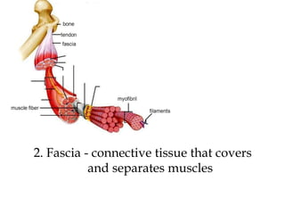
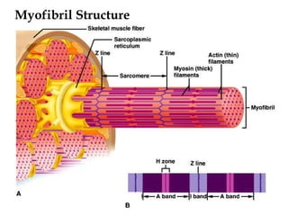
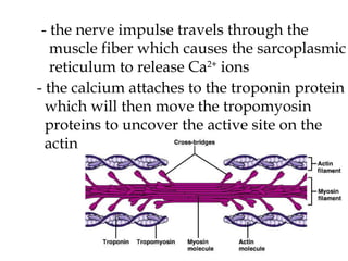
CHAPTER 8: MUSCULAR SYSTEM
3 Types of Muscles 1. Skeletal 2. Smooth 3. cardiac
bone tendon fascia muscle fiber myofibril filaments Structure of a Skeletal Muscle muscle fascicle
1. Tendon – holds the muscle to the bone
2. Fascia - connective tissue that covers    and separates muscles
3. Muscle fiber = the muscle cell
4. Myofibril – the contractile fibers in a    muscle fiber
2 Types a) actin - thin b) myosin - thick  5. Filaments
Muscle Fiber Structure
1. Sarcoplasmic Reticulum - tubes run parallel to the myofibrils - stores calcium & functions as the ER
2. Transverse Tubules - run at right angles to myofibrils - activate muscle contraction when    stimulated
3. Sarcolemma - the “cell membrane”
Myofibril Structure
Neuromuscular Junction - place where nerve fiber & muscle fiber meet
- each muscle fiber is connected to a motor neuron - nerve fiber meets at the motor end plate
- end of motor neurons contains synaptic vesicles that have neurotransmitters in  them - one  motor neuron can control many  muscle fibers    motor unit
The myogram
Skeletal Muscle Contraction    a specific sequence of events needs to  occur in order for a muscle fiber to contract - it starts with a nerve impulse traveling down a motor neuron axon - the nerve impulse causes the synaptic  vesicles to release the neurotransmitter ACh - the ACh attaches to ACh receptors on  sarcolemma then is broken down by acetylcholinesterase Neurotransmitter release
- the nerve impulse travels through the  muscle fiber which causes the sarcoplasmic reticulum to release Ca 2+  ions - the calcium attaches to the troponin protein which will then move the tropomyosin  proteins to uncover the active site on the  actin
- the myosin heads attach to the active site on the actin filament - ATP is used, and the myosin pulls on the actin by the myosin cross-bridges - the muscle shortens as a contraction occurs Muscle contraction - the muscle will stay contracted until the  Ca 2+  is reabsorbed by the SR - the active site will be covered up - actin slides back into its original position Muscle contraction 1
 
Energy Sources for Contraction - ATP supplies the energy - ADP needs to be regenerated into ATP - can borrow a phosphate from  creatine phosphate - regenerate through the process of  cell respiration
 
-  myoglobin  - can store O 2  in muscles for   cell respiration   - if O 2  levels are low and  oxygen debt  occurs glucose  pyruvic acid  lactic acid  cramps
- can lead to  muscle fatigue    muscle loses ability to   contract
Muscular Response Threshold Stimulus  - minimal strength needed to start a contraction All - or - None Response  - a muscle fiber will contract or it will not - no partial contractions Recruitment  - the greater the stimulus, the more motor units will be  activated, more muscle fibers will  contract
The myogram latent period contraction period relaxation period recovery period
latent period  - time between stimulus and contraction contraction period  - time for the muscle to contract relaxation period  - actin slides back into place recovery period  - O 2  resupplied to muscles
Muscular hypertrophy  - muscles get larger due to increased use Muscular atrophy  - muscles get smaller  due to less use
Smooth Muscle - thin - contains actin and myosin - are randomly arranged    no striations 2 Types a) Multiunit - found in eye and blood vessels - contracts only after receives a nerve   impulse
b) Visceral - found in internal organs - displays rhythmic patterns - muscles able to stimulate each other - no continuous stimulus needed - Contraction is almost the same as in  skeletal muscles - besides ACh, can be stimulated by norepinephrin - contraction occurs more slowly - muscles are able to stretch
Cardiac Muscles - only found in the heart - has actin and myosin - contains many mitochondria
Muscular Dystrophy    caused by a defective gene    the muscle fibers are unusually susceptible to damage    muscles become weaker and may be replaced by fat and connective tissue    there are over 40 types of MD    there is no cure
 
Anabolic Steroids Anabolic steroids change muscle mass and  strength by three processes. 1. Increases the production of proteins 2. Blocks the hormone  cortisol , which  decreases muscle breakdown 3. Leads cells to differentiate into muscle  more readily than fat


Physiology CH 8 lecture notes

  • 1.
  • 2.
    3 Types ofMuscles 1. Skeletal 2. Smooth 3. cardiac
  • 3.
    bone tendon fasciamuscle fiber myofibril filaments Structure of a Skeletal Muscle muscle fascicle
  • 4.
    1. Tendon –holds the muscle to the bone
  • 5.
    2. Fascia -connective tissue that covers and separates muscles
  • 6.
    3. Muscle fiber= the muscle cell
  • 7.
    4. Myofibril –the contractile fibers in a muscle fiber
  • 8.
    2 Types a)actin - thin b) myosin - thick 5. Filaments
  • 9.
  • 10.
    1. Sarcoplasmic Reticulum- tubes run parallel to the myofibrils - stores calcium & functions as the ER
  • 11.
    2. Transverse Tubules- run at right angles to myofibrils - activate muscle contraction when stimulated
  • 12.
    3. Sarcolemma -the “cell membrane”
  • 13.
  • 14.
    Neuromuscular Junction -place where nerve fiber & muscle fiber meet
  • 15.
    - each musclefiber is connected to a motor neuron - nerve fiber meets at the motor end plate
  • 16.
    - end ofmotor neurons contains synaptic vesicles that have neurotransmitters in them - one motor neuron can control many muscle fibers  motor unit
  • 17.
  • 18.
    Skeletal Muscle Contraction a specific sequence of events needs to occur in order for a muscle fiber to contract - it starts with a nerve impulse traveling down a motor neuron axon - the nerve impulse causes the synaptic vesicles to release the neurotransmitter ACh - the ACh attaches to ACh receptors on sarcolemma then is broken down by acetylcholinesterase Neurotransmitter release
  • 19.
    - the nerveimpulse travels through the muscle fiber which causes the sarcoplasmic reticulum to release Ca 2+ ions - the calcium attaches to the troponin protein which will then move the tropomyosin proteins to uncover the active site on the actin
  • 20.
    - the myosinheads attach to the active site on the actin filament - ATP is used, and the myosin pulls on the actin by the myosin cross-bridges - the muscle shortens as a contraction occurs Muscle contraction - the muscle will stay contracted until the Ca 2+ is reabsorbed by the SR - the active site will be covered up - actin slides back into its original position Muscle contraction 1
  • 21.
  • 22.
    Energy Sources forContraction - ATP supplies the energy - ADP needs to be regenerated into ATP - can borrow a phosphate from creatine phosphate - regenerate through the process of cell respiration
  • 23.
  • 24.
    - myoglobin - can store O 2 in muscles for cell respiration - if O 2 levels are low and oxygen debt occurs glucose  pyruvic acid  lactic acid  cramps
  • 25.
    - can leadto muscle fatigue  muscle loses ability to contract
  • 26.
    Muscular Response ThresholdStimulus - minimal strength needed to start a contraction All - or - None Response - a muscle fiber will contract or it will not - no partial contractions Recruitment - the greater the stimulus, the more motor units will be activated, more muscle fibers will contract
  • 27.
    The myogram latentperiod contraction period relaxation period recovery period
  • 28.
    latent period - time between stimulus and contraction contraction period - time for the muscle to contract relaxation period - actin slides back into place recovery period - O 2 resupplied to muscles
  • 29.
    Muscular hypertrophy - muscles get larger due to increased use Muscular atrophy - muscles get smaller due to less use
  • 30.
    Smooth Muscle -thin - contains actin and myosin - are randomly arranged  no striations 2 Types a) Multiunit - found in eye and blood vessels - contracts only after receives a nerve impulse
  • 31.
    b) Visceral -found in internal organs - displays rhythmic patterns - muscles able to stimulate each other - no continuous stimulus needed - Contraction is almost the same as in skeletal muscles - besides ACh, can be stimulated by norepinephrin - contraction occurs more slowly - muscles are able to stretch
  • 32.
    Cardiac Muscles -only found in the heart - has actin and myosin - contains many mitochondria
  • 33.
    Muscular Dystrophy  caused by a defective gene  the muscle fibers are unusually susceptible to damage  muscles become weaker and may be replaced by fat and connective tissue  there are over 40 types of MD  there is no cure
  • 34.
  • 35.
    Anabolic Steroids Anabolicsteroids change muscle mass and strength by three processes. 1. Increases the production of proteins 2. Blocks the hormone cortisol , which decreases muscle breakdown 3. Leads cells to differentiate into muscle more readily than fat
  • 36.
  • 37.
  • 38.