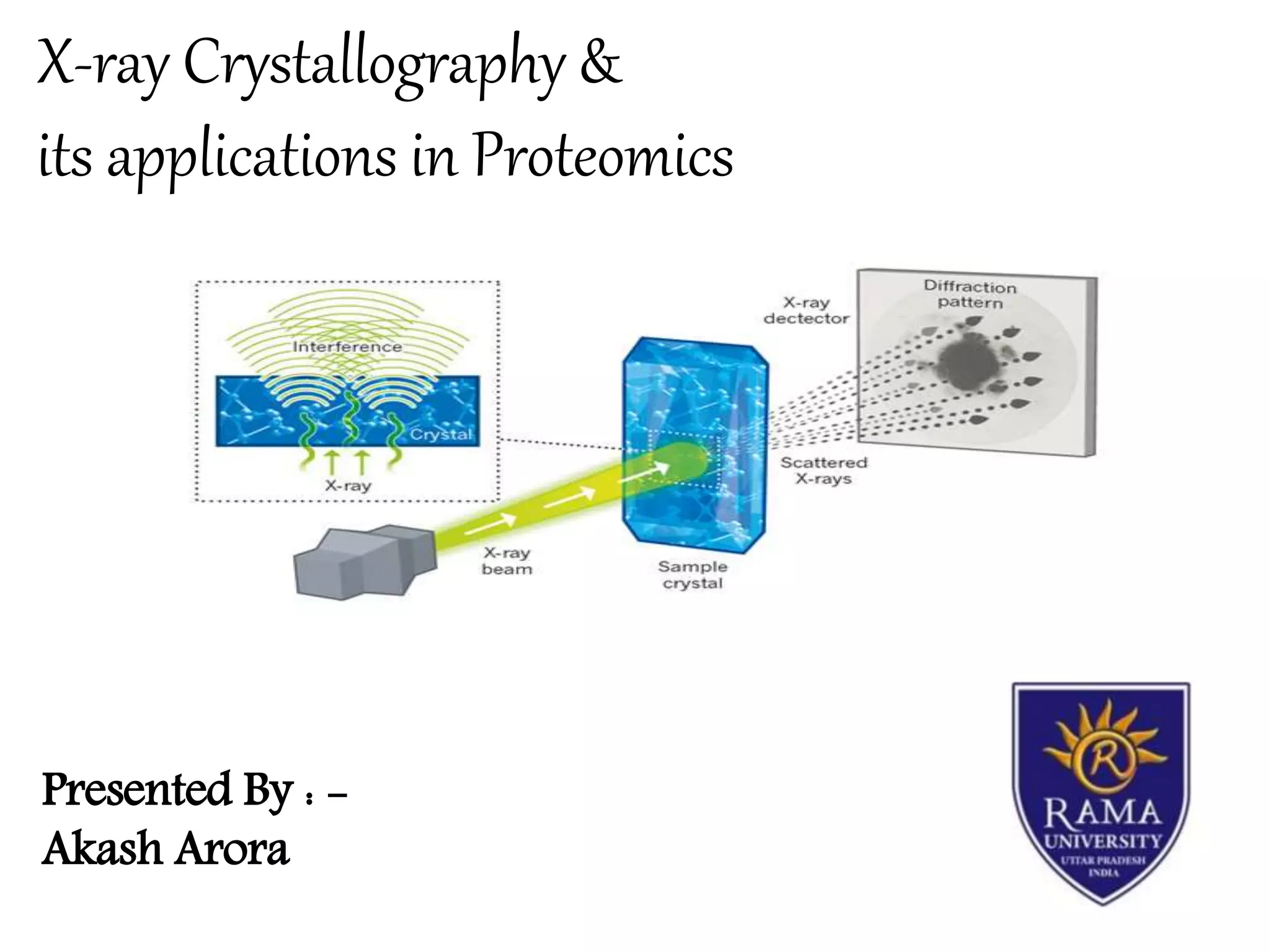
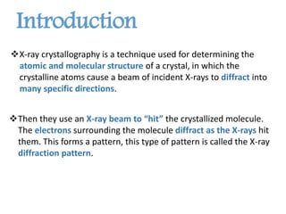
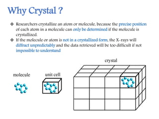
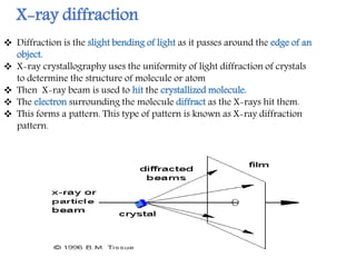
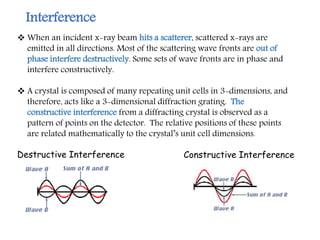
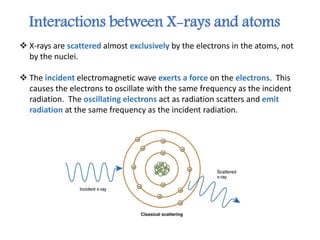
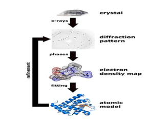
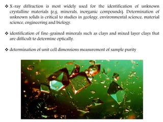
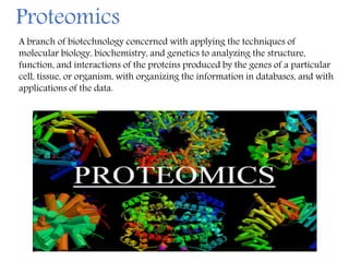
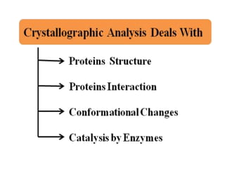
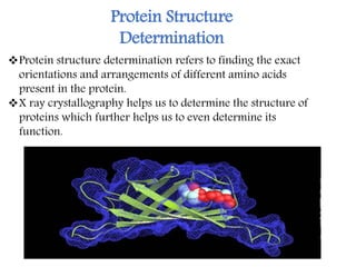
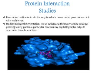
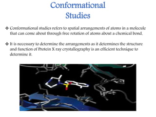
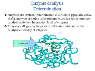
X-ray crystallography is a technique that uses X-rays to determine the atomic structure of crystals. It involves crystallizing molecules and bombarding them with X-rays, which produce a diffraction pattern. This pattern is used to deduce the molecular structure. X-ray crystallography has many applications in proteomics, including determining protein structures, studying protein interactions, and elucidating enzyme catalysis mechanisms. It provides atomic-level insights that advance understanding of protein function.
X-Ray Crystallography is a technique used to determine the atomic and molecular structure of a crystal, in which the crystalline atoms cause a beam of incident X-rays to diffract into many specific directions.
X-ray crystallography is a technique used for determining the atomic and molecular structure of a crystal, in which the crystalline atoms cause a beam of incident X-rays to diffract into many specific directions.
In silico drug designing is the drug design which can be carried out in silicon chip,i.e., within computers. The slides are helpful to know a brief description about in silico drug designing.
Prediction of the three dimensional structure of a given protein sequence i.e. target protein from the amino acid sequence of a homologous (template) protein for which an X-ray or NMR structure is available based on an alignment to one or more known protein structures
Introduction to Applications of Proteomics Science,
Proteomics- Techniques, Applications of proteomics
Presented by
A. Harsha Vardhan Naidu
Department of Pharmacology
X-ray crystallography is the experimental science determining the atomic and molecular structure of a crystal, in which the crystalline structure causes a beam of incident X-rays to diffract into many specific directions. By measuring the angles and intensities of these diffracted beams, a crystallographer can produce a three-dimensional picture of the density of electrons within the crystal. From this electron density, the mean positions of the atoms in the crystal can be determined, as well as their chemical bonds, their crystallographic disorder, and various other information.
X-ray Crystallography is a scientific method used to determine the arrangement of atoms of a crystalline solid in three dimension. It is based on x ray diffraction. Reveals structure of a crystal at atomic level.
X ray crystallography and X ray DiffractionFaisal Hussain
This is the short description about x ray crystallography.
simplest and easy to understand.
Procedure of X ray Diffraction.
Pros and Cons of X ray Crystallography
X ray crystallography and X ray DiffractionFaisal Hussain
This is the short description about x ray crystallography.
simplest and easy to understand.
Procedure of X ray Diffraction.
Advantages and Disadvantages of X ray Crystallography
X-ray diffraction (XRD) is a versatile non-destructive analytical technique used to analyze physical properties such as phase composition, crystal structure and orientation of powder, solid and liquid samples. Many materials are made up of tiny crystallites. The chemical composition and structural type of these crystals is called their 'phase'. Materials can be single phase or multiphase mixtures and may contain crystalline and non-crystalline components. In an X-ray diffractometer, different crystalline phases give different diffraction patterns. Phase identification can be performed by comparing X-ray diffraction patterns obtained from unknown samples to patterns in reference databases.
principles:
X-Ray Diffraction is the result of constructive interference between X-rays and a crystalline sample. The wavelength of the X-rays used is of the same order of magnitude of the distance between the atoms in a crystalline lattice. This gives rise to a diffraction pattern that can be analysed in a number of ways, the most popular being applying the famous Bragg’s Law (nλ=2d sin θ) which is used in the measurement of crystals and their phases.
Applictions:
Many researchers, in industrial as well as in scientific laboratories, rely on X-ray diffraction (XRD) as a tool to develop new materials or to improve production efficiency. Innovations in X-ray diffraction closely follow the research on new materials, such as in semiconductor technologies or pharmaceutical investigations. Industrial research is directed toward the ever-increasing speed and efficiency of production processes. Fully automated X-ray diffraction analyses in mining and building materials production sites result in more cost-effective solutions for production control.
The main uses of X-ray diffraction are:
Qualitative and quantitative phase analysis of pure substances and mixtures. The most common method for phase analysis is often called 'X-ray powder diffraction' (XRPD).
Report Back from SGO 2024: What’s the Latest in Cervical Cancer?bkling
Are you curious about what’s new in cervical cancer research or unsure what the findings mean? Join Dr. Emily Ko, a gynecologic oncologist at Penn Medicine, to learn about the latest updates from the Society of Gynecologic Oncology (SGO) 2024 Annual Meeting on Women’s Cancer. Dr. Ko will discuss what the research presented at the conference means for you and answer your questions about the new developments.
Tom Selleck Health: A Comprehensive Look at the Iconic Actor’s Wellness Journeygreendigital
Tom Selleck, an enduring figure in Hollywood. has captivated audiences for decades with his rugged charm, iconic moustache. and memorable roles in television and film. From his breakout role as Thomas Magnum in Magnum P.I. to his current portrayal of Frank Reagan in Blue Bloods. Selleck's career has spanned over 50 years. But beyond his professional achievements. fans have often been curious about Tom Selleck Health. especially as he has aged in the public eye.
Follow us on: Pinterest
Introduction
Many have been interested in Tom Selleck health. not only because of his enduring presence on screen but also because of the challenges. and lifestyle choices he has faced and made over the years. This article delves into the various aspects of Tom Selleck health. exploring his fitness regimen, diet, mental health. and the challenges he has encountered as he ages. We'll look at how he maintains his well-being. the health issues he has faced, and his approach to ageing .
Early Life and Career
Childhood and Athletic Beginnings
Tom Selleck was born on January 29, 1945, in Detroit, Michigan, and grew up in Sherman Oaks, California. From an early age, he was involved in sports, particularly basketball. which played a significant role in his physical development. His athletic pursuits continued into college. where he attended the University of Southern California (USC) on a basketball scholarship. This early involvement in sports laid a strong foundation for his physical health and disciplined lifestyle.
Transition to Acting
Selleck's transition from an athlete to an actor came with its physical demands. His first significant role in "Magnum P.I." required him to perform various stunts and maintain a fit appearance. This role, which he played from 1980 to 1988. necessitated a rigorous fitness routine to meet the show's demands. setting the stage for his long-term commitment to health and wellness.
Fitness Regimen
Workout Routine
Tom Selleck health and fitness regimen has evolved. adapting to his changing roles and age. During his "Magnum, P.I." days. Selleck's workouts were intense and focused on building and maintaining muscle mass. His routine included weightlifting, cardiovascular exercises. and specific training for the stunts he performed on the show.
Selleck adjusted his fitness routine as he aged to suit his body's needs. Today, his workouts focus on maintaining flexibility, strength, and cardiovascular health. He incorporates low-impact exercises such as swimming, walking, and light weightlifting. This balanced approach helps him stay fit without putting undue strain on his joints and muscles.
Importance of Flexibility and Mobility
In recent years, Selleck has emphasized the importance of flexibility and mobility in his fitness regimen. Understanding the natural decline in muscle mass and joint flexibility with age. he includes stretching and yoga in his routine. These practices help prevent injuries, improve posture, and maintain mobilit
Pulmonary Thromboembolism - etilogy, types, medical- Surgical and nursing man...VarunMahajani
Disruption of blood supply to lung alveoli due to blockage of one or more pulmonary blood vessels is called as Pulmonary thromboembolism. In this presentation we will discuss its causes, types and its management in depth.
MANAGEMENT OF ATRIOVENTRICULAR CONDUCTION BLOCK.pdfJim Jacob Roy
Cardiac conduction defects can occur due to various causes.
Atrioventricular conduction blocks ( AV blocks ) are classified into 3 types.
This document describes the acute management of AV block.
These lecture slides, by Dr Sidra Arshad, offer a quick overview of physiological basis of a normal electrocardiogram.
Learning objectives:
1. Define an electrocardiogram (ECG) and electrocardiography
2. Describe how dipoles generated by the heart produce the waveforms of the ECG
3. Describe the components of a normal electrocardiogram of a typical bipolar leads (limb II)
4. Differentiate between intervals and segments
5. Enlist some common indications for obtaining an ECG
Study Resources:
1. Chapter 11, Guyton and Hall Textbook of Medical Physiology, 14th edition
2. Chapter 9, Human Physiology - From Cells to Systems, Lauralee Sherwood, 9th edition
3. Chapter 29, Ganong’s Review of Medical Physiology, 26th edition
4. Electrocardiogram, StatPearls - https://www.ncbi.nlm.nih.gov/books/NBK549803/
5. ECG in Medical Practice by ABM Abdullah, 4th edition
6. ECG Basics, http://www.nataliescasebook.com/tag/e-c-g-basics
Title: Sense of Taste
Presenter: Dr. Faiza, Assistant Professor of Physiology
Qualifications:
MBBS (Best Graduate, AIMC Lahore)
FCPS Physiology
ICMT, CHPE, DHPE (STMU)
MPH (GC University, Faisalabad)
MBA (Virtual University of Pakistan)
Learning Objectives:
Describe the structure and function of taste buds.
Describe the relationship between the taste threshold and taste index of common substances.
Explain the chemical basis and signal transduction of taste perception for each type of primary taste sensation.
Recognize different abnormalities of taste perception and their causes.
Key Topics:
Significance of Taste Sensation:
Differentiation between pleasant and harmful food
Influence on behavior
Selection of food based on metabolic needs
Receptors of Taste:
Taste buds on the tongue
Influence of sense of smell, texture of food, and pain stimulation (e.g., by pepper)
Primary and Secondary Taste Sensations:
Primary taste sensations: Sweet, Sour, Salty, Bitter, Umami
Chemical basis and signal transduction mechanisms for each taste
Taste Threshold and Index:
Taste threshold values for Sweet (sucrose), Salty (NaCl), Sour (HCl), and Bitter (Quinine)
Taste index relationship: Inversely proportional to taste threshold
Taste Blindness:
Inability to taste certain substances, particularly thiourea compounds
Example: Phenylthiocarbamide
Structure and Function of Taste Buds:
Composition: Epithelial cells, Sustentacular/Supporting cells, Taste cells, Basal cells
Features: Taste pores, Taste hairs/microvilli, and Taste nerve fibers
Location of Taste Buds:
Found in papillae of the tongue (Fungiform, Circumvallate, Foliate)
Also present on the palate, tonsillar pillars, epiglottis, and proximal esophagus
Mechanism of Taste Stimulation:
Interaction of taste substances with receptors on microvilli
Signal transduction pathways for Umami, Sweet, Bitter, Sour, and Salty tastes
Taste Sensitivity and Adaptation:
Decrease in sensitivity with age
Rapid adaptation of taste sensation
Role of Saliva in Taste:
Dissolution of tastants to reach receptors
Washing away the stimulus
Taste Preferences and Aversions:
Mechanisms behind taste preference and aversion
Influence of receptors and neural pathways
Impact of Sensory Nerve Damage:
Degeneration of taste buds if the sensory nerve fiber is cut
Abnormalities of Taste Detection:
Conditions: Ageusia, Hypogeusia, Dysgeusia (parageusia)
Causes: Nerve damage, neurological disorders, infections, poor oral hygiene, adverse drug effects, deficiencies, aging, tobacco use, altered neurotransmitter levels
Neurotransmitters and Taste Threshold:
Effects of serotonin (5-HT) and norepinephrine (NE) on taste sensitivity
Supertasters:
25% of the population with heightened sensitivity to taste, especially bitterness
Increased number of fungiform papillae
HOT NEW PRODUCT! BIG SALES FAST SHIPPING NOW FROM CHINA!! EU KU DB BK substit...GL Anaacs
Contact us if you are interested:
Email / Skype : kefaya1771@gmail.com
Threema: PXHY5PDH
New BATCH Ku !!! MUCH IN DEMAND FAST SALE EVERY BATCH HAPPY GOOD EFFECT BIG BATCH !
Contact me on Threema or skype to start big business!!
Hot-sale products:
NEW HOT EUTYLONE WHITE CRYSTAL!!
5cl-adba precursor (semi finished )
5cl-adba raw materials
ADBB precursor (semi finished )
ADBB raw materials
APVP powder
5fadb/4f-adb
Jwh018 / Jwh210
Eutylone crystal
Protonitazene (hydrochloride) CAS: 119276-01-6
Flubrotizolam CAS: 57801-95-3
Metonitazene CAS: 14680-51-4
Payment terms: Western Union,MoneyGram,Bitcoin or USDT.
Deliver Time: Usually 7-15days
Shipping method: FedEx, TNT, DHL,UPS etc.Our deliveries are 100% safe, fast, reliable and discreet.
Samples will be sent for your evaluation!If you are interested in, please contact me, let's talk details.
We specializes in exporting high quality Research chemical, medical intermediate, Pharmaceutical chemicals and so on. Products are exported to USA, Canada, France, Korea, Japan,Russia, Southeast Asia and other countries.
Anti ulcer drugs and their Advance pharmacology ||
Anti-ulcer drugs are medications used to prevent and treat ulcers in the stomach and upper part of the small intestine (duodenal ulcers). These ulcers are often caused by an imbalance between stomach acid and the mucosal lining, which protects the stomach lining.
||Scope: Overview of various classes of anti-ulcer drugs, their mechanisms of action, indications, side effects, and clinical considerations.
ARTIFICIAL INTELLIGENCE IN HEALTHCARE.pdfAnujkumaranit
Artificial intelligence (AI) refers to the simulation of human intelligence processes by machines, especially computer systems. It encompasses tasks such as learning, reasoning, problem-solving, perception, and language understanding. AI technologies are revolutionizing various fields, from healthcare to finance, by enabling machines to perform tasks that typically require human intelligence.
2. X-ray crystallography is a technique used for determining the
atomic and molecular structure of a crystal, in which the
crystalline atoms cause a beam of incident X-rays to diffract into
many specific directions.
Then they use an X-ray beam to “hit” the crystallized molecule.
The electrons surrounding the molecule diffract as the X-rays hit
them. This forms a pattern, this type of pattern is called the X-ray
diffraction pattern.
Introduction
3. The English physicist Sir William Henry Bragg pioneered the
determination of crustal structure by X-ray diffraction
methods
X-ray crystallography is a complex field that has been
associated with several of science’s major breakthroughs in
the 20th century
Using X-ray crystal data, Dr. James Watson and Dr. Francis
Crick were able to determine the helix structure of DNA in
1953.
History
5. Why X rays ?
An electromagnetic wave of high energy and very short wavelength, which is able
to pass through many materials opaque to light.(wavelength 1 angstrom)
The wavelength of X-ray photons is on the order of the distance between
atomic nuclei in solids (bonds are roughly 1.5-2.5 Å). You can think of it like
the waves fit nice between the atoms and "fill" the crystal.
6. Why Crystal ?
Researchers crystallize an atom or molecule, because the precise position
of each atom in a molecule can only be determined if the molecule is
crystallized.
If the molecule or atom is not in a crystallized form, the X-rays will
diffract unpredictably and the data retrieved will be too difficult if not
impossible to understand
7.
8. X-ray diffraction
Diffraction is the slight bending of light as it passes around the edge of an
object.
X-ray crystallography uses the uniformity of light diffraction of crystals
to determine the structure of molecule or atom
Then X-ray beam is used to hit the crystallized molecule.
The electron surrounding the molecule diffract as the X-rays hit them.
This forms a pattern. This type of pattern is known as X-ray diffraction
pattern.
9. Bragg’s Law
There is a definite relationship between the angle at which a beam of X rays
must fall on the parallel planes of atoms in a crystal in order that there be
strong reflection, the wavelength of the X rays, and the distance between the
crystal planes
2d sinƟ = nλ
Here d is the spacing between diffracting planes, Ɵ is the incident angle, n is
any integer, and λ is the wavelength of the beam.
10. When an incident x-ray beam hits a scatterer, scattered x-rays are
emitted in all directions. Most of the scattering wave fronts are out of
phase interfere destructively. Some sets of wave fronts are in phase and
interfere constructively.
A crystal is composed of many repeating unit cells in 3-dimensions, and
therefore, acts like a 3-dimensional diffraction grating. The
constructive interference from a diffracting crystal is observed as a
pattern of points on the detector. The relative positions of these points
are related mathematically to the crystal’s unit cell dimensions.
Destructive Interference Constructive Interference
Interference
11. X-rays are scattered almost exclusively by the electrons in the atoms, not
by the nuclei.
The incident electromagnetic wave exerts a force on the electrons. This
causes the electrons to oscillate with the same frequency as the incident
radiation. The oscillating electrons act as radiation scatters and emit
radiation at the same frequency as the incident radiation.
Interactions between X-rays and atoms
12. The first-and often most difficult-step is to obtain
an adequate crystal of the material under study.
The crystal should be sufficiently large (typically
larger than 0.1 mm in all dimensions), pure in
composition and regular in structure, with no
significant internal imperfections such as cracks.
Researchers crystallize an atom or molecule,
because the precise position of each atom in a
molecule can only be determined if the molecule
is crystallized.
Procedure
13. The crystal is placed in an intense beam of X-rays,
usually of a single wavelength (monochromatic X-
rays), producing the regular pattern of reflections.
As the crystal is gradually rotated, previous
reflections disappear and new ones appear; the
intensity of every spot is recorded at every
orientation of the crystal.
Multiple data sets may have to be collected, with
each set covering slightly more than half a full
rotation of the crystal and typically containing tens
of thousands of reflections.
14. In the third step, these data are combined
computationally with complementary chemical
information to produce and refine a model of the
arrangement of atoms within the crystal. The final,
refined model of the atomic arrangement-now called
a crystal structure is usually stored in a public
database.
After the diffraction pattern is obtained, the data is
then processed by a computer and the structure of the
atom or molecule is deduced and visualized.
17. Generally a typical x-ray diffraction contain below parts:
X-ray source
Detector
Crystal on the end of mounting needle
Liquid nitrogen steam to keep crystal cold
Movable mount to rotate crystal
Collimator
18. X-ray Tube: the source of X Rays
Incident-beam optics: condition the X-ray beam
before it hits the sample
The goniometer: the platform that holds and
moves the sample, optics, detector, and/or tube
The sample & sample holder
Receiving-side optics: condition the X-ray beam
after it has encountered the sample
Detector: count the number of X Rays scattered
by the sample.
19.
20. Limitations
Small-molecule crystallography typically involves crystals with fewer than
100 atoms in their asymmetric unit; such crystal structures are usually so
well resolved that the atoms can be discerned as isolated "blobs." of
electron density.
By contrast, macromolecular crystallography often involves tens of
thousands of atoms in the unit cell. Such crystal structures are generally
less well-resolved (more "smeared out"); the atoms and chemical bonds
appear as tubes of electron density, rather than as isolated atoms.
In general, small molecules are also easier to crystallize than
macromolecules; however, X-ray crystallography has proven possible even
for viruses with hundreds of thousands of atoms.
21. Application of X ray
Crystallography
XRD is a nondestructive technique
To identify crystalline phases and orientation
To determine structural properties.
To measure thickness of thin films and multi-layers
To determine atomic arrangement
22. X-ray diffraction is most widely used for the identification of unknown
crystalline materials (e.g. minerals, inorganic compounds). Determination of
unknown solids is critical to studies in geology, environmental science, material
science, engineering and biology.
identification of fine-grained minerals such as clays and mixed layer clays that
are difficult to determine optically.
determination of unit cell dimensions measurement of sample purity
24. A branch of biotechnology concerned with applying the techniques of
molecular biology, biochemistry, and genetics to analyzing the structure,
function, and interactions of the proteins produced by the genes of a particular
cell, tissue, or organism, with organizing the information in databases, and with
applications of the data.
Proteomics
25.
26. Protein Structure
Determination
Protein structure determination refers to finding the exact
orientations and arrangements of different amino acids
present in the protein.
X ray crystallography helps us to determine the structure of
proteins which further helps us to even determine its
function.
27. Protein Interaction
Studies
Protein interaction refers to the way in which two or more proteins interact
with each other.
Studies include the orientation, site of action and the major amino acids (of
protein) taking part in a particular reaction ray crystallography helps to
determine there Interactions.
28. Conformational
Studies
It is necessary to determine the arrangements as it determines the structure
and function of Protein X ray crystallography is an efficient technique to
determine it.
Conformational studies refers to spatial arrangements of atoms in a molecule
that can come about through free rotation of atoms about a chemical bond.
29. Enzyme catalysis
Determination
Enzymes are protein. Determination of structure (specially active
site’s) and type of amino acids present in active sites determines
catalytic activities, Interaction level of enzymes.
X ray crystallography helps us to determine and predict the
catalytic efficiency of enzymes.
30. X-ray crystallography is essentially a form of very high resolution microscopy.
It enables us to visualize protein structures at the atomic level and enhances our
understanding of protein function.
Specifically we can study how proteins interact with other molecules, how they
undergo conformational changes, and how they perform catalysis in the case of
enzymes.
Armed with this information we can design novel drugs that target a particular
protein, or rationally engineer an enzyme for a specific industrial process.
Conclusion