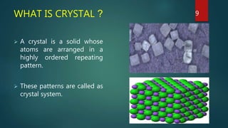
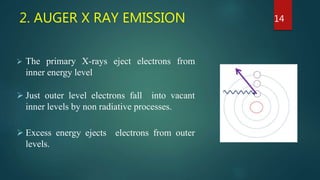
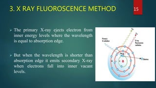
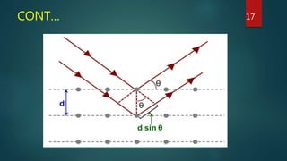
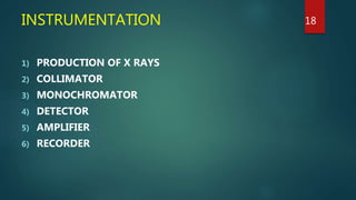
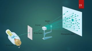
X-RAY CRYSTALLOGRAPHY
DIFFERENT X RAY METHODS AND PRODUCTION OF X RAYS
PRESENTED BY:
SHUBHAM KANAUJIYA
M.PHARM 1ST SEM PHARMACEUTICS
DEPARTMENT OF PHARMACEUTICAL SCIENCES
BBAU LUCKNOW
.
GUIDED BY:
DR. SUDIPTA SAHA
ASSISTANT PROFESSOR
DEPARTMENT OF PHARMACEUTICAL SCIENCES
BBAU LUCKNOW
CONTENTS
 INTRODUCTION
 PRINCIPLE
 WHAT IS X RAY
 WHAT IS CRYSTAL
 WHY X RAY IS USED
 WHY CRYSTALS ARE USED
 DIFFERENT X RAY METHODS
 INSTRUMENTATION
 PRODUCTION OF X RAY
 APPLICATION
2
INTRODUCTION
 X-ray crystallography is a technique used for determining the atomic
and molecular structure of a crystal
 The crystalline atoms cause a beam of incident X-rays to diffract into
many specific directions.
 By measuring the angles and intensities of these diffracted beams, a
crystallographer can produce a three-dimensional picture of the
density of electrons within the crystal.
3
CONT…
 From this electron density, the mean positions of the atoms in the
crystal can be determined, as well as their chemical bonds, their
disorder, and various other information.
4
PRINCIPLE OF X RAY CRYSTALLOGRAPGY
 Principle of X ray crystallography is that the crystalline atoms
cause beam of X rays to diffract into many specific directions.
 A crystal can be described with the aid of grid or lattice, defined by
three axis and angles between them.
 X ray crystallography uses the principle of X ray diffraction to analyze
the sample, but is done in many different directions so that the 3D
structure can be buildup.
5
CONT…
 It is a technique that has helped to deduce the 3D crystal
structure of many materials, especially biological materials.
6
WHAT IS X RAY ?
 X rays are the beam of electromagnetic radiation.
 These are of smaller wavelength than visible light.
Wavelength of X ray - 0.01-10 nm
Analytical purpose - 0.07-0.2 nm
 X rays have higher energy.
 They are more penetrative.
7
. 8
WHAT IS CRYSTAL ?
 A crystal is a solid whose
atoms are arranged in a
highly ordered repeating
pattern.
 These patterns are called as
crystal system.
9
WHY X RAYS ARE USED ?
 X rays have wavelength similar to the size of an atom.
 X rays have smaller wavelength than visible light so they have
higher energy and can penetrate the matter more easily than
the visible light.
10
WHY CRYSTALS ARE USED ?
 When sample is present in liquid form , the bond angle keep
changing , the position of atoms keep changing in every
moment so it is very hard to determine the structure of that
sample.
 So when we change biological liquid sample in crystalline form
(solid form) , the conformation of molecule is fixed in a single
place , so it makes easy to determine the structure of that
crystal.
11
DIFFERENT X RAY METHODS
1) X ray absorption method
2) Auger X ray emission method
3) X ray fluorescence method
4) X ray diffraction method (XRD)
12
1. X RAY ABSORPTION METHOD
 This method is similar to absorption in other regions of
electromagnetic spectra like UV-Visible / IR spectroscopy etc.
 Wavelength at which a sudden change in absorption occurs is used
to identify an element present in a sample, and the magnitude of
the change determines the amount of particular element present.
13
2. AUGER X RAY EMISSION
 The primary X-rays eject electrons from
inner energy level
 Just outer level electrons fall into vacant
inner levels by non radiative processes.
 Excess energy ejects electrons from outer
levels.
14
3. X RAY FLUOROSCENCE METHOD
 The primary X-ray ejects electron from
inner energy levels where the wavelength
is equal to absorption edge.
 But when the wavelength is shorter than
absorption edge it emits secondary X-ray
when electrons fall into inner vacant
levels.
15
4. X RAY DIFFRACTION MRTHOD(XRD)
 X ray diffraction works by irradiating a material with incident x
ray and then measuring the intensities and scattering angles of
the x ray that leave the material.
 When a beam of monochromatic X ray is directed at a crystalline
material, one observes reflection or diffraction of the X-rays at a
various angle with respect to the primary beam
 The relationship between the X-ray , angle of diffraction and
distance between each set of atomic planes of crystal lattice is given
by Braggs equation-
nλ= 2dsinθ
16
CONT… 17
INSTRUMENTATION
1) PRODUCTION OF X RAYS
2) COLLIMATOR
3) MONOCHROMATOR
4) DETECTOR
5) AMPLIFIER
6) RECORDER
18
19
1) PRODUCTION OF X RAYS
 X rays are produced inside the X ray tube when high energy
projectile electrons from the filament interact with the atoms of
the anode.
 X ray tube (Coolidge tube) : It is a large vacuum tube
containing a heated cathode of tungsten filament and copper or
molybdenum target metal anode.
• Operated at higher voltage up to 60 KV.
20
.
Figure. X ray tube
21
22
23
24
CONT…
 There is cathode which is a filament of tungsten metal heated by a
battery to emit the thermionic electrons.
 This beam of electrons moves towards anode target and attain
the kinetic energy and 99% of energy is converted into heat via
collision and remaining 0.5-1% is converted to X rays.
 Generally the target gets very hot in use so this problem has been
solved to some extent by cooling the tube with water.
25
WHAT HAPPENS ACTUALLY ?
 If sufficiently energetic electrons are available, the transfer of energy
from the impinging electron beam may eject an electron from one of
the inner levels of the target atoms.
 Within each atom, the place of the ejected electron is promptly filled
by an electron from an outer level whose place, in turn is taken by an
electron coming from still farther out.
 Thus the ionized atom returns to its normal state in as series of steps,
in each of which an X-ray photon of definite energy is emitted or
excess energy is released by ejection of a second electron with
characteristic energy.
26
CONT…
 The k series of line is observed when
an electron in the innermost K level
(n=1) is dislodged and electrons drop
down from the L (n=2) or M (n=3)
levels into the vacancy in k level.
 Corresponding vacancies in the L
levels are filled by electron
transitions from outer levels and give
rise to L series.
27
CONT…
 Disadvantage of most of X-ray tubes is that there is lack of focusing
of electrons so that whole surface becomes a source of X-rays.
28
2) COLLIMATOR
 It used to get the narrow beam of X rays.
 To get a narrow beam of X-rays, the X-rays
generated by the target material are allowed to
pass through a collimator which consists of two
sets of closely packed metal plates separated by a
small gap.
 The collimator absorbs all the X-rays except the
narrow beam that passes between the gaps.
29
3) MONOCHROMATOR
I. FILTER MONOCHROMATOR
II. CRYSTAL MONOCHROMATOR
30
.
.
31
CONT…
I. FILTER MONOCHROMATOR-
 A filter is a window of material that absorbs undesirable radiation but
allows the radiation of required wavelength to pass.
II. CRYSTAL MONOCHROMATOR-
 A crystal monochromator is made up of a suitable crystalline material
positioned in the X-ray beam so that the angle of reflecting planes
satisfied Bragg’s equation for the required wavelength.
32
4) DETECTOR
I. PHOTOGRAPHIC METHOD-
 In order to record the position and intensity of X-ray beam a plane or
cylindrical film is used.
 The film after exposing to X-rays is developed. The blackening of the
developed film is expressed in terms of density units D given by
 D = log I˳/I
33
CONT…
II. COUNTER METHODS-
 The Geiger tube is filled with an inert gas like argon and the central
wire anode is maintained at a positive potential of 800 to 2500V.
 When an X-ray is entering the Geiger tube, this ray undergoes
collision with the filling gas, resulting in the production of an ion
pair: the electron produced moves towards the central anode while
the positive ions move towards outer electrode.
34
CONT…
 The electron is accelerated by the potential gradient and causes
the ionisation of large number of argon atoms, resulting
production of an avalanche of electrons that are travelling
towards the central anode.
 The Geiger tube is in expensive and is relatively trouble free
detector. This tube gives the highest signal for given X-ray
intensity.
35
APPLICATION OF CRYSTALLOGRAPHY
 Characterization of crystalline materials.
 Identification of fine-grained minerals such as clays and mixed layer
clays that are difficult to determine optically.
 Determination of unit cell dimensions measurement of sample purity.
 Determination of Cis- trans isomerism.
 Differentiation of sugar.
36
.
 X-ray analysis of milk powder.
 Structure of protein and DNA
37
REFERENCES
 https://www.ncbi.nlm.nih.gov/pmc/articles/PMC1186895/
 https://www.sciencedirect.com/topics/biochemistry-genetics-and-
molecular-biology/x-ray-crystallography
 https://www.azom.com/article.aspx?ArticleID=18684
 https://www.slideshare.net/HasanulKarim2/x-ray-crystallograpy
 https://www.creativebiomart.net/resource/principle-protocol-x-ray-
crystallography-393.htm
38
39

X ray crystallography slideshare

  • 1.
    X-RAY CRYSTALLOGRAPHY DIFFERENT XRAY METHODS AND PRODUCTION OF X RAYS PRESENTED BY: SHUBHAM KANAUJIYA M.PHARM 1ST SEM PHARMACEUTICS DEPARTMENT OF PHARMACEUTICAL SCIENCES BBAU LUCKNOW . GUIDED BY: DR. SUDIPTA SAHA ASSISTANT PROFESSOR DEPARTMENT OF PHARMACEUTICAL SCIENCES BBAU LUCKNOW
  • 2.
    CONTENTS  INTRODUCTION  PRINCIPLE WHAT IS X RAY  WHAT IS CRYSTAL  WHY X RAY IS USED  WHY CRYSTALS ARE USED  DIFFERENT X RAY METHODS  INSTRUMENTATION  PRODUCTION OF X RAY  APPLICATION 2
  • 3.
    INTRODUCTION  X-ray crystallographyis a technique used for determining the atomic and molecular structure of a crystal  The crystalline atoms cause a beam of incident X-rays to diffract into many specific directions.  By measuring the angles and intensities of these diffracted beams, a crystallographer can produce a three-dimensional picture of the density of electrons within the crystal. 3
  • 4.
    CONT…  From thiselectron density, the mean positions of the atoms in the crystal can be determined, as well as their chemical bonds, their disorder, and various other information. 4
  • 5.
    PRINCIPLE OF XRAY CRYSTALLOGRAPGY  Principle of X ray crystallography is that the crystalline atoms cause beam of X rays to diffract into many specific directions.  A crystal can be described with the aid of grid or lattice, defined by three axis and angles between them.  X ray crystallography uses the principle of X ray diffraction to analyze the sample, but is done in many different directions so that the 3D structure can be buildup. 5
  • 6.
    CONT…  It isa technique that has helped to deduce the 3D crystal structure of many materials, especially biological materials. 6
  • 7.
    WHAT IS XRAY ?  X rays are the beam of electromagnetic radiation.  These are of smaller wavelength than visible light. Wavelength of X ray - 0.01-10 nm Analytical purpose - 0.07-0.2 nm  X rays have higher energy.  They are more penetrative. 7
  • 8.
  • 9.
    WHAT IS CRYSTAL?  A crystal is a solid whose atoms are arranged in a highly ordered repeating pattern.  These patterns are called as crystal system. 9
  • 10.
    WHY X RAYSARE USED ?  X rays have wavelength similar to the size of an atom.  X rays have smaller wavelength than visible light so they have higher energy and can penetrate the matter more easily than the visible light. 10
  • 11.
    WHY CRYSTALS AREUSED ?  When sample is present in liquid form , the bond angle keep changing , the position of atoms keep changing in every moment so it is very hard to determine the structure of that sample.  So when we change biological liquid sample in crystalline form (solid form) , the conformation of molecule is fixed in a single place , so it makes easy to determine the structure of that crystal. 11
  • 12.
    DIFFERENT X RAYMETHODS 1) X ray absorption method 2) Auger X ray emission method 3) X ray fluorescence method 4) X ray diffraction method (XRD) 12
  • 13.
    1. X RAYABSORPTION METHOD  This method is similar to absorption in other regions of electromagnetic spectra like UV-Visible / IR spectroscopy etc.  Wavelength at which a sudden change in absorption occurs is used to identify an element present in a sample, and the magnitude of the change determines the amount of particular element present. 13
  • 14.
    2. AUGER XRAY EMISSION  The primary X-rays eject electrons from inner energy level  Just outer level electrons fall into vacant inner levels by non radiative processes.  Excess energy ejects electrons from outer levels. 14
  • 15.
    3. X RAYFLUOROSCENCE METHOD  The primary X-ray ejects electron from inner energy levels where the wavelength is equal to absorption edge.  But when the wavelength is shorter than absorption edge it emits secondary X-ray when electrons fall into inner vacant levels. 15
  • 16.
    4. X RAYDIFFRACTION MRTHOD(XRD)  X ray diffraction works by irradiating a material with incident x ray and then measuring the intensities and scattering angles of the x ray that leave the material.  When a beam of monochromatic X ray is directed at a crystalline material, one observes reflection or diffraction of the X-rays at a various angle with respect to the primary beam  The relationship between the X-ray , angle of diffraction and distance between each set of atomic planes of crystal lattice is given by Braggs equation- nλ= 2dsinθ 16
  • 17.
  • 18.
    INSTRUMENTATION 1) PRODUCTION OFX RAYS 2) COLLIMATOR 3) MONOCHROMATOR 4) DETECTOR 5) AMPLIFIER 6) RECORDER 18
  • 19.
  • 20.
    1) PRODUCTION OFX RAYS  X rays are produced inside the X ray tube when high energy projectile electrons from the filament interact with the atoms of the anode.  X ray tube (Coolidge tube) : It is a large vacuum tube containing a heated cathode of tungsten filament and copper or molybdenum target metal anode. • Operated at higher voltage up to 60 KV. 20
  • 21.
  • 22.
  • 23.
  • 24.
  • 25.
    CONT…  There iscathode which is a filament of tungsten metal heated by a battery to emit the thermionic electrons.  This beam of electrons moves towards anode target and attain the kinetic energy and 99% of energy is converted into heat via collision and remaining 0.5-1% is converted to X rays.  Generally the target gets very hot in use so this problem has been solved to some extent by cooling the tube with water. 25
  • 26.
    WHAT HAPPENS ACTUALLY?  If sufficiently energetic electrons are available, the transfer of energy from the impinging electron beam may eject an electron from one of the inner levels of the target atoms.  Within each atom, the place of the ejected electron is promptly filled by an electron from an outer level whose place, in turn is taken by an electron coming from still farther out.  Thus the ionized atom returns to its normal state in as series of steps, in each of which an X-ray photon of definite energy is emitted or excess energy is released by ejection of a second electron with characteristic energy. 26
  • 27.
    CONT…  The kseries of line is observed when an electron in the innermost K level (n=1) is dislodged and electrons drop down from the L (n=2) or M (n=3) levels into the vacancy in k level.  Corresponding vacancies in the L levels are filled by electron transitions from outer levels and give rise to L series. 27
  • 28.
    CONT…  Disadvantage ofmost of X-ray tubes is that there is lack of focusing of electrons so that whole surface becomes a source of X-rays. 28
  • 29.
    2) COLLIMATOR  Itused to get the narrow beam of X rays.  To get a narrow beam of X-rays, the X-rays generated by the target material are allowed to pass through a collimator which consists of two sets of closely packed metal plates separated by a small gap.  The collimator absorbs all the X-rays except the narrow beam that passes between the gaps. 29
  • 30.
    3) MONOCHROMATOR I. FILTERMONOCHROMATOR II. CRYSTAL MONOCHROMATOR 30
  • 31.
  • 32.
    CONT… I. FILTER MONOCHROMATOR- A filter is a window of material that absorbs undesirable radiation but allows the radiation of required wavelength to pass. II. CRYSTAL MONOCHROMATOR-  A crystal monochromator is made up of a suitable crystalline material positioned in the X-ray beam so that the angle of reflecting planes satisfied Bragg’s equation for the required wavelength. 32
  • 33.
    4) DETECTOR I. PHOTOGRAPHICMETHOD-  In order to record the position and intensity of X-ray beam a plane or cylindrical film is used.  The film after exposing to X-rays is developed. The blackening of the developed film is expressed in terms of density units D given by  D = log I˳/I 33
  • 34.
    CONT… II. COUNTER METHODS- The Geiger tube is filled with an inert gas like argon and the central wire anode is maintained at a positive potential of 800 to 2500V.  When an X-ray is entering the Geiger tube, this ray undergoes collision with the filling gas, resulting in the production of an ion pair: the electron produced moves towards the central anode while the positive ions move towards outer electrode. 34
  • 35.
    CONT…  The electronis accelerated by the potential gradient and causes the ionisation of large number of argon atoms, resulting production of an avalanche of electrons that are travelling towards the central anode.  The Geiger tube is in expensive and is relatively trouble free detector. This tube gives the highest signal for given X-ray intensity. 35
  • 36.
    APPLICATION OF CRYSTALLOGRAPHY Characterization of crystalline materials.  Identification of fine-grained minerals such as clays and mixed layer clays that are difficult to determine optically.  Determination of unit cell dimensions measurement of sample purity.  Determination of Cis- trans isomerism.  Differentiation of sugar. 36
  • 37.
    .  X-ray analysisof milk powder.  Structure of protein and DNA 37
  • 38.
    REFERENCES  https://www.ncbi.nlm.nih.gov/pmc/articles/PMC1186895/  https://www.sciencedirect.com/topics/biochemistry-genetics-and- molecular-biology/x-ray-crystallography https://www.azom.com/article.aspx?ArticleID=18684  https://www.slideshare.net/HasanulKarim2/x-ray-crystallograpy  https://www.creativebiomart.net/resource/principle-protocol-x-ray- crystallography-393.htm 38
  • 39.