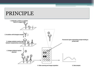
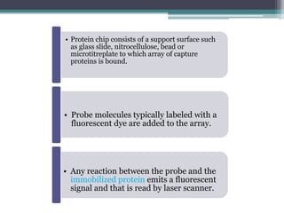
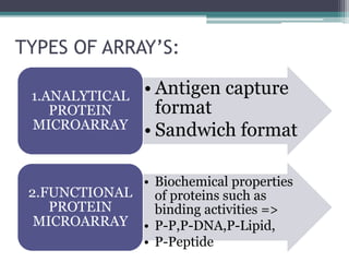
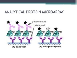
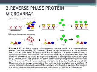
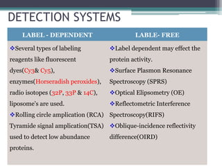
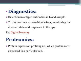
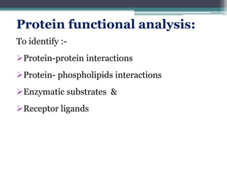
Protein microarrays allow high-throughput analysis of protein interactions and functions. They consist of large numbers of capture proteins immobilized on a surface to which labeled probe molecules are added to detect reactions by fluorescence. There are analytical arrays to study protein binding properties and functional arrays containing full-length proteins to assay enzymatic activity and detect antibodies. Protein microarrays have applications in diagnostics, proteomics, analyzing protein interactions and functions, antibody characterization, and treatment development.
it will help you to understand how the protein microarrays are made, what are the different types and what all purposes they are used for. its very useful ppt
Introduction to Applications of Proteomics Science,
Proteomics- Techniques, Applications of proteomics
Presented by
A. Harsha Vardhan Naidu
Department of Pharmacology
protein structure prediction methods. homology modelling, fold recognition, threading, ab initio methods. in short and easy form slides. after one time read you can easily understand methods for protein structure prediction.
Yeast two-hybrid is based on the reconstitution of a functional transcription factor (TF) when two proteins or polypeptides of interest interact. Upon interaction between the bait and the prey, the DBD and AD are brought in close proximity and a functional TF is reconstituted upstream of the reporter gene.
This is technique used widely for protein separation from a mixture and is very easy and less costly method. Slides cover all essential points about EMSA and it is quite interesting to know that how it detect and separate different proteins and their mobility shift assay.
Creation of a cDNA library starts with mRNA instead of DNA. Messenger RNA carries encoded information from DNA to ribosomes for translation into protein. To create a cDNA library, these mRNA molecules are treated with the enzyme reverse transcriptase, which is used to make a DNA copy of an mRNA (i.e., cDNA). A cDNA library represents a sampling of the transcribed genes, but a genomic library includes untranscribed regions.
Protein microarray Preparation of protein microarray Different methods of arr...naveed ul mushtaq
Protein microarray
Preparation of protein microarray
Different methods of arraying the proteins.FUNCTIONAL PROTEIN MICROARRAYSAnalytical microarrays:-
3.REVERSE PHASE PROTEIN MICROARRAY APPLICATIONS:-
Prediction of the three dimensional structure of a given protein sequence i.e. target protein from the amino acid sequence of a homologous (template) protein for which an X-ray or NMR structure is available based on an alignment to one or more known protein structures
protein microarray_k.b institute (m.pharm pharmacology) .pptxNittalVekaria
1: Introduction
Welcome to our presentation on Protein Microarrays.
Discover the revolutionary technology transforming protein analysis and biomolecular research
2: What are Protein Microarrays?
Protein microarrays are high-throughput platforms for studying protein-protein interactions, protein function, and biomarker discovery.
They consist of thousands of immobilized proteins on a solid surface, allowing for simultaneous analysis of multiple proteins.
3Components of Protein Microarrays
Substrate: Glass slides, membranes, or beads.
Proteins: Target proteins immobilized on the substrate.
Detection System: Fluorescent dyes, antibodies, or other probes.
Imaging System: Scanners or cameras for data acquisition.
4: Types of Protein Microarrays
Analytical Microarrays: Used for studying protein-protein interactions, protein expression profiling, and protein function analysis.
Antibody Microarrays: Utilized for detecting and quantifying specific proteins or antibodies in biological samples.
Reverse-Phase Protein Arrays (RPPAs): Designed for high-throughput protein expression profiling and signaling pathway analysis.
5:Applications of Protein Microarrays
Biomarker Discovery: Identification of disease-specific biomarkers for diagnosis, prognosis, and treatment monitoring.
Drug Discovery: High-throughput screening of drug candidates and target validation.
Functional Proteomics: Mapping protein-protein interactions, post-translational modifications, and protein function analysis.
Clinical Diagnostics: Detection of infectious diseases, cancer biomarkers, and autoimmune disorders.
6: Workflow of Protein Microarray Experiment
Protein immobilization: Spotting or printing target proteins onto the microarray substrate.
Sample incubation: Incubating the microarray with biological samples containing proteins of interest.
Detection and analysis: Using fluorescent probes or antibodies to detect bound proteins and quantifying the signals.
Data interpretation: Analyzing and interpreting the results to extract meaningful biological insights.
7: Advantages of Protein Microarrays
-High-throughput analysis of thousands of proteins in parallel.
Small sample volume requirement.
Enables multiplexed assays for comprehensive protein profiling.
Facilitates rapid biomarker discovery and validation.
8: Challenges and Considerations
Standardization of protocols and reagents.
Optimization of protein immobilization and detection methods.
Data analysis and interpretation complexities.
Cost and accessibility of microarray platforms.
9: Future Perspectives
Integration with other omics technologies for holistic biological insights.
Development of miniaturized and portable microarray platforms for point-of-care diagnostics.
Advancements in data analysis algorithms and bioinformatics tools.
Expanding applications in personalized medicine and precision healthcare
10: Conclusion
Protein microarrays offer a powerful and versatile tool for protein analysis and biomarker discover
NMR spectroscopy is the use of NMR phenomena to study the physical, chemical, and biological properties of matter. Chemists use it to determine molecular identity and structure.
Proteomics and its applications in phytopathologyAbhijeet Kashyap
Dear friends, I Abhijeet kashyap presenting the basics of proteomics to you all . Proteomics is the large-scale study of proteins, particularly their structures and functions.Proteomics helps in understanding the structure and function of different proteins as well as protein-protein interactions of an organism.
THE IMPORTANCE OF MARTIAN ATMOSPHERE SAMPLE RETURN.Sérgio Sacani
The return of a sample of near-surface atmosphere from Mars would facilitate answers to several first-order science questions surrounding the formation and evolution of the planet. One of the important aspects of terrestrial planet formation in general is the role that primary atmospheres played in influencing the chemistry and structure of the planets and their antecedents. Studies of the martian atmosphere can be used to investigate the role of a primary atmosphere in its history. Atmosphere samples would also inform our understanding of the near-surface chemistry of the planet, and ultimately the prospects for life. High-precision isotopic analyses of constituent gases are needed to address these questions, requiring that the analyses are made on returned samples rather than in situ.
This presentation explores a brief idea about the structural and functional attributes of nucleotides, the structure and function of genetic materials along with the impact of UV rays and pH upon them.
Multi-source connectivity as the driver of solar wind variability in the heli...Sérgio Sacani
The ambient solar wind that flls the heliosphere originates from multiple
sources in the solar corona and is highly structured. It is often described
as high-speed, relatively homogeneous, plasma streams from coronal
holes and slow-speed, highly variable, streams whose source regions are
under debate. A key goal of ESA/NASA’s Solar Orbiter mission is to identify
solar wind sources and understand what drives the complexity seen in the
heliosphere. By combining magnetic feld modelling and spectroscopic
techniques with high-resolution observations and measurements, we show
that the solar wind variability detected in situ by Solar Orbiter in March
2022 is driven by spatio-temporal changes in the magnetic connectivity to
multiple sources in the solar atmosphere. The magnetic feld footpoints
connected to the spacecraft moved from the boundaries of a coronal hole
to one active region (12961) and then across to another region (12957). This
is refected in the in situ measurements, which show the transition from fast
to highly Alfvénic then to slow solar wind that is disrupted by the arrival of
a coronal mass ejection. Our results describe solar wind variability at 0.5 au
but are applicable to near-Earth observatories.
Observation of Io’s Resurfacing via Plume Deposition Using Ground-based Adapt...Sérgio Sacani
Since volcanic activity was first discovered on Io from Voyager images in 1979, changes
on Io’s surface have been monitored from both spacecraft and ground-based telescopes.
Here, we present the highest spatial resolution images of Io ever obtained from a groundbased telescope. These images, acquired by the SHARK-VIS instrument on the Large
Binocular Telescope, show evidence of a major resurfacing event on Io’s trailing hemisphere. When compared to the most recent spacecraft images, the SHARK-VIS images
show that a plume deposit from a powerful eruption at Pillan Patera has covered part
of the long-lived Pele plume deposit. Although this type of resurfacing event may be common on Io, few have been detected due to the rarity of spacecraft visits and the previously low spatial resolution available from Earth-based telescopes. The SHARK-VIS instrument ushers in a new era of high resolution imaging of Io’s surface using adaptive
optics at visible wavelengths.
Earliest Galaxies in the JADES Origins Field: Luminosity Function and Cosmic ...Sérgio Sacani
We characterize the earliest galaxy population in the JADES Origins Field (JOF), the deepest
imaging field observed with JWST. We make use of the ancillary Hubble optical images (5 filters
spanning 0.4−0.9µm) and novel JWST images with 14 filters spanning 0.8−5µm, including 7 mediumband filters, and reaching total exposure times of up to 46 hours per filter. We combine all our data
at > 2.3µm to construct an ultradeep image, reaching as deep as ≈ 31.4 AB mag in the stack and
30.3-31.0 AB mag (5σ, r = 0.1” circular aperture) in individual filters. We measure photometric
redshifts and use robust selection criteria to identify a sample of eight galaxy candidates at redshifts
z = 11.5 − 15. These objects show compact half-light radii of R1/2 ∼ 50 − 200pc, stellar masses of
M⋆ ∼ 107−108M⊙, and star-formation rates of SFR ∼ 0.1−1 M⊙ yr−1
. Our search finds no candidates
at 15 < z < 20, placing upper limits at these redshifts. We develop a forward modeling approach to
infer the properties of the evolving luminosity function without binning in redshift or luminosity that
marginalizes over the photometric redshift uncertainty of our candidate galaxies and incorporates the
impact of non-detections. We find a z = 12 luminosity function in good agreement with prior results,
and that the luminosity function normalization and UV luminosity density decline by a factor of ∼ 2.5
from z = 12 to z = 14. We discuss the possible implications of our results in the context of theoretical
models for evolution of the dark matter halo mass function.
A brief information about the SCOP protein database used in bioinformatics.
The Structural Classification of Proteins (SCOP) database is a comprehensive and authoritative resource for the structural and evolutionary relationships of proteins. It provides a detailed and curated classification of protein structures, grouping them into families, superfamilies, and folds based on their structural and sequence similarities.
Deep Behavioral Phenotyping in Systems Neuroscience for Functional Atlasing a...Ana Luísa Pinho
Functional Magnetic Resonance Imaging (fMRI) provides means to characterize brain activations in response to behavior. However, cognitive neuroscience has been limited to group-level effects referring to the performance of specific tasks. To obtain the functional profile of elementary cognitive mechanisms, the combination of brain responses to many tasks is required. Yet, to date, both structural atlases and parcellation-based activations do not fully account for cognitive function and still present several limitations. Further, they do not adapt overall to individual characteristics. In this talk, I will give an account of deep-behavioral phenotyping strategies, namely data-driven methods in large task-fMRI datasets, to optimize functional brain-data collection and improve inference of effects-of-interest related to mental processes. Key to this approach is the employment of fast multi-functional paradigms rich on features that can be well parametrized and, consequently, facilitate the creation of psycho-physiological constructs to be modelled with imaging data. Particular emphasis will be given to music stimuli when studying high-order cognitive mechanisms, due to their ecological nature and quality to enable complex behavior compounded by discrete entities. I will also discuss how deep-behavioral phenotyping and individualized models applied to neuroimaging data can better account for the subject-specific organization of domain-general cognitive systems in the human brain. Finally, the accumulation of functional brain signatures brings the possibility to clarify relationships among tasks and create a univocal link between brain systems and mental functions through: (1) the development of ontologies proposing an organization of cognitive processes; and (2) brain-network taxonomies describing functional specialization. To this end, tools to improve commensurability in cognitive science are necessary, such as public repositories, ontology-based platforms and automated meta-analysis tools. I will thus discuss some brain-atlasing resources currently under development, and their applicability in cognitive as well as clinical neuroscience.
2. INTRODUCTION
• Protein micro array(or Protein chip) is a high-
throughput method used to track the interactions
& activities of proteins, to determinate their
functions.
• Protein micro array are rapid, automated,
economical and highly sensitive consuming only
small quantities of samples and reagents.
3. HISTORY
• Micro array technology was first developed from
an earlier concept called “Ambient analyte
immunoassay” by Roger Ekins in 1989.
• Later transformed into DNA microarray.
• Due to some limitations in DNA microarray ,
this protein microarray was developed to
overcome those limitations.
5. • Protein chip consists of a support surface such
as glass slide, nitrocellulose, bead or
microtitreplate to which array of capture
proteins is bound.
• Probe molecules typically labeled with a
fluorescent dye are added to the array.
• Any reaction between the probe and the
immobilized protein emits a fluorescent
signal and that is read by laser scanner.
6. TYPES OF ARRAY’S:
• Antigen capture
format
• Sandwich format
1.ANALYTICAL
PROTEIN
MICROARRAY
• Biochemical properties
of proteins such as
binding activities =>
• P-P,P-DNA,P-Lipid,
• P-Peptide
2.FUNCTIONAL
PROTEIN
MICROARRAY
9. • Functional protein microarray (target protein
array) are constructed by immobilizing large
num of proteins to assay enzymatic activity and
to detect antibodies & their functions.
• They differ from analytical protein microarray
by containing full length functional proteins or
protein domains.
10. • These are used to study the biochemical
activities of entire proteome in a single
experiment.
• The proteins must retain their native or original
structure so that meaningful functional
interactions takes place on the array surface.
12. DETECTION SYSTEMS
LABEL - DEPENDENT LABLE- FREE
Several types of labeling
reagents like fluorescent
dyes(Cy3& Cy5),
enzymes(Horseradish peroxides),
radio isotopes (32P, 33P & 14C),
liposome's are used.
Rolling circle amplication (RCA)
Tyramide signal amplication(TSA)
used to detect low abundance
proteins.
Label dependent may effect the
protein activity.
Surface Plasmon Resonance
Spectroscopy (SPRS)
Optical Elipsometry (OE)
Reflectometric Interference
Spectroscopy(RIFS)
Oblique-incidence reflectivity
difference(OIRD)
13. APPLICATIONS
• Mainly used in 5 major areas
• Diagnostics
• Proteomics
• Protein functional analysis
• Antibody characterization &
• Treatment development
14. •Diagnostics:
Detection in antigen antibodies in blood sample
To discover new disease biomarkers; monitoring the
diseased state and responses to therapy.
Ex: Digital bioassay
Proteomics:
Protein expression profiling i.e., which proteins are
expressed in a particular cell.
15. Protein functional analysis:
To identify :-
Protein-protein interactions
Protein- phospholipids interactions
Enzymatic substrates &
Receptor ligands
16. Antibody characterization:
characterization of cross reactivity, specificity and
mapping epitopes.
Treatment development:
Development of antigen- specificity therapies for
autoimmunity , cancer and allergies.
Identification of small molecules that could potentially
used to be as new drugs