White paper "SMI – a new technique for the analysis of the microvascular tree in reactive and suspected malignant lymphadenopathy in advanced stages of malignant melanoma."
1) SMI is a new ultrasound technique that can visualize microvessels better than conventional Doppler imaging methods by removing signals from tissue motion.
2) In an initial study of 10 melanoma patients, SMI provided additional information in 3 cases compared to conventional techniques, changing the primary diagnosis which was confirmed with further testing.
3) SMI allowed for better assessment of microvascular architecture in reactive lymph nodes and metastases compared to other techniques, aiding in evaluation and biopsy of small lesions and subcutaneous structures.
Introduction
Ultrasound technology to visualise the microvas- culature continues to improve where power and colour Doppler ultrasound remains a quick and non-invasive method of assessing the vascularity of tumours and tissue. The advent of contrast enhanced ultrasound (CEUS) imaging using micro- bubbles improved the sensitivity and resolution
of the microvessels which can be imaged but requires an intravenous administration of contrast agents.
A radiological insight into various musculoskeletal complications in patients suffering from AIDS and how it'll affect the management of the patient. A must know for all Radiologists.
Superb microvascular imaging A Sonographic Technical AdvanceDr. Muhammad Bin ...Dr. Muhammad Bin Zulfiqar
Superb microvascular imaging A Sonographic Technical AdvanceDr. Muhammad Bin Zulfiqar Services Institute of Medical Sciences / Services Hospital Lahore
Details about New Technical Advances in Sonography.
Similar to White paper "SMI – a new technique for the analysis of the microvascular tree in reactive and suspected malignant lymphadenopathy in advanced stages of malignant melanoma."
SWI , high susceptibility for blood products, iron depositions, and calcifications
makes susceptibility-weighted imaging an important additional sequence for the diagnostic
workup of pediatric brain pathologic abnormalities. Compared with conventional MRI
sequences, susceptibility-weighted imaging may show lesions in better detail or with higher
sensitivity
Optical coherence tomography-guided algorithm for percutaneous coronary intervention. Vessel diameter should be assessed using the external elastic lamina (EEL)-EEL diameter at the reference segments, and rounded down to select interventional devices (balloons, stents). If the EEL cannot be identified, luminal measures are used and rounded up to 0.5 mm larger for selection of the devices. Optical coherence tomography (OCT)-guided optimisation strategies post stent implantation per EEL-based diameter measurement and per lumen-based diameter measurement are shown. For instance, if the distal EEL-EEL diameter measures 3.2 mm×3.1 mm (i.e., the mean EEL-based diameter is 3.15 mm), this number is rounded down to the next available stent size and post-dilation balloon to be used at the distal segment. Thus, a 3.0 mm stent and non-compliant balloon diameter is selected. If the proximal EEL cannot be visualised, the mean lumen diameter should be used for device sizing. For instance, if the mean proximal lumen diameter measures 3.4 mm, this number is rounded up to the next available balloon diameter (within up to 0.5 mm larger) for post-dilation. MLA: minimal lumen area; MSA: minimal stent area;NC: non-compliant
Austin Journal of Obstetrics and Gynecology is an open access, peer reviewed, scholarly journal dedicated to publication of medical and surgical area of expertise that focuses on the research prospectus of reproductive system in females.
The journal aims to publish most complete and consistent resource of information on the discoveries and present developments in all areas of the field and making them freely obtainable through online without any limitations or any other subscriptions to researchers worldwide.
Austin Journal of Obstetrics and Gynecology accepts original research articles, personal perspectives, editorials, letters, review articles, case reports, clinical images and rapid communication on all the aspects of diagnosis and treatment of Gynecology.
Despite the recent developments that have been made in the field of percutaneous left main (LM) intervention, the
treatment of distal LM bifurcation remains challenging. The provisional one-stent approach for LM bifurcation has
shown more favorable outcomes than the two-stent technique, making the former the preferred strategy in most
types of LM bifurcation stenosis. However, elective two-stent techniques, none of which has been proven superior
to the others, are still used in patients with severely diseased large side branches to avoid acute hemodynamic
compromise. Selecting the proper bifurcation treatment strategy using meticulous intravascular ultrasound evaluation
for side branch ostium is crucial for reducing the risk of side branch occlusion and for improving patient outcomes. In
addition, unnecessary complex intervention can be avoided by measuring fractional flow reserve in angiographically
isolated side branches. Most importantly, good long-term clinical outcomes are more related to the successful
procedure itself than to the type of stenting technique, emphasizing the greater importance of optimizing
the chosen technique than the choice of metho
1 enfermedad cv ateroesclerótica figura 1 (1)sagita28
Similar to White paper "SMI – a new technique for the analysis of the microvascular tree in reactive and suspected malignant lymphadenopathy in advanced stages of malignant melanoma." (20)
VISIONS magazine is a publication of Canon Medical Europe and is offered free of charge to health professionals.
The magazine is published twice a year. Registration to access full, previously published, digital editions can be done via the website: https://eu.medical.canon/visions-magazine
VISIONS magazine is covering Canon Medical’s European region and as such reflects products, technologies and
services for this particular area. The mentioned products may not be available in other geographic regions.
Please consult your Canon Medical representative sales office in case of any questions.
VISIONS magazine is Canon Medical’s customer magazine about innovative technologies and applications in medical imaging. Read all about diagnostic imaging in the fields of CT, MRI, X-Ray, Ultrasound, HII, Eye Care and stay up-to-date with the latest developments in your clinical environment.
This Veterinary Special edition of VISIONS magazine is a publication of Canon Medical Europe and is offered
free of charge to health professionals. To download the digital edition of this Veterinary Special, please visit:
https://eu.medical.canon/visions.
VISIONS magazine is covering Canon Medical’s European region and as such reflects products, technologies
and services for this particular area. The mentioned products may not be available in other geographic regions.
Please consult your Canon Medical representative sales office in case of any questions.
No part of this publication may be reproduced in whole or in part, stored in an automated storage and retrieval
system or transmitted in any manner whatsoever without written permission of the publisher. The opinions
expressed in this publication are solely those of the authors and not necessarily those of Canon Medical.
Canon Medical does not guarantee the accuracy or reliability of the information provided herein.
News, articles and the full edition of VISIONS magazine are announced firstly, as pre-publication, via the
dedicated VISIONS LinkedIn Group: https://www.linkedin.com/groups/3698045. In this group you can
actively participate in discussions about the content and future direction of the magazine.
Canon Medical Systems VISIONS Magazine - issue 35
VISIONS magazine is a publication of Canon Medical Europe and is offered free of charge to medical and health professionals. The magazine is published twice a year.
Registration to access full, previously published, digital editions can be done via the web site: https://nl.medical.canon/visions-magazine.
VISIONS magazine is covering Canon Medical’s European region and as such reflects products, technologies and services for this particular area. The mentioned products may not be available in other geographic regions. Please consult your Canon Medical representative sales office in case of any questions.
No part of this publication may be reproduced in whole or in part, stored in an automated storage and retrieval system or transmitted in any manner whatsoever without written permission of the publisher. The opinions expressed in this publication are solely those of the authors and not necessarily those of Canon Medical. Canon Medical does not guarantee the accuracy or reliability of the information provided herein.
News items and articles are announced firstly, as pre-publication, via the dedicated VISIONS LinkedIn Group: https://www.linkedin.com/groups/3698045. In this group you can actively participate in discussions about the content and future direction of the magazine.
This MR Special edition of VISIONS magazine is a publication of Canon Medical Europe and is offered
free of charge to health professionals. To download the digital edition of this MR Special, please visit:
https://eu.medical.canon/visions.
VISIONS magazine is covering Canon Medical’s European region and as such reflects products, technologies
and services for this particular area. The mentioned products may not be available in other geographic regions.
Please consult your Canon Medical representative sales office in case of any questions.
VISIONS magazine is a publication of Canon Medical Europe and is offered free of charge to health professionals.
The magazine is published twice a year. Registration to access full, previously published, digital editions can
be done via the web site: https://nl.medical.canon/visions-magazine. Canon Medical stores and uses personal
data of the registration to send out the magazine and inform members about new developments. Members can
customize preferences or opt-out, after registration, in the online VISIONS profile.
Canon Medical Systems VISIONS Magazine - issue 31
VISIONS magazine is a publication of Canon Medical Europe and is offered free of charge to medical and health professionals. The magazine is published twice a year.
Registration to access full, previously published, digital editions can be done via the web site: https://nl.medical.canon/visions-magazine.
VISIONS magazine is covering Canon Medical’s European region and as such reflects products, technologies and services for this particular area. The mentioned products may not be available in other geographic regions. Please consult your Canon Medical representative sales office in case of any questions.
No part of this publication may be reproduced in whole or in part, stored in an automated storage and retrieval system or transmitted in any manner whatsoever without written permission of the publisher. The opinions expressed in this publication are solely those of the authors and not necessarily those of Canon Medical. Canon Medical does not guarantee the accuracy or reliability of the information provided herein.
News items and articles are announced firstly, as pre-publication, via the dedicated VISIONS LinkedIn Group: https://www.linkedin.com/groups/3698045. In this group you can actively participate in discussions about the content and future direction of the magazine.
The assessment of cardiac function is essential in the athletic population not only as part of the screening process for underlying cardiac disease, but also to longitudinally assess performance and training adaptations - Source: Toshiba's VISIONS Magazine #26 | www.toshiba-medical.eu/visions
Post contrast iodine maps were introduced as part of dual-energy imaging over 10 years ago but these have never become part of routine practice in most centres for the investigation of pulmonary thromboembolic disease.
CASE: 58 year old male presented with a liver lesion seen on prior imaging. A dual energy scan was performed to further characterize the lesion. Volumetric Dual Energy scans were performed following the injection of 80mls of contrast. Scans were performed during Arterial Phase, early Portal Phase and Delayed at 3mins. Monochromatic images, iodine maps and virtual non contrast images were generated for review.
Computed tomography angiography (CTA) of the coronary arteries is a useful noninvasive tool to rule out significant coronary artery disease (CAD) in many clinical situations. Recent guidelines of stable CAD and non-ST segment elevation myocardial infarction endorse the use of CTA in symptomatic patients with low to intermediate likelihood of the disease, given the particularly high negative predictive value of the technique. However, in patients with high pre-test likelihood of CAD, the technique is not recommended, and one of the reasons is the high probability of coronary calcification in these patients, which interferes with the analysis of the images and reduces the specificity and negative predictive value of CTA.
(TOSHIBA CTEU140095) - Article from Toshiba's VISIONS Magazine#25, March 2015
Couples presenting to the infertility clinic- Do they really have infertility...Sujoy Dasgupta
Dr Sujoy Dasgupta presented the study on "Couples presenting to the infertility clinic- Do they really have infertility? – The unexplored stories of non-consummation" in the 13th Congress of the Asia Pacific Initiative on Reproduction (ASPIRE 2024) at Manila on 24 May, 2024.
These lecture slides, by Dr Sidra Arshad, offer a quick overview of physiological basis of a normal electrocardiogram.
Learning objectives:
1. Define an electrocardiogram (ECG) and electrocardiography
2. Describe how dipoles generated by the heart produce the waveforms of the ECG
3. Describe the components of a normal electrocardiogram of a typical bipolar leads (limb II)
4. Differentiate between intervals and segments
5. Enlist some common indications for obtaining an ECG
Study Resources:
1. Chapter 11, Guyton and Hall Textbook of Medical Physiology, 14th edition
2. Chapter 9, Human Physiology - From Cells to Systems, Lauralee Sherwood, 9th edition
3. Chapter 29, Ganong’s Review of Medical Physiology, 26th edition
4. Electrocardiogram, StatPearls - https://www.ncbi.nlm.nih.gov/books/NBK549803/
5. ECG in Medical Practice by ABM Abdullah, 4th edition
6. ECG Basics, http://www.nataliescasebook.com/tag/e-c-g-basics
HOT NEW PRODUCT! BIG SALES FAST SHIPPING NOW FROM CHINA!! EU KU DB BK substit...GL Anaacs
Contact us if you are interested:
Email / Skype : kefaya1771@gmail.com
Threema: PXHY5PDH
New BATCH Ku !!! MUCH IN DEMAND FAST SALE EVERY BATCH HAPPY GOOD EFFECT BIG BATCH !
Contact me on Threema or skype to start big business!!
Hot-sale products:
NEW HOT EUTYLONE WHITE CRYSTAL!!
5cl-adba precursor (semi finished )
5cl-adba raw materials
ADBB precursor (semi finished )
ADBB raw materials
APVP powder
5fadb/4f-adb
Jwh018 / Jwh210
Eutylone crystal
Protonitazene (hydrochloride) CAS: 119276-01-6
Flubrotizolam CAS: 57801-95-3
Metonitazene CAS: 14680-51-4
Payment terms: Western Union,MoneyGram,Bitcoin or USDT.
Deliver Time: Usually 7-15days
Shipping method: FedEx, TNT, DHL,UPS etc.Our deliveries are 100% safe, fast, reliable and discreet.
Samples will be sent for your evaluation!If you are interested in, please contact me, let's talk details.
We specializes in exporting high quality Research chemical, medical intermediate, Pharmaceutical chemicals and so on. Products are exported to USA, Canada, France, Korea, Japan,Russia, Southeast Asia and other countries.
TEST BANK for Operations Management, 14th Edition by William J. Stevenson, Ve...kevinkariuki227
TEST BANK for Operations Management, 14th Edition by William J. Stevenson, Verified Chapters 1 - 19, Complete Newest Version.pdf
Pulmonary Thromboembolism - etilogy, types, medical- Surgical and nursing man...VarunMahajani
Disruption of blood supply to lung alveoli due to blockage of one or more pulmonary blood vessels is called as Pulmonary thromboembolism. In this presentation we will discuss its causes, types and its management in depth.
Lung Cancer: Artificial Intelligence, Synergetics, Complex System Analysis, S...Oleg Kshivets
RESULTS: Overall life span (LS) was 2252.1±1742.5 days and cumulative 5-year survival (5YS) reached 73.2%, 10 years – 64.8%, 20 years – 42.5%. 513 LCP lived more than 5 years (LS=3124.6±1525.6 days), 148 LCP – more than 10 years (LS=5054.4±1504.1 days).199 LCP died because of LC (LS=562.7±374.5 days). 5YS of LCP after bi/lobectomies was significantly superior in comparison with LCP after pneumonectomies (78.1% vs.63.7%, P=0.00001 by log-rank test). AT significantly improved 5YS (66.3% vs. 34.8%) (P=0.00000 by log-rank test) only for LCP with N1-2. Cox modeling displayed that 5YS of LCP significantly depended on: phase transition (PT) early-invasive LC in terms of synergetics, PT N0—N12, cell ratio factors (ratio between cancer cells- CC and blood cells subpopulations), G1-3, histology, glucose, AT, blood cell circuit, prothrombin index, heparin tolerance, recalcification time (P=0.000-0.038). Neural networks, genetic algorithm selection and bootstrap simulation revealed relationships between 5YS and PT early-invasive LC (rank=1), PT N0—N12 (rank=2), thrombocytes/CC (3), erythrocytes/CC (4), eosinophils/CC (5), healthy cells/CC (6), lymphocytes/CC (7), segmented neutrophils/CC (8), stick neutrophils/CC (9), monocytes/CC (10); leucocytes/CC (11). Correct prediction of 5YS was 100% by neural networks computing (area under ROC curve=1.0; error=0.0).
CONCLUSIONS: 5YS of LCP after radical procedures significantly depended on: 1) PT early-invasive cancer; 2) PT N0--N12; 3) cell ratio factors; 4) blood cell circuit; 5) biochemical factors; 6) hemostasis system; 7) AT; 8) LC characteristics; 9) LC cell dynamics; 10) surgery type: lobectomy/pneumonectomy; 11) anthropometric data. Optimal diagnosis and treatment strategies for LC are: 1) screening and early detection of LC; 2) availability of experienced thoracic surgeons because of complexity of radical procedures; 3) aggressive en block surgery and adequate lymph node dissection for completeness; 4) precise prediction; 5) adjuvant chemoimmunoradiotherapy for LCP with unfavorable prognosis.
Ozempic: Preoperative Management of Patients on GLP-1 Receptor Agonists Saeid Safari
Preoperative Management of Patients on GLP-1 Receptor Agonists like Ozempic and Semiglutide
ASA GUIDELINE
NYSORA Guideline
2 Case Reports of Gastric Ultrasound
New Directions in Targeted Therapeutic Approaches for Older Adults With Mantl...i3 Health
i3 Health is pleased to make the speaker slides from this activity available for use as a non-accredited self-study or teaching resource.
This slide deck presented by Dr. Kami Maddocks, Professor-Clinical in the Division of Hematology and
Associate Division Director for Ambulatory Operations
The Ohio State University Comprehensive Cancer Center, will provide insight into new directions in targeted therapeutic approaches for older adults with mantle cell lymphoma.
STATEMENT OF NEED
Mantle cell lymphoma (MCL) is a rare, aggressive B-cell non-Hodgkin lymphoma (NHL) accounting for 5% to 7% of all lymphomas. Its prognosis ranges from indolent disease that does not require treatment for years to very aggressive disease, which is associated with poor survival (Silkenstedt et al, 2021). Typically, MCL is diagnosed at advanced stage and in older patients who cannot tolerate intensive therapy (NCCN, 2022). Although recent advances have slightly increased remission rates, recurrence and relapse remain very common, leading to a median overall survival between 3 and 6 years (LLS, 2021). Though there are several effective options, progress is still needed towards establishing an accepted frontline approach for MCL (Castellino et al, 2022). Treatment selection and management of MCL are complicated by the heterogeneity of prognosis, advanced age and comorbidities of patients, and lack of an established standard approach for treatment, making it vital that clinicians be familiar with the latest research and advances in this area. In this activity chaired by Michael Wang, MD, Professor in the Department of Lymphoma & Myeloma at MD Anderson Cancer Center, expert faculty will discuss prognostic factors informing treatment, the promising results of recent trials in new therapeutic approaches, and the implications of treatment resistance in therapeutic selection for MCL.
Target Audience
Hematology/oncology fellows, attending faculty, and other health care professionals involved in the treatment of patients with mantle cell lymphoma (MCL).
Learning Objectives
1.) Identify clinical and biological prognostic factors that can guide treatment decision making for older adults with MCL
2.) Evaluate emerging data on targeted therapeutic approaches for treatment-naive and relapsed/refractory MCL and their applicability to older adults
3.) Assess mechanisms of resistance to targeted therapies for MCL and their implications for treatment selection
Tom Selleck Health: A Comprehensive Look at the Iconic Actor’s Wellness Journeygreendigital
Tom Selleck, an enduring figure in Hollywood. has captivated audiences for decades with his rugged charm, iconic moustache. and memorable roles in television and film. From his breakout role as Thomas Magnum in Magnum P.I. to his current portrayal of Frank Reagan in Blue Bloods. Selleck's career has spanned over 50 years. But beyond his professional achievements. fans have often been curious about Tom Selleck Health. especially as he has aged in the public eye.
Follow us on: Pinterest
Introduction
Many have been interested in Tom Selleck health. not only because of his enduring presence on screen but also because of the challenges. and lifestyle choices he has faced and made over the years. This article delves into the various aspects of Tom Selleck health. exploring his fitness regimen, diet, mental health. and the challenges he has encountered as he ages. We'll look at how he maintains his well-being. the health issues he has faced, and his approach to ageing .
Early Life and Career
Childhood and Athletic Beginnings
Tom Selleck was born on January 29, 1945, in Detroit, Michigan, and grew up in Sherman Oaks, California. From an early age, he was involved in sports, particularly basketball. which played a significant role in his physical development. His athletic pursuits continued into college. where he attended the University of Southern California (USC) on a basketball scholarship. This early involvement in sports laid a strong foundation for his physical health and disciplined lifestyle.
Transition to Acting
Selleck's transition from an athlete to an actor came with its physical demands. His first significant role in "Magnum P.I." required him to perform various stunts and maintain a fit appearance. This role, which he played from 1980 to 1988. necessitated a rigorous fitness routine to meet the show's demands. setting the stage for his long-term commitment to health and wellness.
Fitness Regimen
Workout Routine
Tom Selleck health and fitness regimen has evolved. adapting to his changing roles and age. During his "Magnum, P.I." days. Selleck's workouts were intense and focused on building and maintaining muscle mass. His routine included weightlifting, cardiovascular exercises. and specific training for the stunts he performed on the show.
Selleck adjusted his fitness routine as he aged to suit his body's needs. Today, his workouts focus on maintaining flexibility, strength, and cardiovascular health. He incorporates low-impact exercises such as swimming, walking, and light weightlifting. This balanced approach helps him stay fit without putting undue strain on his joints and muscles.
Importance of Flexibility and Mobility
In recent years, Selleck has emphasized the importance of flexibility and mobility in his fitness regimen. Understanding the natural decline in muscle mass and joint flexibility with age. he includes stretching and yoga in his routine. These practices help prevent injuries, improve posture, and maintain mobilit
Report Back from SGO 2024: What’s the Latest in Cervical Cancer?bkling
Are you curious about what’s new in cervical cancer research or unsure what the findings mean? Join Dr. Emily Ko, a gynecologic oncologist at Penn Medicine, to learn about the latest updates from the Society of Gynecologic Oncology (SGO) 2024 Annual Meeting on Women’s Cancer. Dr. Ko will discuss what the research presented at the conference means for you and answer your questions about the new developments.
NVBDCP.pptx Nation vector borne disease control programSapna Thakur
NVBDCP was launched in 2003-2004 . Vector-Borne Disease: Disease that results from an infection transmitted to humans and other animals by blood-feeding arthropods, such as mosquitoes, ticks, and fleas. Examples of vector-borne diseases include Dengue fever, West Nile Virus, Lyme disease, and malaria.
These simplified slides by Dr. Sidra Arshad present an overview of the non-respiratory functions of the respiratory tract.
Learning objectives:
1. Enlist the non-respiratory functions of the respiratory tract
2. Briefly explain how these functions are carried out
3. Discuss the significance of dead space
4. Differentiate between minute ventilation and alveolar ventilation
5. Describe the cough and sneeze reflexes
Study Resources:
1. Chapter 39, Guyton and Hall Textbook of Medical Physiology, 14th edition
2. Chapter 34, Ganong’s Review of Medical Physiology, 26th edition
3. Chapter 17, Human Physiology by Lauralee Sherwood, 9th edition
4. Non-respiratory functions of the lungs https://academic.oup.com/bjaed/article/13/3/98/278874
263778731218 Abortion Clinic /Pills In Harare ,sisternakatoto
263778731218 Abortion Clinic /Pills In Harare ,ABORTION WOMEN’S CLINIC +27730423979 IN women clinic we believe that every woman should be able to make choices in her pregnancy. Our job is to provide compassionate care, safety,affordable and confidential services. That’s why we have won the trust from all generations of women all over the world. we use non surgical method(Abortion pills) to terminate…Dr.LISA +27730423979women Clinic is committed to providing the highest quality of obstetrical and gynecological care to women of all ages. Our dedicated staff aim to treat each patient and her health concerns with compassion and respect.Our dedicated group ABORTION WOMEN’S CLINIC +27730423979 IN women clinic we believe that every woman should be able to make choices in her pregnancy. Our job is to provide compassionate care, safety,affordable and confidential services. That’s why we have won the trust from all generations of women all over the world. we use non surgical method(Abortion pills) to terminate…Dr.LISA +27730423979women Clinic is committed to providing the highest quality of obstetrical and gynecological care to women of all ages. Our dedicated staff aim to treat each patient and her health concerns with compassion and respect.Our dedicated group of receptionists, nurses, and physicians have worked together as a teamof receptionists, nurses, and physicians have worked together as a team wwww.lisywomensclinic.co.za/
The prostate is an exocrine gland of the male mammalian reproductive system
It is a walnut-sized gland that forms part of the male reproductive system and is located in front of the rectum and just below the urinary bladder
Function is to store and secrete a clear, slightly alkaline fluid that constitutes 10-30% of the volume of the seminal fluid that along with the spermatozoa, constitutes semen
A healthy human prostate measures (4cm-vertical, by 3cm-horizontal, 2cm ant-post ).
It surrounds the urethra just below the urinary bladder. It has anterior, median, posterior and two lateral lobes
It’s work is regulated by androgens which are responsible for male sex characteristics
Generalised disease of the prostate due to hormonal derangement which leads to non malignant enlargement of the gland (increase in the number of epithelial cells and stromal tissue)to cause compression of the urethra leading to symptoms (LUTS
- Video recording of this lecture in English language: https://youtu.be/lK81BzxMqdo
- Video recording of this lecture in Arabic language: https://youtu.be/Ve4P0COk9OI
- Link to download the book free: https://nephrotube.blogspot.com/p/nephrotube-nephrology-books.html
- Link to NephroTube website: www.NephroTube.com
- Link to NephroTube social media accounts: https://nephrotube.blogspot.com/p/join-nephrotube-on-social-media.html
White paper "SMI – a new technique for the analysis of the microvascular tree in reactive and suspected malignant lymphadenopathy in advanced stages of malignant melanoma."
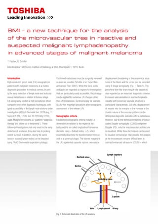
1. Fig. 1: Schematic illustration of the LN anatomy
Cortex
Cortical sinus
Lymph capillary
Medullary sinus
Capsule
Vein Lymph vessel Artery
T. Fischer, G. Schäfer
Interdisciplinary US Centre, Institute of Radiology at CC6, Charitéplatz 1, 10117 Berlin
SMI – a new technique for the analysis
of the microvascular tree in reactive and
suspected malignant lymphadenopathy
in advanced stages of malignant melanoma
Introduction
High-resolution lymph node (LN) sonography in
patients with malignant melanoma is a routine
diagnostic procedure in medical centres. Its aim
is the early detection of lymph node and subcuta-
neous metastases in relation to tumour stage.
LN sonography exhibits a high acceptance when
compared with other diagnostic techniques, with
good accessibility of the lymph node stations under
investigation (J Dtsch Dermatol Ges. 2013 Aug; 11
Suppl 6:1-116, 1-126. doi: 10.1111/ddg.12113_
suppl. Malignant melanoma S3-guideline “diagnosis,
therapy and follow-up of melanoma”). These
follow-up investigations not only result in the early
detection of a relapse, they also help to prolong
overall survival. In addition, during the same
session suspect lymph nodes can be biopsied
using FNAC (fine-needle aspiration cytology).
Confirmed metastases must be surgically removed
as soon as possible (Schäfer et al. Expert Rev
Anticancer Ther. 2007). While the neck, axilla
and groin are regarded as regions for investigation
that are particularly easily accessible, this strategy
can be applied to numerous LN changes other
than LN metastases. Sentinel biopsy for example
is a further important procedure after sonographic
assessment of the relevant LN.
Sonographic criteria
Established sonographic criteria include LN
dimensions in relation to the region of the
body and the so-called longitudinal/transverse
diameter ratio (= Solbiati index, >2), which
essentially describes the transformation from an
oval to a spherical shape. The blurred margins of
the LN, a potential capsular rupture, necrosis or
displacement/broadening of the anatomical struc-
tures of the hilum and the cortex can be recorded
using B-image sonography (Fig. 1, Table 1). The
peripheral tree-like branching of hilar vessels is
also regarded as an important diagnostic criterion.
Increased vascularisation in reactive lymphade-
nopathy with preserved vascular structure is
particularly characteristic. Cut-offs, displacement
of vessels into the margins or the increase in the
chaotic nature of the vascularpattern can be
differential diagnostic indicators of LN metastases.
However, due to the technical limitations of colour-
coded Doppler sonography (CCDS) and power
Doppler (PD), only the macrovascular architecture
is visualized. While these techniques can be used
to visualize normal larger hilar vessels, the analysis
of the microvessels remains difficult even in
contrast-enhanced ultrasound (CEUS) – which
2. Benign Mallgnant
Form elongated oval round to round-oval, spherical
Solbiati index
(longitudinal/transverse diameter)
> 2 < 2
Margins sharp partially blurred, partially sharp
Cortex narrow with low echogenicity absent or asymmetrically hypoechoic
Centre relatively homogeneous, high echogenicity inhomogeneous, hypoechoic
Hilum usually present on image absent/asymmetric
Table 1: Differentiation criteria for lymph nodes that have undergone inflammatory or metastatic change
Doppler signals
Conventional Doppler Imaging
Superb Microvascular Imaging (SMI)
intensity
velocity
intensity
velocity
intensity
velocity
moreover incurs additional costs. Analysis of the
vascular architecture is particularly complicated
in the case of small LNs (< 5mm) (Cui, XW et al.
Worl J Gastroenterol 2013). Superb Microvascular
Imaging (SMI), whose basic principles will be briefly
explained below, is a novel sonographic technique
to complement CEUS and elastography. Drawing
on an initial group of patients with malignant
melanoma, typical examples are described where
B-mode imaging, CCDS, PD and SMI, as well as
elastography were performed.
Superb Microvascular Imaging (SMI)
SMI is based on a powerful intelligent algorithm,
applying a similar concept to that used for
ultrasonic Doppler signals. SMI effectively
separates flow signals from overlaying tissue
motion artefacts, preserving even the subtlest
low-flow components with unmatched detail
and definition.
1. Doppler signals
Both blood flow and tissue motion (clutter) produce
ultrasonic Doppler signals. The strong clutter signals
overlap the low-velocity blood flow components.
2. Conventional Doppler imaging
Conventional Doppler imaging applies a wall filter
to remove clutter and motion artefacts, resulting
in a loss of low-flow components.
3. SMI
SMI does not primarily focus on Doppler shift or
the strength of a specific signal, but on its local
distribution across the region of interest. While
tissue motion occurs simultaneously across a
region, blood flow always occurs locally. High-
density ultrasound system architecture with
real-time application capability allows the system
to identify and remove global motion signals in real
time, while preserving subtle low-flow components.
Non-interventional study
To date, 10 patients have been included in an initial
non-interventional study. The SMI technique was
used in addition to the standard programme
involving B-image sonography, CCDS, PD and
FNAC on the Aplio 500 ultrasound system.
All patients had a confirmed Clark’s Level III-IV
melanoma and were treated as part of follow-up.
In 3 out of 10 cases, the primary diagnosis that
2 SMI – a new technique for the analysis of the microvascular tree in reactive and suspected malignant lymphadenopathy
in advanced stages of malignant melanoma
3. B
C
A
D
E
had been made previously was changed based
on vascular detection using SMI and this change
was confirmed by FNAC and surgical excision.
In 7 out of 10 cases, the images of the finest
vascular networks generated with SMI were
subjectively superior to those generated by the
standard techniques, CCDS and PD. This applied
both to reactively enlarged LNs (Fig. 2 A – E) and to
metastases (Fig. 3). The vascular architecture of
superficial and small LNs, in particular, was easier
to assess using SMI (Fig. 4). Up to a penetration
depth of 2.5 cm and, in particular, in subcutaneous
locations, SMI exhibited clear advantages in
delineating the hilar vessels and the smallest of
peripheral branches. Surprisingly, SMI detected
microvessel in portions that had previously been
classified as avascular or necrotic, and a targeted
FNAC was immediately performed (Fig. 4).
A subcutaneous manifestation was found in
2 of the 10 patients in the region of the scar.
The assessment was initially difficult due to
postoperative seroma but detection of vessels
was particularly important in these cases in
order to be able to carry out a FNAC as quickly
as possible. The investigation was performed in
the frequency range of between 14 and 18 MHz,
depending on penetration depth. In the case of
suspected malignancy, elastography was carried
out on the suspect area of the affected LN. It
revealed a good correlation between the absence
of elasticity and the presence of bizarre vascular
patterns produced by the melanoma metastases
(Fig. 5). Furthermore, SMI was able to visualize
vessels that pass through the capsule; another
important detail for the assessment of potential
metastatic spread that conventional techniques
did not offer.
In summary, SMI, which can be used with or
without contrast agents, constitutes a novel and
promising technique for visualizing microcirculation.
In an initial non-interventional study, additional
information was found in 3 of 10 cases that had
not been detected previously using the conventional
techniques CCDS and PD. In particular, SMI
provided relevant information that allowed the
evaluation of small lesions, subcutaneous masses
and structures in the region of the scar.
Figure 2 A – E: Right axillary reactive lymphadenopathy (A). Unremarkable finding in FNAC. Illustration of the full vascular tree with its microarchitecture in SMI mode (B, C).
The large vessels close to the hilum are seen in CCDS (D) and power Doppler (E), the vascular tree is not free of artefacts and its detection is limited.
SMI – a new technique for the analysis of the microvascular tree in reactive and suspected malignant lymphadenopathy 3
in advanced stages of malignant melanoma
4. A
B
C
D
E
B
CA
Figure 3 A – E: Large right inguinal LN metastasis that has been histologically confirmed (A). The pathological vascular pattern and the vessels
passing through the LN capsule are visualized only by SMI (B). Power Doppler (C) and CCDS (D) only allow delineation of the macrovessels. In this
case, no change in the diagnosis was to be expected based on the clear situation. Of note, however, is the level of detail visible in the microvessels
and the fact that very fine vessels have broken through the capsule (E). The sensitive, artefact-free image produced by SMI of pathological vascular
networks is well documented in this figure.
Figure 4 A – C: 6 mm metastasis adjacent to the region of the scar on the right thigh. Vessels with branching in the margins are visualized by SMI,
while the lesion appears avascular in power Doppler. Metastasis confirmed with FNAC (C).
SMI – a new technique for the analysis of the microvascular tree in reactive and suspected malignant lymphadenopathy 4
in advanced stages of malignant melanoma
5. B
C
A
Figure 5 A – C: Correlation between SMI (A), colour Doppler (B) and elastography (C) for a large LN metastasis. SMI visualizes the finest tumour vascular networks.
SMI – a new technique for the analysis of the microvascular tree in reactive and suspected malignant lymphadenopathy 5
in advanced stages of malignant melanoma