This document discusses Superb Microvascular Imaging (SMI), a new ultrasound technique developed by Toshiba Medical Systems that allows for detailed imaging of microvasculature without the need for contrast agents. Three case studies are presented that illustrate how SMI can detect low-grade inflammation not seen with conventional power Doppler. SMI provided clearer depiction of microvascular branching patterns in the tendon attachment of a patient's knee, in the synovium of a patient's foot with psoriatic arthritis, and in the synovium of a patient's sternoclavicular joint with rheumatoid arthritis. The increased sensitivity of SMI appears beneficial for identifying subtle inflammation and guiding clinical management decisions.
Introduction
High-resolution lymph node (LN) sonography in patients with malignant melanoma is a routine diagnostic procedure in medical centres. Its aim is the early detection of lymph node and subcuta- neous metastases in relation to tumour stage.
LN sonography exhibits a high acceptance when compared with other diagnostic techniques, with good accessibility of the lymph node stations under investigation (J Dtsch Dermatol Ges. 2013 Aug; 11 Suppl 6:1-116, 1-126. doi: 10.1111/ddg.12113_ suppl. Malignant melanoma S3-guideline “diagnosis, therapy and follow-up of melanoma”). These follow-up investigations not only result in the early detection of a relapse, they also help to prolong overall survival. In addition, during the same session suspect lymph nodes can be biopsied using FNAC (fine-needle aspiration cytology).
Confirmed metastases must be surgically removed as soon as possible (Schäfer et al. Expert Rev Anticancer Ther. 2007). While the neck, axilla and groin are regarded as regions for investigation that are particularly easily accessible, this strategy can be applied to numerous LN changes other than LN metastases. Sentinel biopsy for example is a further important procedure after sonographic assessment of the relevant LN.
A radiological insight into various musculoskeletal complications in patients suffering from AIDS and how it'll affect the management of the patient. A must know for all Radiologists.
High-Resolution Three-Dimensional Weight-Bearing Imaging of Lower Extremity U...Carestream
This paper addresses the benefits of a prototype (INVESTIGATIONAL – NOT FOR COMMERCIAL SALE) cone beam computed tomography system (hereafter referred to as the “CBCT system”) dedicated to extremity imaging. The CBCT system was co-developed by scientists at Carestream Health and John Hopkins University. The CBCT system has demonstrated spatial and contrast resolution beyond the limits of conventional multi-detector CT (MDCT) at a reduced radiation exposure1. The CBCT system was designed to image both upper and lower extremities, with the lower extremities also capable of being imaged in a weight-bearing configuration. This unique capability can unveil and better characterize certain pathologies in the knee and ankle joints such as meniscal extrusion, altered tibiofemoral joint space morphology, flatfoot deformity, and distal tibiofibular syndesmosis insufficiency.
Learn more about Carestream's portfolio of products at http://www.carestream.com/medical
VISIONS magazine is a publication of Canon Medical Europe and is offered free of charge to health professionals.
The magazine is published twice a year. Registration to access full, previously published, digital editions can be done via the website: https://eu.medical.canon/visions-magazine
VISIONS magazine is covering Canon Medical’s European region and as such reflects products, technologies and
services for this particular area. The mentioned products may not be available in other geographic regions.
Please consult your Canon Medical representative sales office in case of any questions.
VISIONS magazine is Canon Medical’s customer magazine about innovative technologies and applications in medical imaging. Read all about diagnostic imaging in the fields of CT, MRI, X-Ray, Ultrasound, HII, Eye Care and stay up-to-date with the latest developments in your clinical environment.
This Veterinary Special edition of VISIONS magazine is a publication of Canon Medical Europe and is offered
free of charge to health professionals. To download the digital edition of this Veterinary Special, please visit:
https://eu.medical.canon/visions.
VISIONS magazine is covering Canon Medical’s European region and as such reflects products, technologies
and services for this particular area. The mentioned products may not be available in other geographic regions.
Please consult your Canon Medical representative sales office in case of any questions.
No part of this publication may be reproduced in whole or in part, stored in an automated storage and retrieval
system or transmitted in any manner whatsoever without written permission of the publisher. The opinions
expressed in this publication are solely those of the authors and not necessarily those of Canon Medical.
Canon Medical does not guarantee the accuracy or reliability of the information provided herein.
News, articles and the full edition of VISIONS magazine are announced firstly, as pre-publication, via the
dedicated VISIONS LinkedIn Group: https://www.linkedin.com/groups/3698045. In this group you can
actively participate in discussions about the content and future direction of the magazine.
Canon Medical Systems VISIONS Magazine - issue 35
VISIONS magazine is a publication of Canon Medical Europe and is offered free of charge to medical and health professionals. The magazine is published twice a year.
Registration to access full, previously published, digital editions can be done via the web site: https://nl.medical.canon/visions-magazine.
VISIONS magazine is covering Canon Medical’s European region and as such reflects products, technologies and services for this particular area. The mentioned products may not be available in other geographic regions. Please consult your Canon Medical representative sales office in case of any questions.
No part of this publication may be reproduced in whole or in part, stored in an automated storage and retrieval system or transmitted in any manner whatsoever without written permission of the publisher. The opinions expressed in this publication are solely those of the authors and not necessarily those of Canon Medical. Canon Medical does not guarantee the accuracy or reliability of the information provided herein.
News items and articles are announced firstly, as pre-publication, via the dedicated VISIONS LinkedIn Group: https://www.linkedin.com/groups/3698045. In this group you can actively participate in discussions about the content and future direction of the magazine.
This MR Special edition of VISIONS magazine is a publication of Canon Medical Europe and is offered
free of charge to health professionals. To download the digital edition of this MR Special, please visit:
https://eu.medical.canon/visions.
VISIONS magazine is covering Canon Medical’s European region and as such reflects products, technologies
and services for this particular area. The mentioned products may not be available in other geographic regions.
Please consult your Canon Medical representative sales office in case of any questions.
VISIONS magazine is a publication of Canon Medical Europe and is offered free of charge to health professionals.
The magazine is published twice a year. Registration to access full, previously published, digital editions can
be done via the web site: https://nl.medical.canon/visions-magazine. Canon Medical stores and uses personal
data of the registration to send out the magazine and inform members about new developments. Members can
customize preferences or opt-out, after registration, in the online VISIONS profile.
Canon Medical Systems VISIONS Magazine - issue 31
VISIONS magazine is a publication of Canon Medical Europe and is offered free of charge to medical and health professionals. The magazine is published twice a year.
Registration to access full, previously published, digital editions can be done via the web site: https://nl.medical.canon/visions-magazine.
VISIONS magazine is covering Canon Medical’s European region and as such reflects products, technologies and services for this particular area. The mentioned products may not be available in other geographic regions. Please consult your Canon Medical representative sales office in case of any questions.
No part of this publication may be reproduced in whole or in part, stored in an automated storage and retrieval system or transmitted in any manner whatsoever without written permission of the publisher. The opinions expressed in this publication are solely those of the authors and not necessarily those of Canon Medical. Canon Medical does not guarantee the accuracy or reliability of the information provided herein.
News items and articles are announced firstly, as pre-publication, via the dedicated VISIONS LinkedIn Group: https://www.linkedin.com/groups/3698045. In this group you can actively participate in discussions about the content and future direction of the magazine.
The assessment of cardiac function is essential in the athletic population not only as part of the screening process for underlying cardiac disease, but also to longitudinally assess performance and training adaptations - Source: Toshiba's VISIONS Magazine #26 | www.toshiba-medical.eu/visions
Post contrast iodine maps were introduced as part of dual-energy imaging over 10 years ago but these have never become part of routine practice in most centres for the investigation of pulmonary thromboembolic disease.
CASE: 58 year old male presented with a liver lesion seen on prior imaging. A dual energy scan was performed to further characterize the lesion. Volumetric Dual Energy scans were performed following the injection of 80mls of contrast. Scans were performed during Arterial Phase, early Portal Phase and Delayed at 3mins. Monochromatic images, iodine maps and virtual non contrast images were generated for review.
Computed tomography angiography (CTA) of the coronary arteries is a useful noninvasive tool to rule out significant coronary artery disease (CAD) in many clinical situations. Recent guidelines of stable CAD and non-ST segment elevation myocardial infarction endorse the use of CTA in symptomatic patients with low to intermediate likelihood of the disease, given the particularly high negative predictive value of the technique. However, in patients with high pre-test likelihood of CAD, the technique is not recommended, and one of the reasons is the high probability of coronary calcification in these patients, which interferes with the analysis of the images and reduces the specificity and negative predictive value of CTA.
(TOSHIBA CTEU140095) - Article from Toshiba's VISIONS Magazine#25, March 2015
Pulmonary Thromboembolism - etilogy, types, medical- Surgical and nursing man...VarunMahajani
Disruption of blood supply to lung alveoli due to blockage of one or more pulmonary blood vessels is called as Pulmonary thromboembolism. In this presentation we will discuss its causes, types and its management in depth.
The prostate is an exocrine gland of the male mammalian reproductive system
It is a walnut-sized gland that forms part of the male reproductive system and is located in front of the rectum and just below the urinary bladder
Function is to store and secrete a clear, slightly alkaline fluid that constitutes 10-30% of the volume of the seminal fluid that along with the spermatozoa, constitutes semen
A healthy human prostate measures (4cm-vertical, by 3cm-horizontal, 2cm ant-post ).
It surrounds the urethra just below the urinary bladder. It has anterior, median, posterior and two lateral lobes
It’s work is regulated by androgens which are responsible for male sex characteristics
Generalised disease of the prostate due to hormonal derangement which leads to non malignant enlargement of the gland (increase in the number of epithelial cells and stromal tissue)to cause compression of the urethra leading to symptoms (LUTS
Flu Vaccine Alert in Bangalore Karnatakaaddon Scans
As flu season approaches, health officials in Bangalore, Karnataka, are urging residents to get their flu vaccinations. The seasonal flu, while common, can lead to severe health complications, particularly for vulnerable populations such as young children, the elderly, and those with underlying health conditions.
Dr. Vidisha Kumari, a leading epidemiologist in Bangalore, emphasizes the importance of getting vaccinated. "The flu vaccine is our best defense against the influenza virus. It not only protects individuals but also helps prevent the spread of the virus in our communities," he says.
This year, the flu season is expected to coincide with a potential increase in other respiratory illnesses. The Karnataka Health Department has launched an awareness campaign highlighting the significance of flu vaccinations. They have set up multiple vaccination centers across Bangalore, making it convenient for residents to receive their shots.
To encourage widespread vaccination, the government is also collaborating with local schools, workplaces, and community centers to facilitate vaccination drives. Special attention is being given to ensuring that the vaccine is accessible to all, including marginalized communities who may have limited access to healthcare.
Residents are reminded that the flu vaccine is safe and effective. Common side effects are mild and may include soreness at the injection site, mild fever, or muscle aches. These side effects are generally short-lived and far less severe than the flu itself.
Healthcare providers are also stressing the importance of continuing COVID-19 precautions. Wearing masks, practicing good hand hygiene, and maintaining social distancing are still crucial, especially in crowded places.
Protect yourself and your loved ones by getting vaccinated. Together, we can help keep Bangalore healthy and safe this flu season. For more information on vaccination centers and schedules, residents can visit the Karnataka Health Department’s official website or follow their social media pages.
Stay informed, stay safe, and get your flu shot today!
Report Back from SGO 2024: What’s the Latest in Cervical Cancer?bkling
Are you curious about what’s new in cervical cancer research or unsure what the findings mean? Join Dr. Emily Ko, a gynecologic oncologist at Penn Medicine, to learn about the latest updates from the Society of Gynecologic Oncology (SGO) 2024 Annual Meeting on Women’s Cancer. Dr. Ko will discuss what the research presented at the conference means for you and answer your questions about the new developments.
TEST BANK for Operations Management, 14th Edition by William J. Stevenson, Ve...kevinkariuki227
TEST BANK for Operations Management, 14th Edition by William J. Stevenson, Verified Chapters 1 - 19, Complete Newest Version.pdf
Anti ulcer drugs and their Advance pharmacology ||
Anti-ulcer drugs are medications used to prevent and treat ulcers in the stomach and upper part of the small intestine (duodenal ulcers). These ulcers are often caused by an imbalance between stomach acid and the mucosal lining, which protects the stomach lining.
||Scope: Overview of various classes of anti-ulcer drugs, their mechanisms of action, indications, side effects, and clinical considerations.
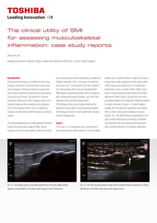
1. Introduction
Ultrasound technology to visualise the microvas-
culature continues to improve where power and
colour Doppler ultrasound remains a quick and
non-invasive method of assessing the vascularity
of tumours and tissue. The advent of contrast
enhanced ultrasound (CEUS) imaging using micro
bubbles improved the sensitivity and resolution
of the microvessels which can be imaged but
requires an intravenous administration of contrast
agents.
The latest development by Toshiba Medical Systems,
Superb Microvascular Imaging (SMI), allows
imaging of the microvasculature without the need
for contrast enhancement employing an advanced
Doppler algorithm with a new level of sensitivity
and frame rate. The sensitivity and finer detail of
the microvessels which can be visualised with
SMI appears significantly better when compared
with conventional power Doppler, and rivals that
depicted with contrast enhancement.
The following three case studies illustrate the
potential clinical value of this advanced Doppler
technology and how it could significantly change
clinical management.
Case 1
This case is in a gentleman who complained of
pain overlying the medial aspect of his left patella
tendon near its tibial insertion. Figure 1a shows a
normal grey-scale appearance with some subtle,
mildly reduced echogenicity but no significant
thickening or tear is evident. When SMI is used,
there is clear hypervascularity within the tendon
attachment itself (Figure 1b) and the fine micro-
vasculature detail of this segment of inflamed tendon
is clearly outlined in Figure 1c. Power Doppler
images did not reveal any significant vascularity
with a mostly noisy power background signal
(Figure 1d). The SMI finding corresponded to the
site of exact tenderness confirming a tendinitis
and therefore this led to appropriate treatment
with a steroid injection for symptom alleviation.
Adrian KP Lim
Imaging Department, Imperial College London and Healthcare NHS Trust, London, United Kingdom
The clinical utility of SMI
for assessing musculoskeletal
inflammation: case study reports
Fig. 1b: The fine microvasculature detail of this inflamed tendon attachment is clearly
illustrated on this SMI mode single view image (arrow).
Fig. 1a: The medial aspect of the tibial attachment of this left patella tendon
appears unremarkable on this grey-scale image at site of tenderness.
2. 2 The Clinical utility of Superb Microvascular Imaging (SMI) for low grade inflammation in MSK
Fig. 2a: Grey-scale image of the left naviculo-cuneiform joint in a patient with psoriatic
arthritis and pain.
Case 2
The following are images of a patient with a history
of psoriatic arthropathy who complained of tender
ness in the left mid-foot region. The grey-scale
ultrasound image (Figure 2a) demonstrates a mildly
thickened synovium with no erosions, of the left
naviculo-cuneiform joint.
Figure 2b shows significant vascularity with
power Doppler consistent with an active synovitis.
When SMI is applied, the branching pattern of
the microvasculature can be readily appreciated
(Figure 2c).
Case 3
This example is of a patient with known rheumatoid
arthritis who complained of pain in the right sterno
clavicular joint. In Figure 3a, there is a modestly
thickened synovium but no definite erosion is evident.
Figure 3b shows that there is no significant vascu-
larity with power Doppler. There is only minimal
Fig. 2b: The thickened synovium of the left naviculo-cuneiform joint is markedly
vascular.
Fig. 1d: There is no significant vascularity with power Doppler, where the gain has
been increased to just above noise.
Fig. 1c: The marked vascularity of the tendon attachment can be detected with
SMI (arrow) corresponding to the site of tenderness and compatible with a tendinitis.
Fig. 2c: With Superb Microvascular Imaging (SMI), the detailed branching pattern of
the microvasculature is better defined.
3. The Clinical utility of Superb Microvascular Imaging (SMI) for low grade inflammation in MSK 3
Fig. 3a: The right sternoclavicular joint of a patient with rheumatoid arthritis
demonstrates a thickened synovium on this grey-scale image.
Fig. 3b: There is only minimal peripheral vascularity with power Doppler (thick arrow).
vascularity seen in the periphery of the joint just
above the noise level. With SMI, the branching pat-
tern of the microvasculature within the inflamed joint
itself can be clearly detected and is illustrated in
Figure 3c. This convincingly confirmed the presence
of an active synovitis and the patient subsequently
had a steroid injection which alleviated her symptoms.
Conclusion
Our early experience with SMI shows that it has
excellent depiction and fine detail of the microvas-
culature not seen with routine Doppler technology.
With significantly increased sensitivity, SMI has
great potential at identifying low-grade inflammation
which was not possible previously. The improved
diagnostic confidence with this technology would
have a significant clinical impact and influence
clinical management of patients.
Fig. 3c: However, with the use of SMI, vascularity within the synovium (thin arrow)
can be detected confirming an active synovitis.