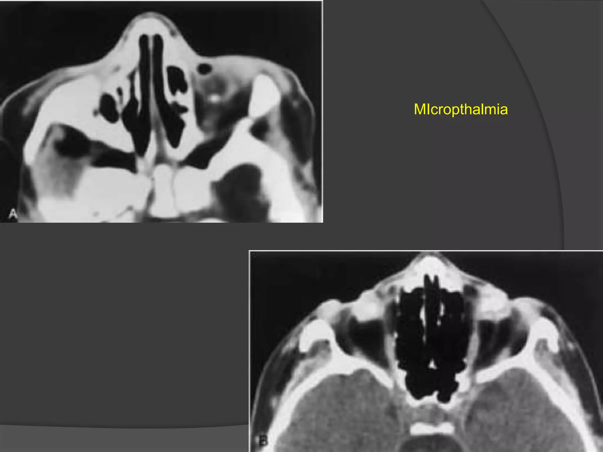
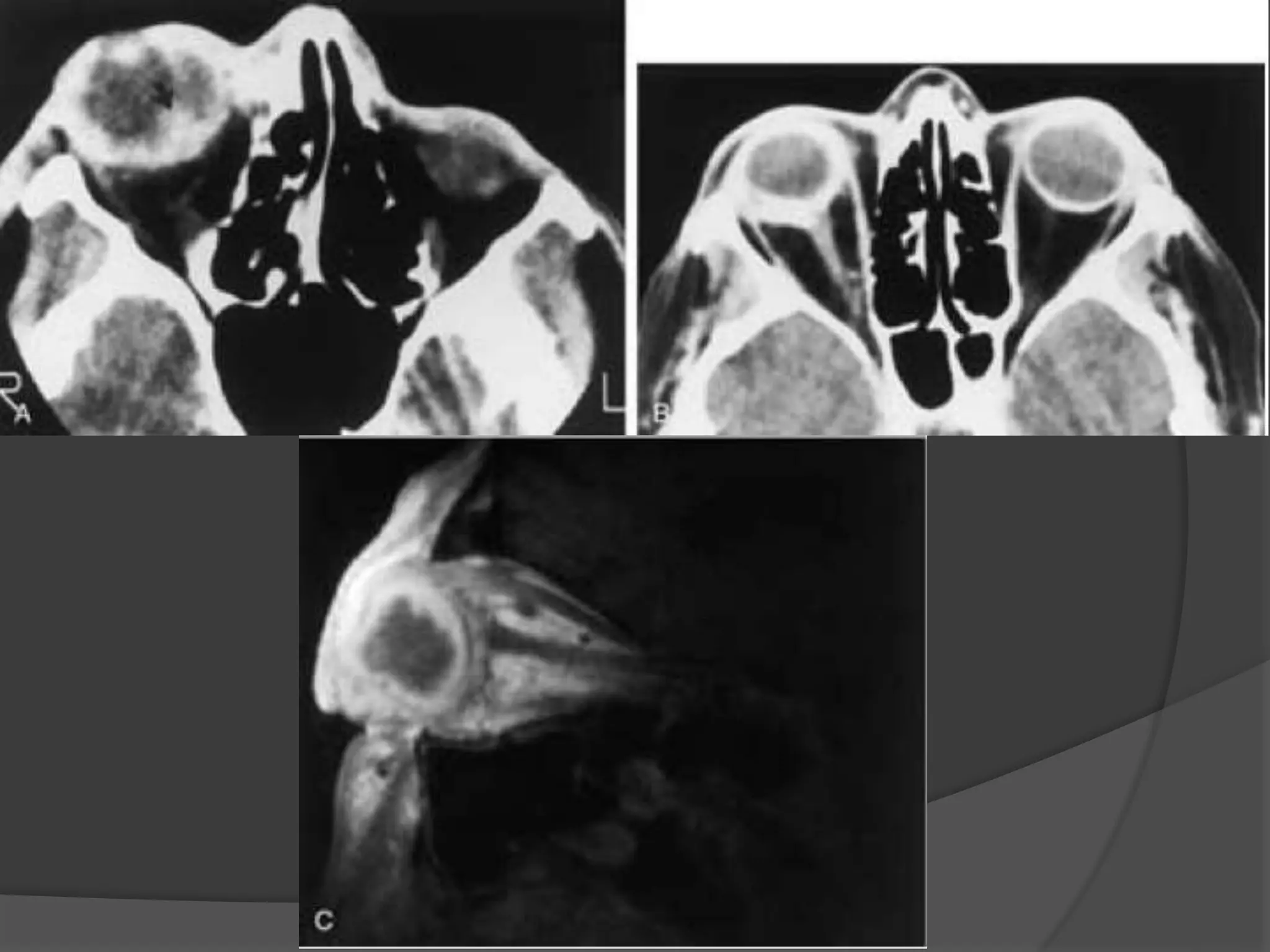
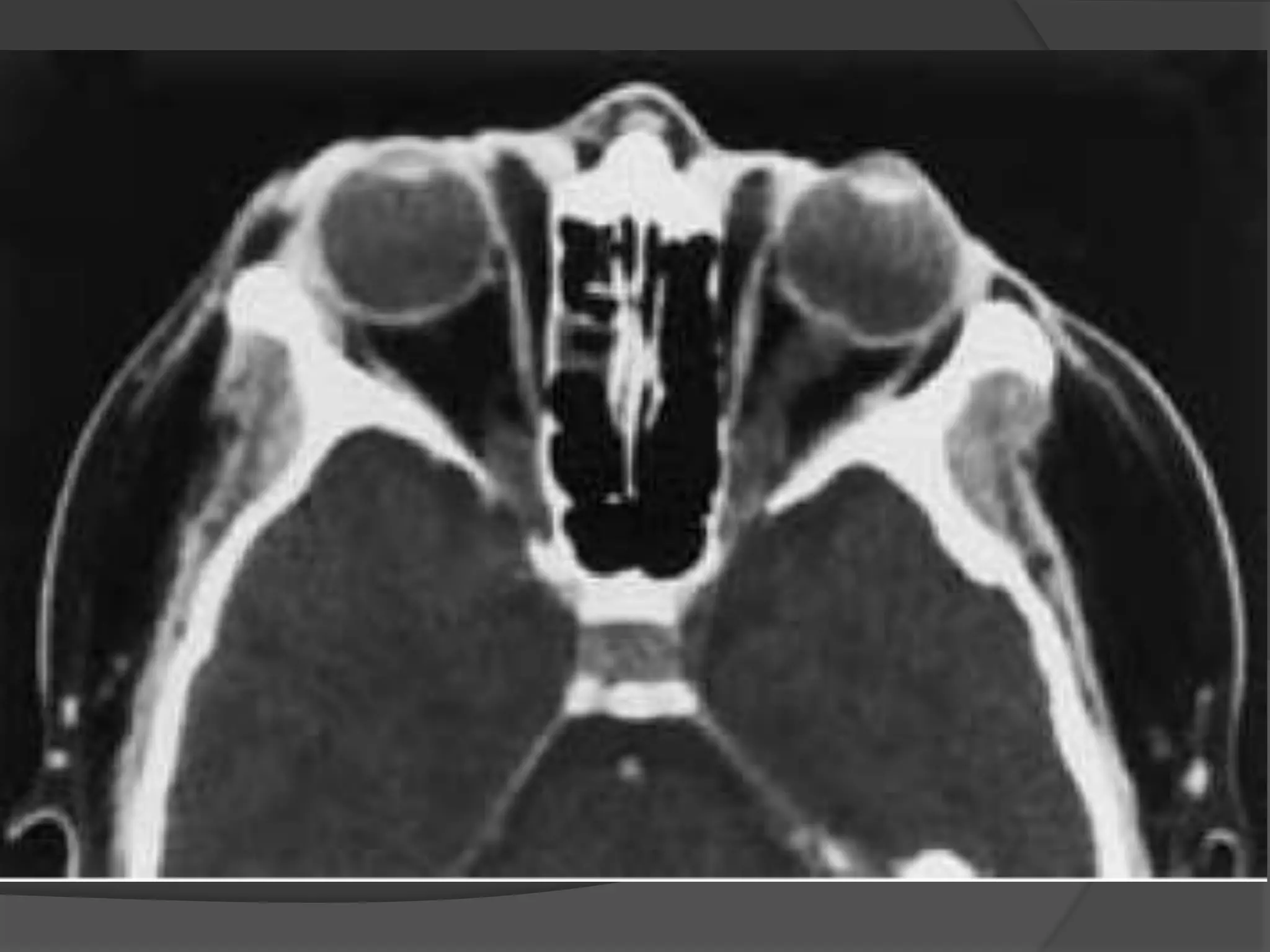
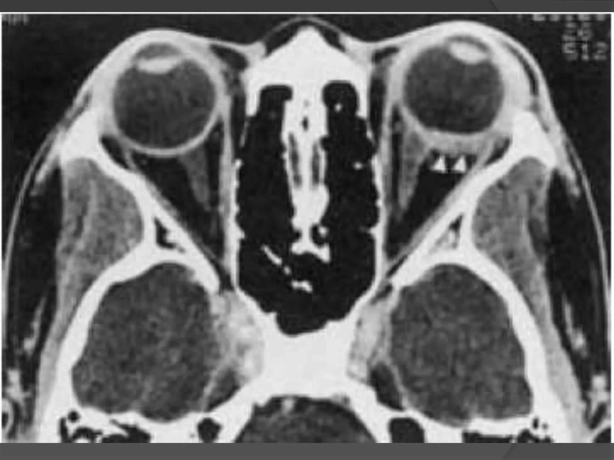
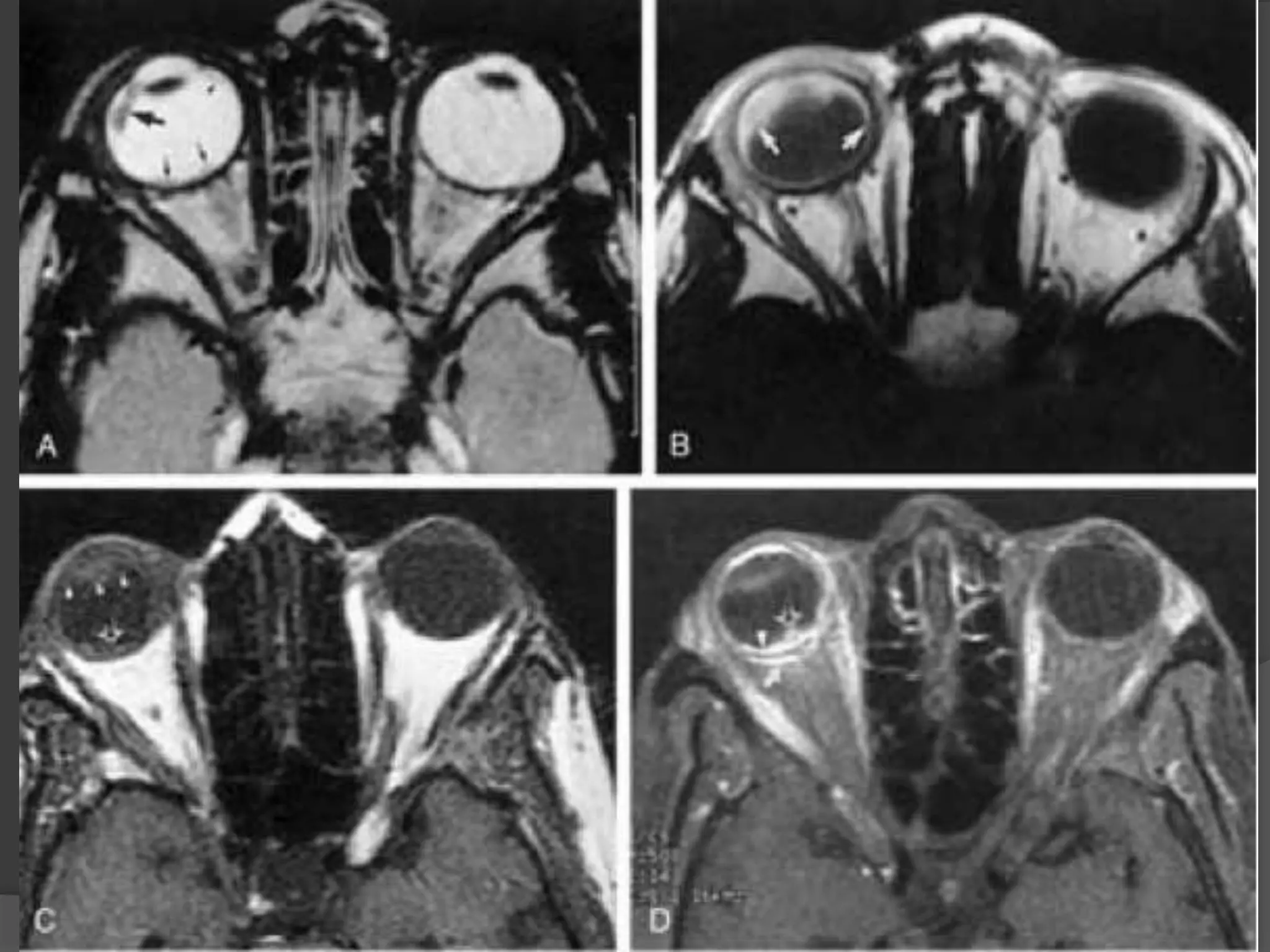
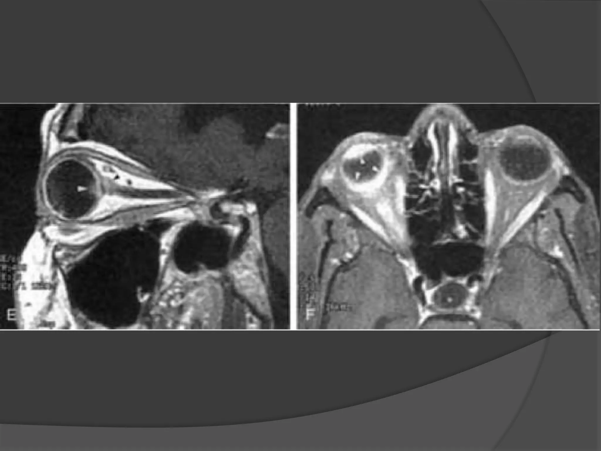
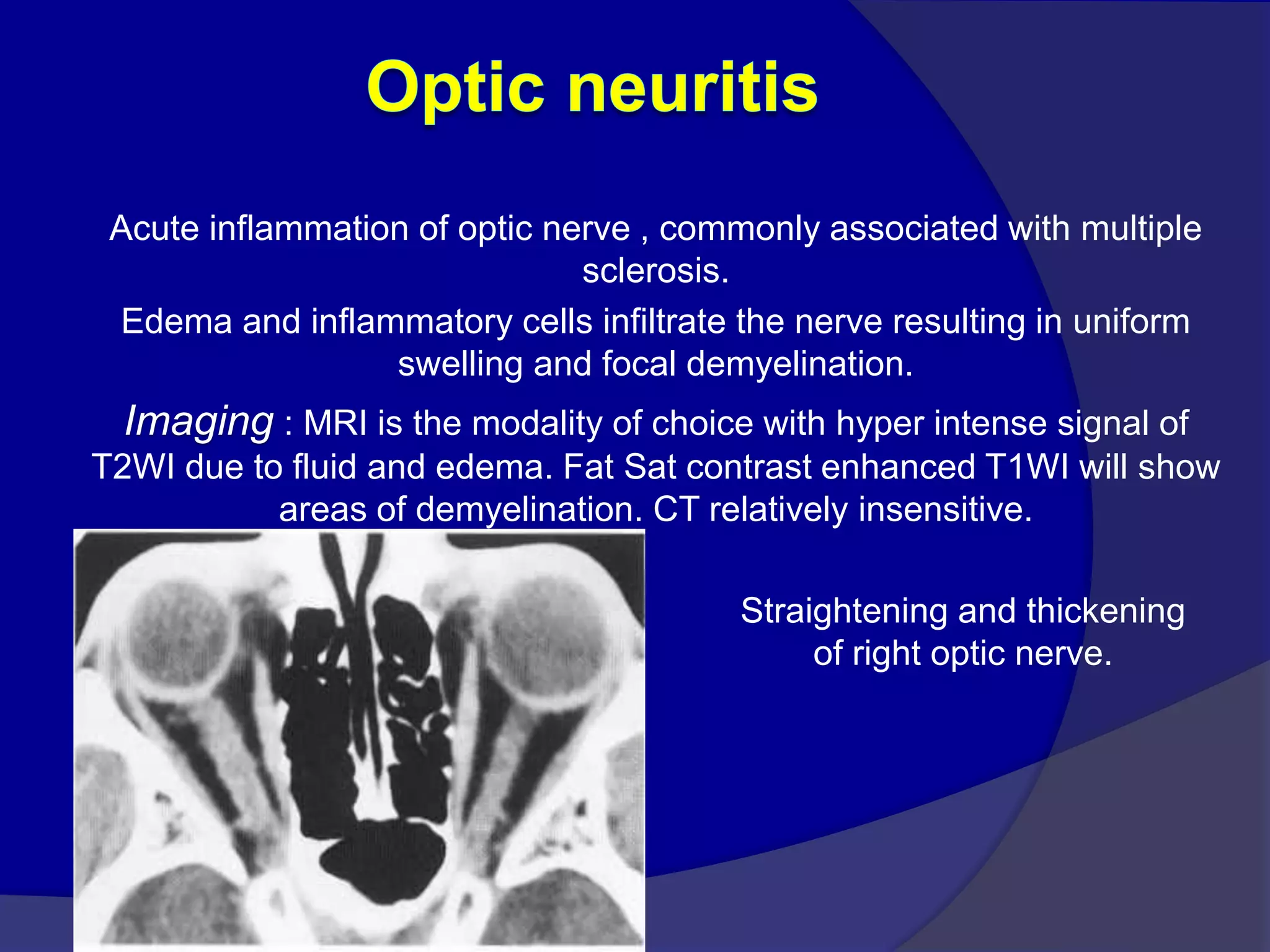
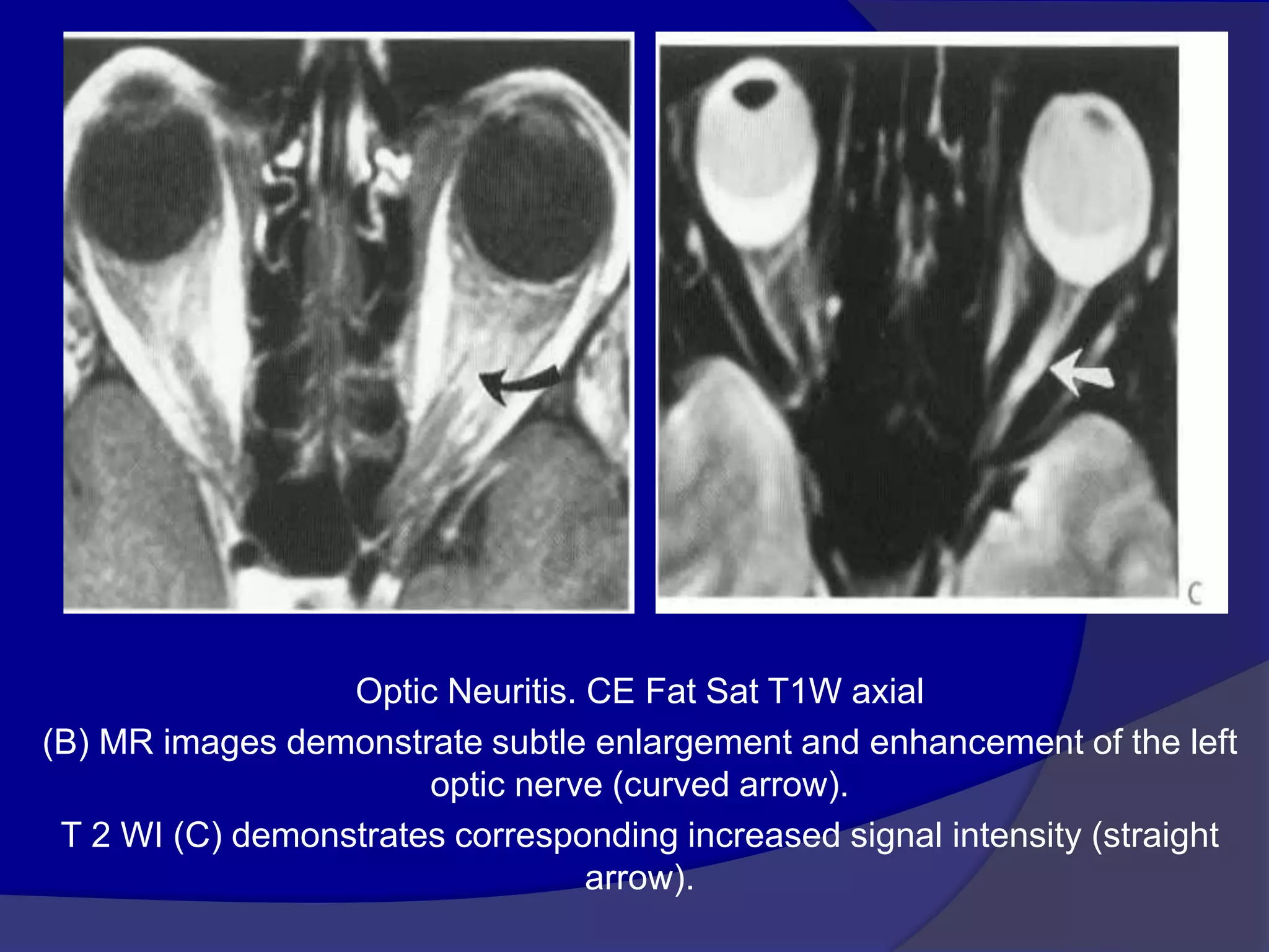
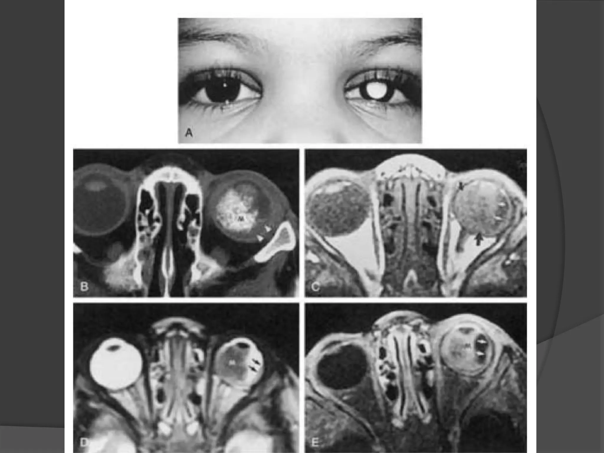
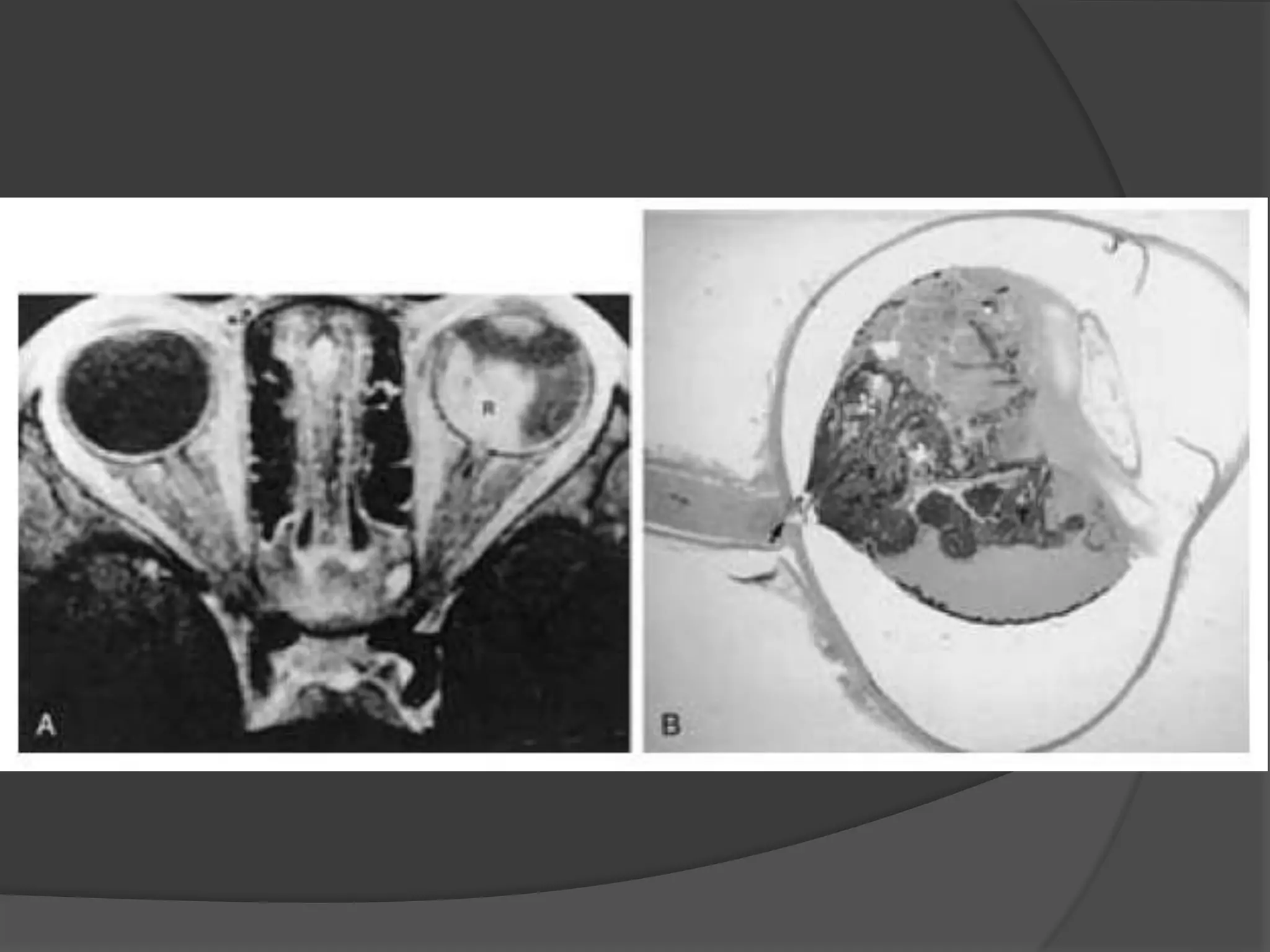
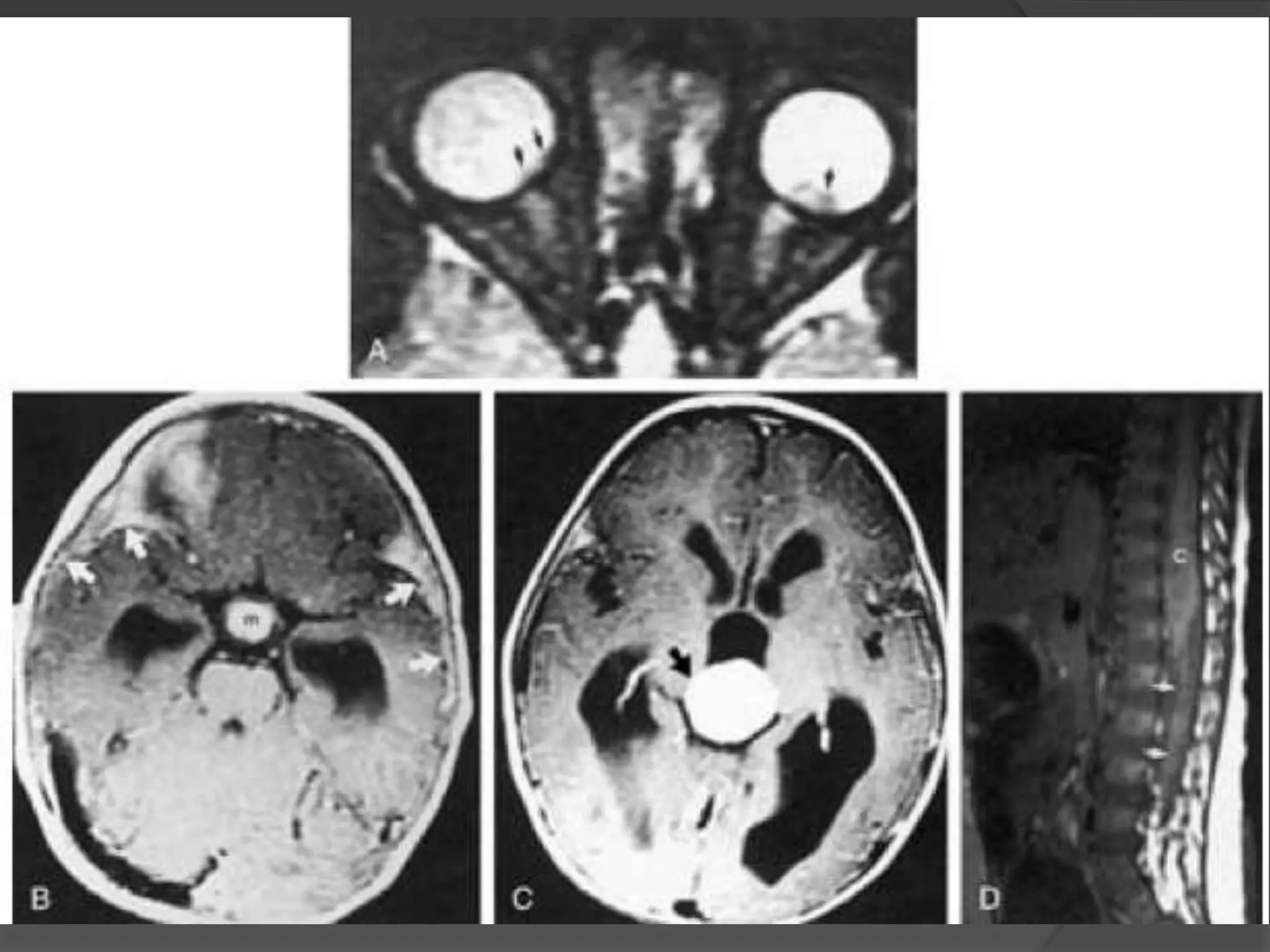
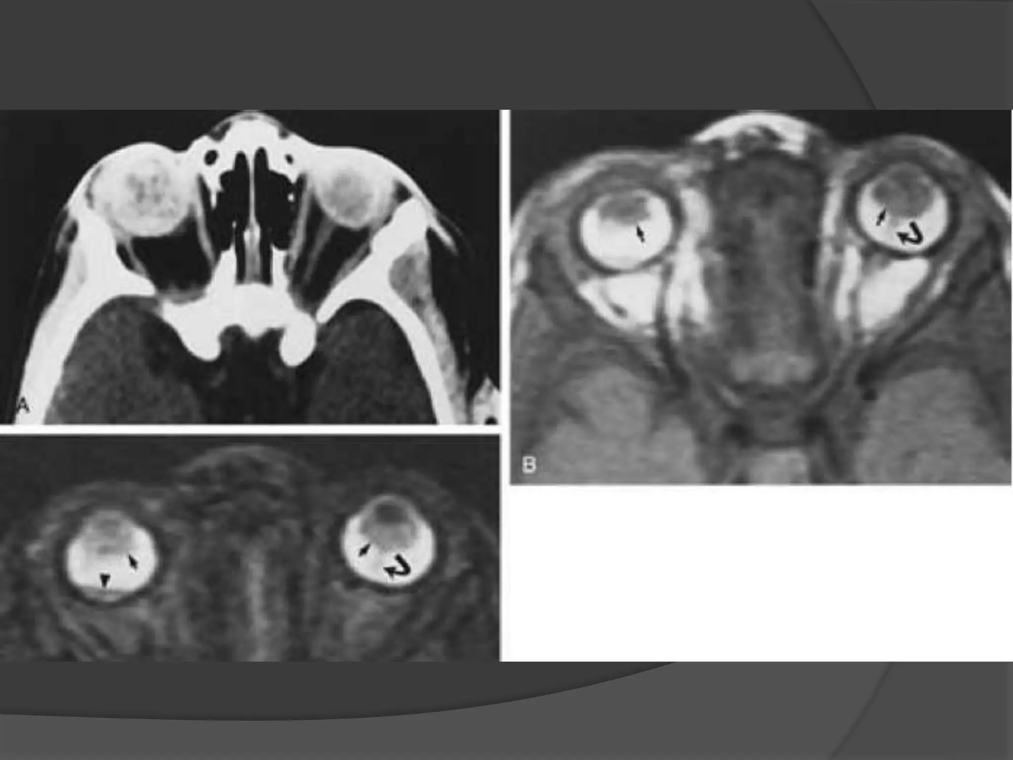
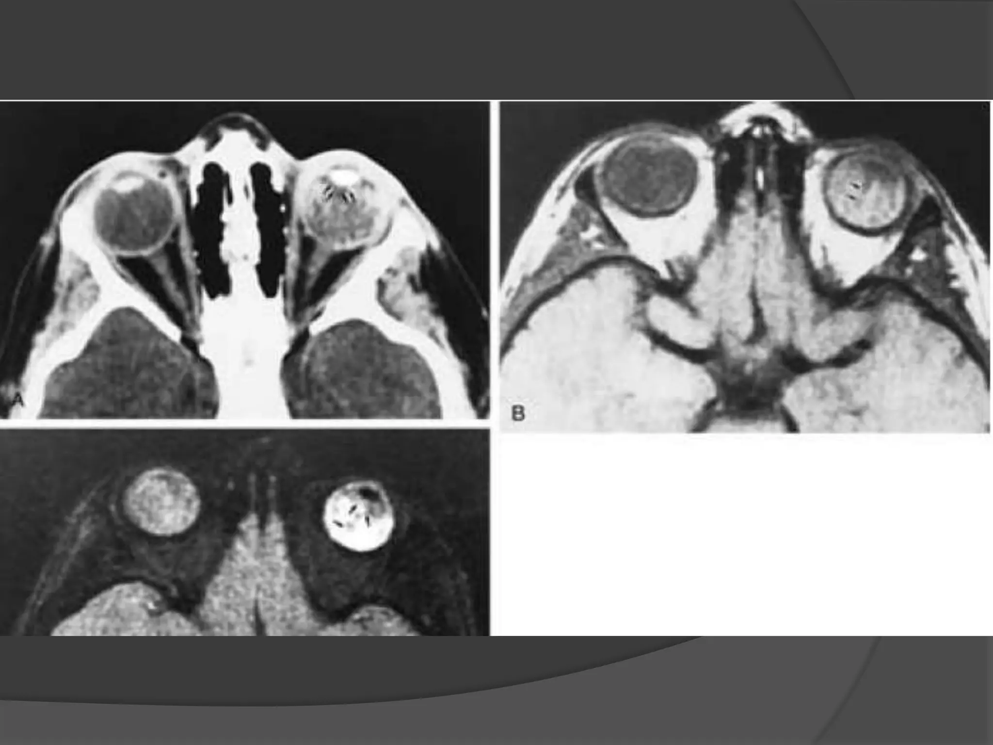
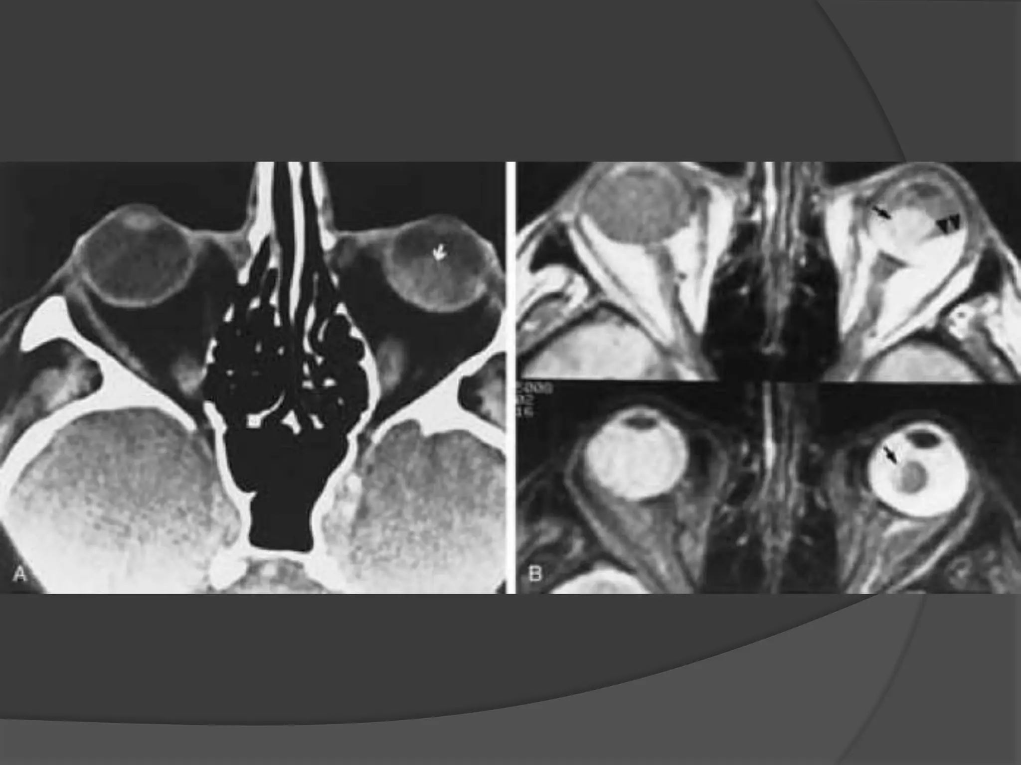
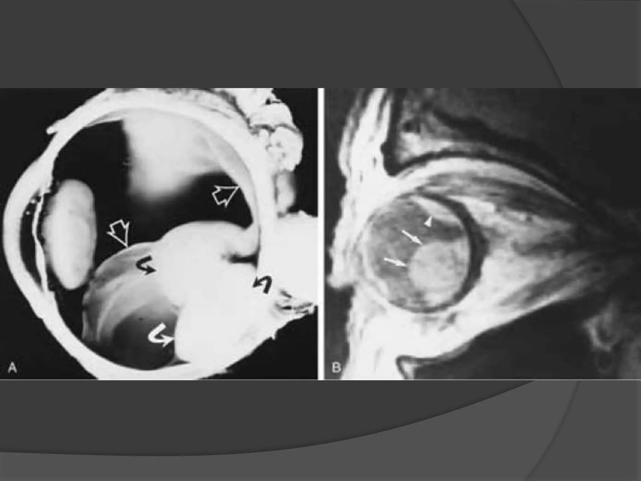
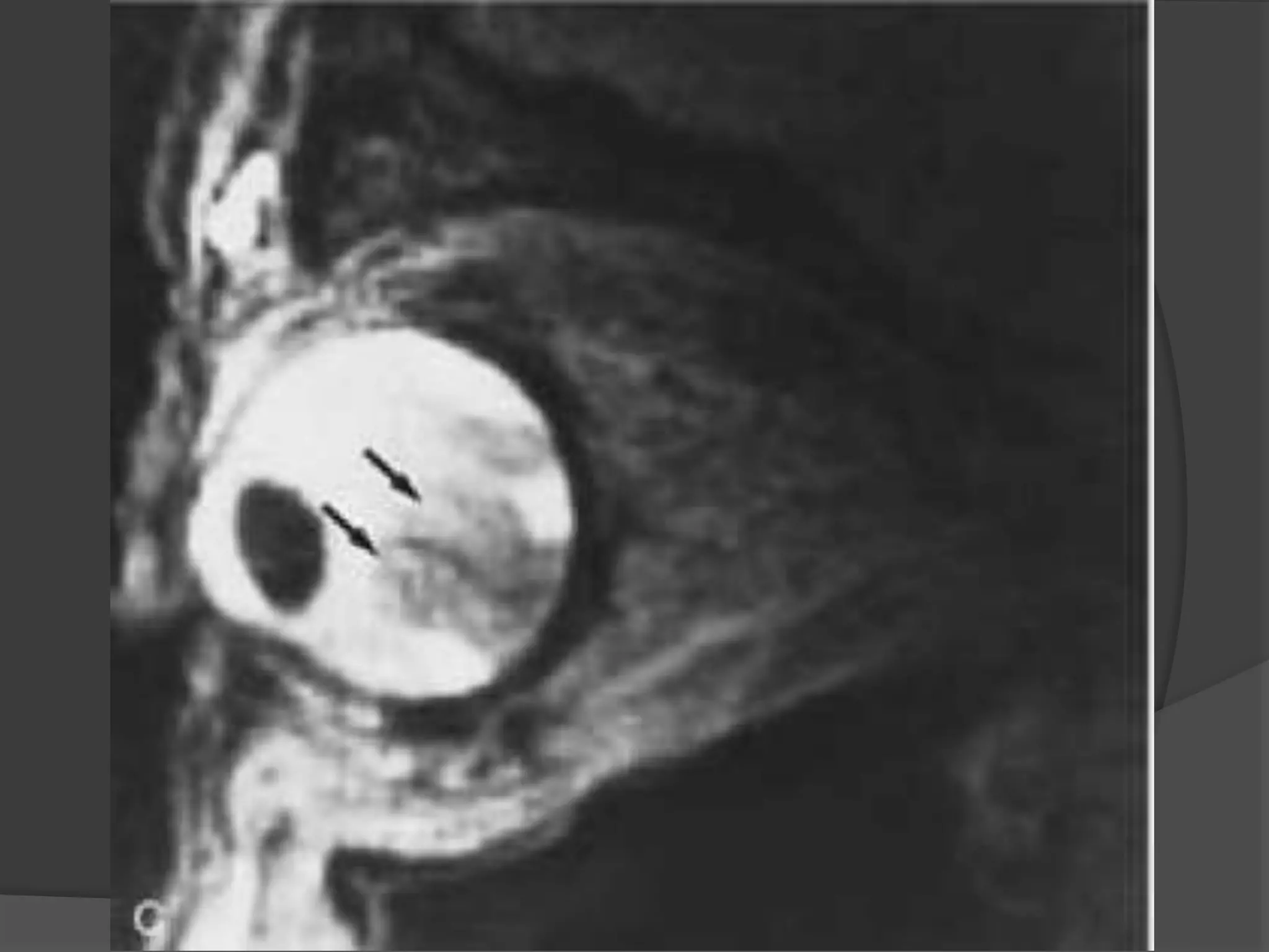
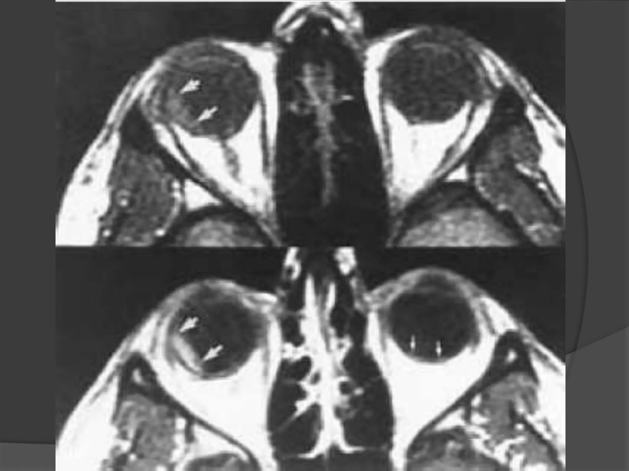
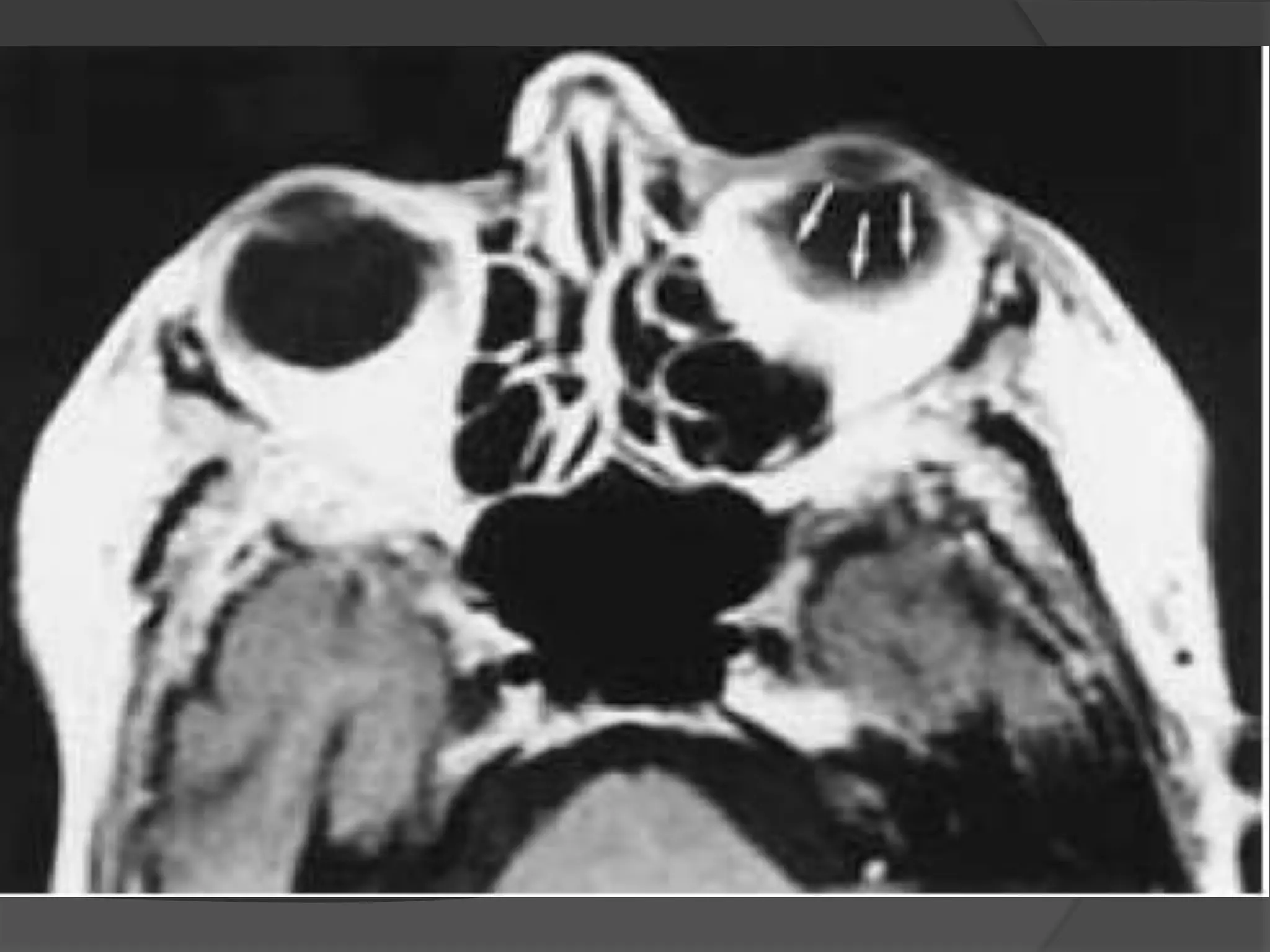
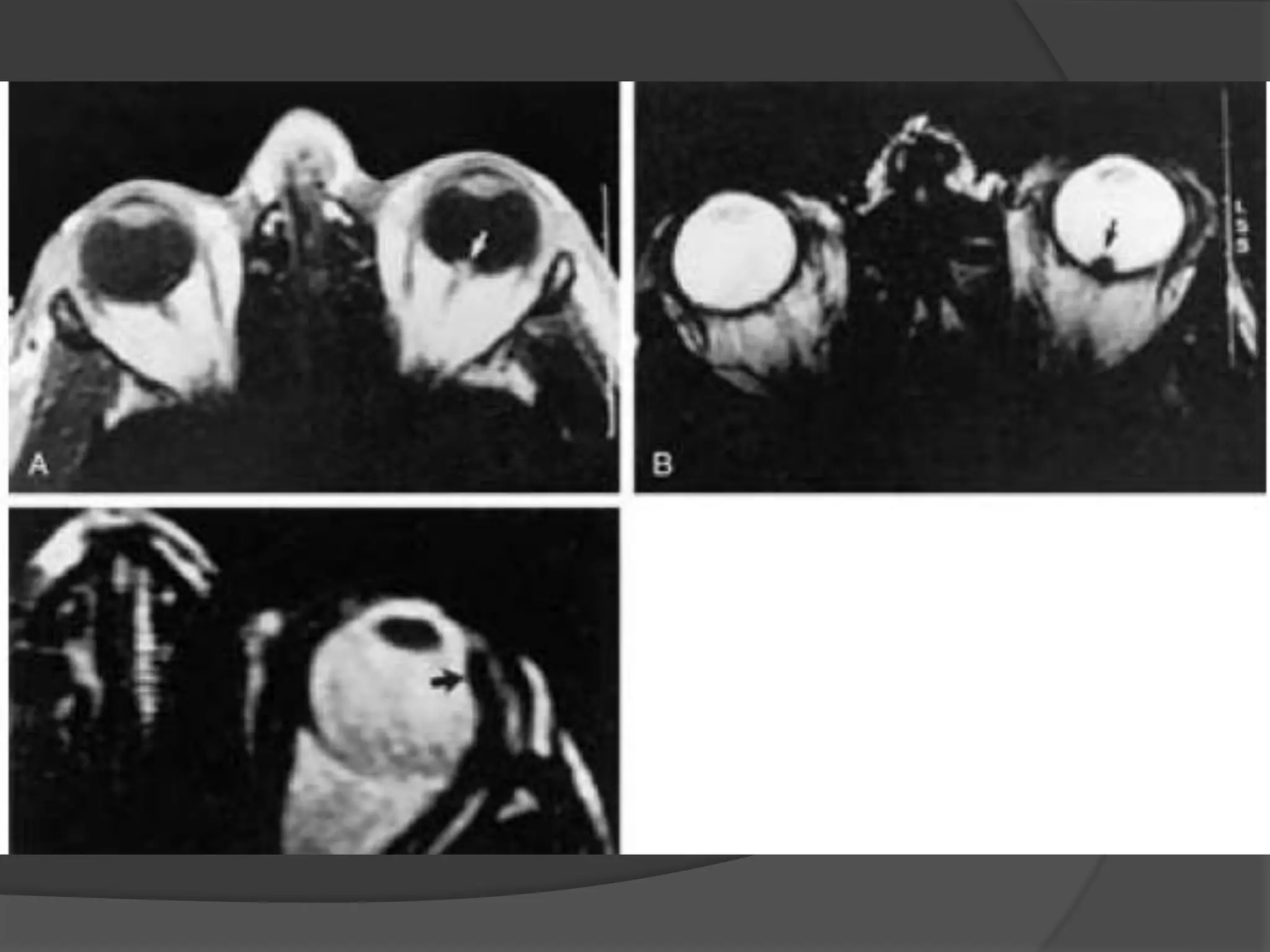
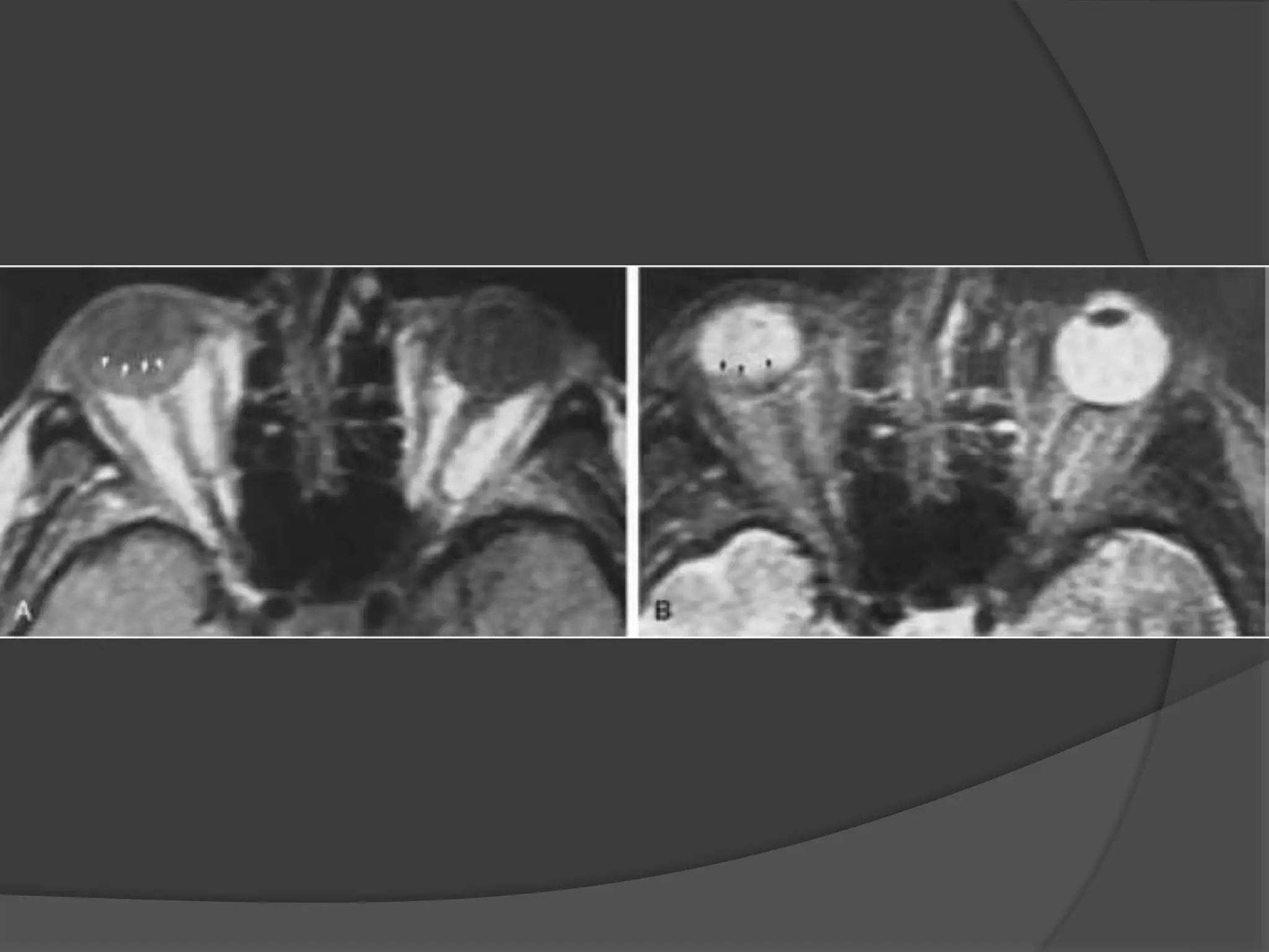
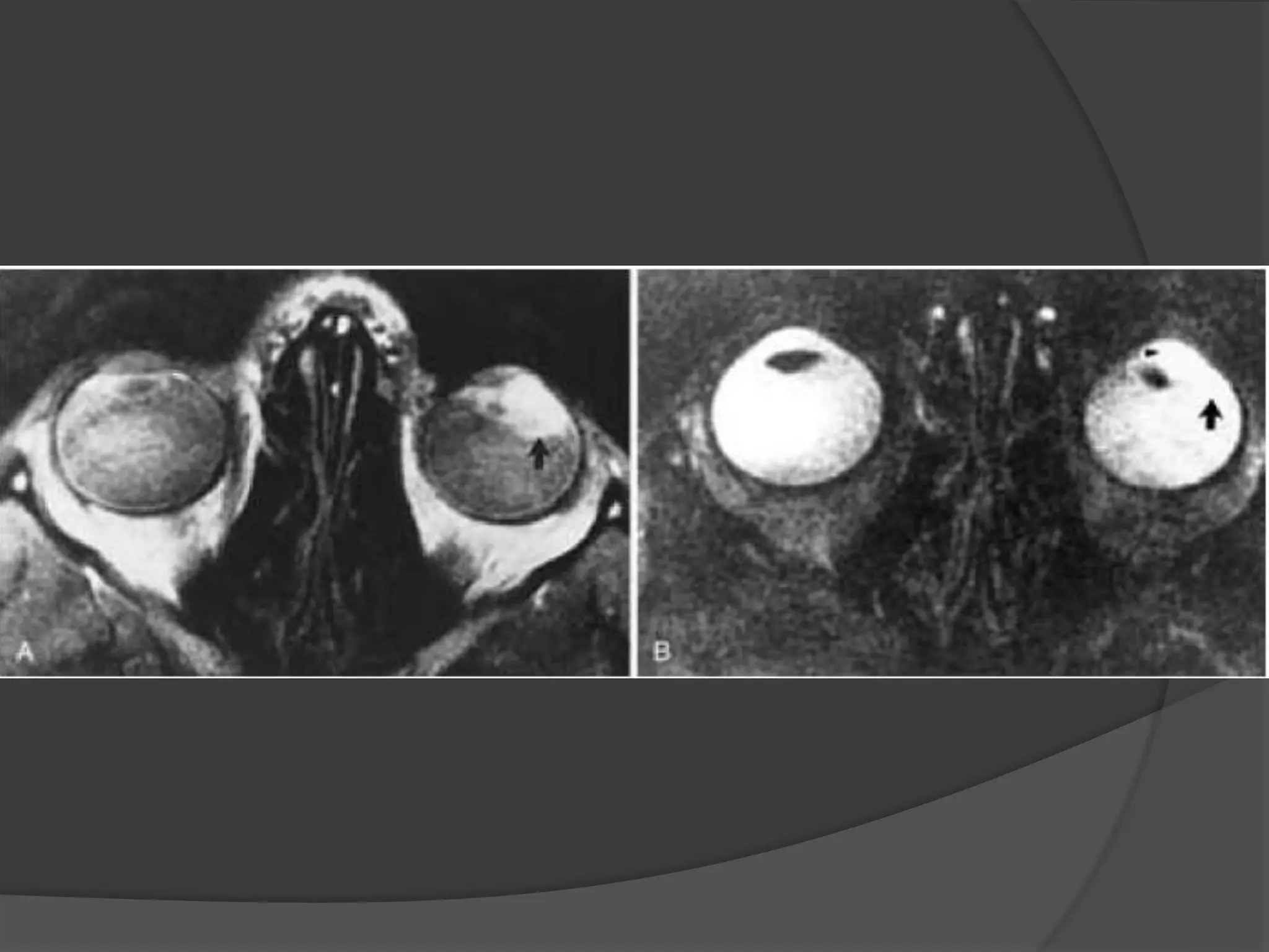
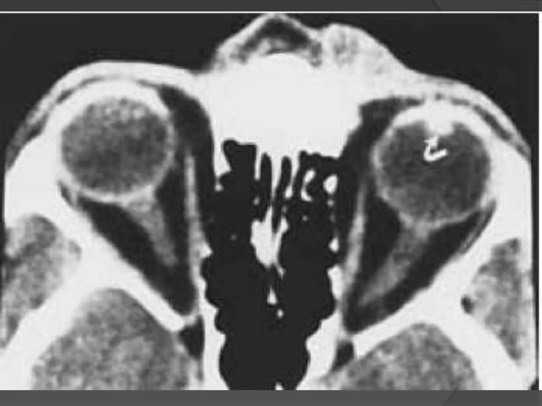
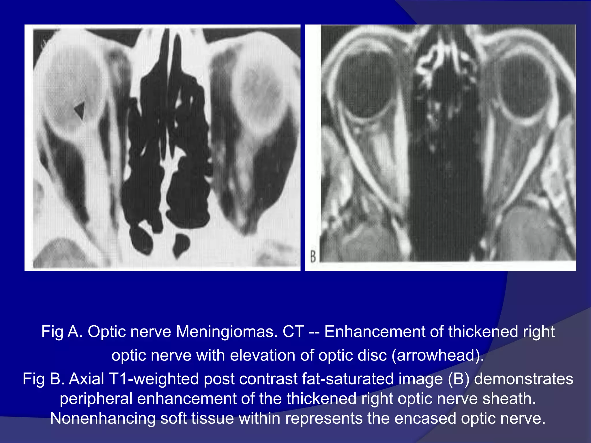
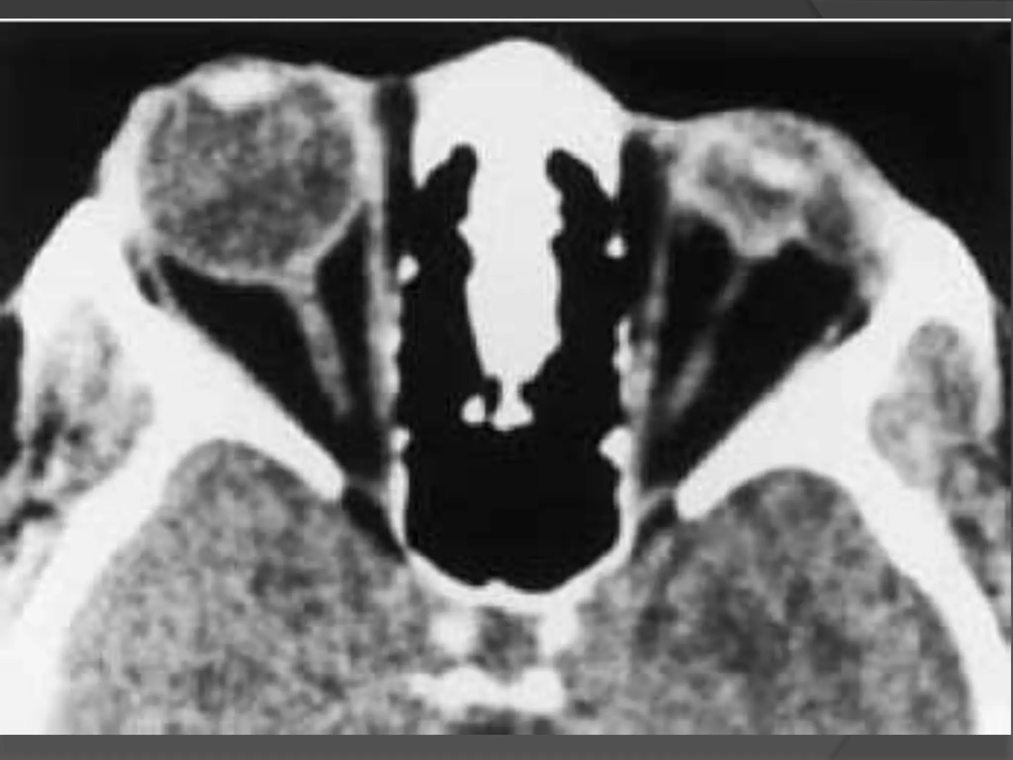
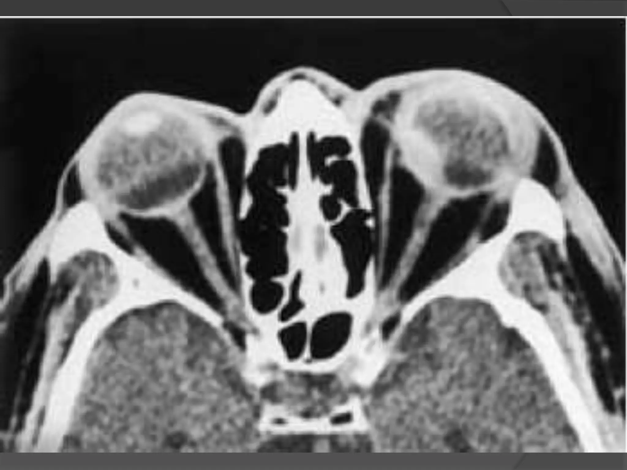
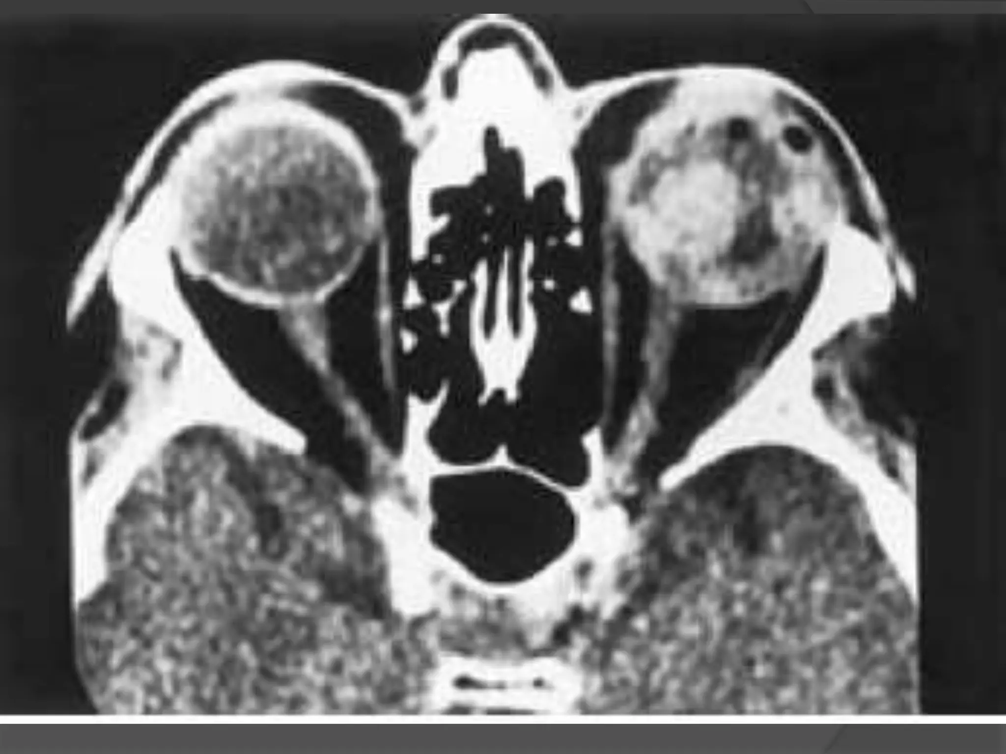
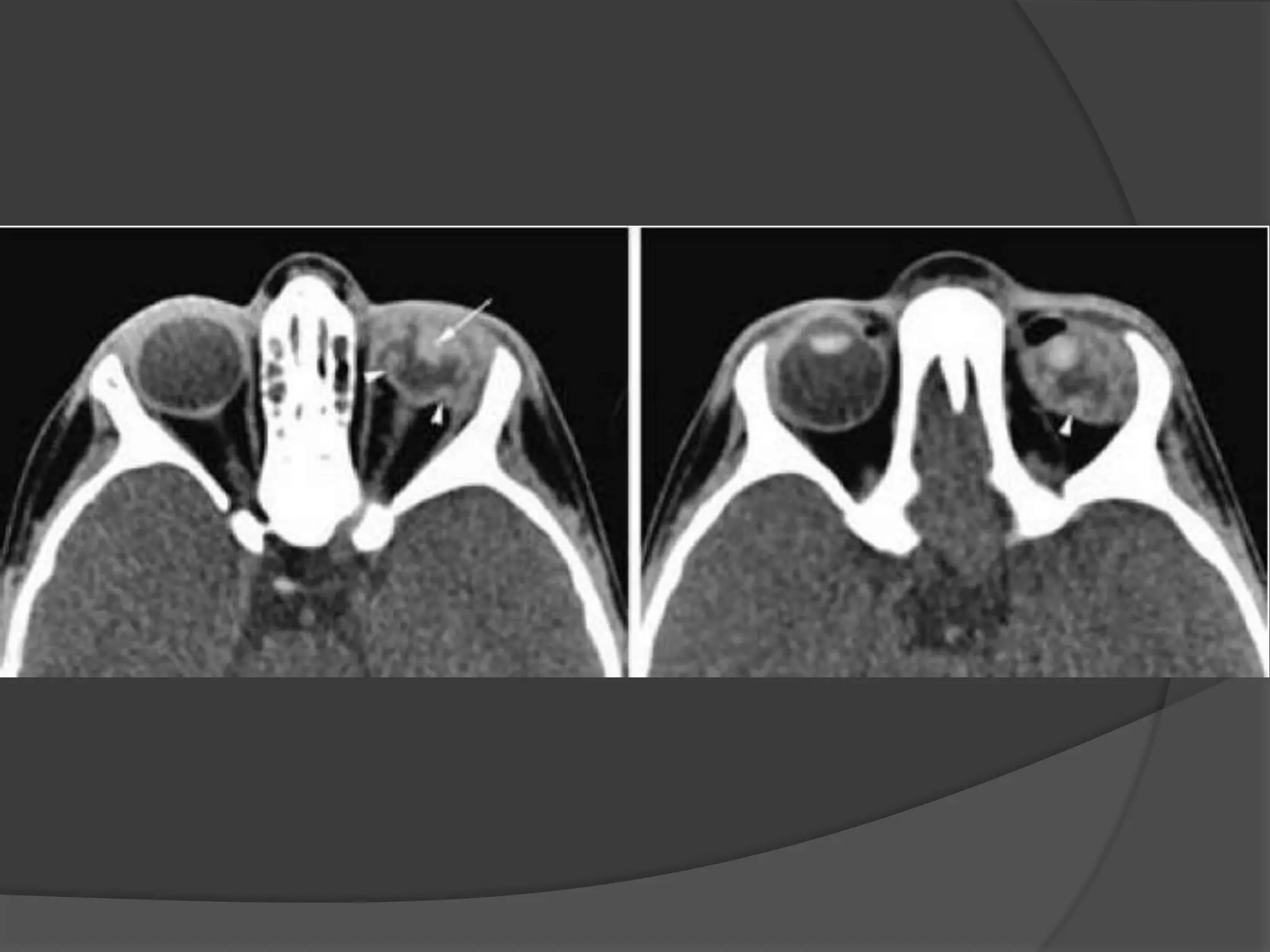
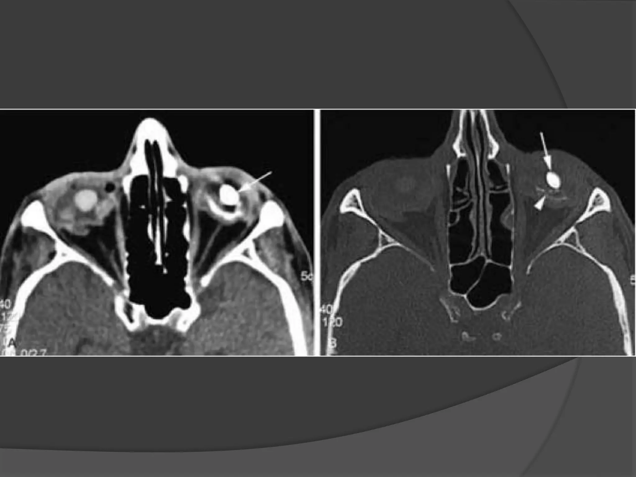
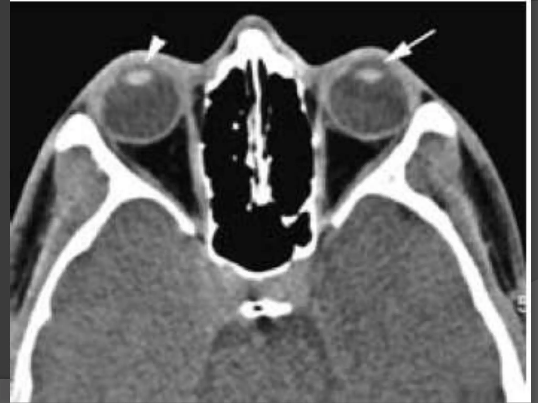
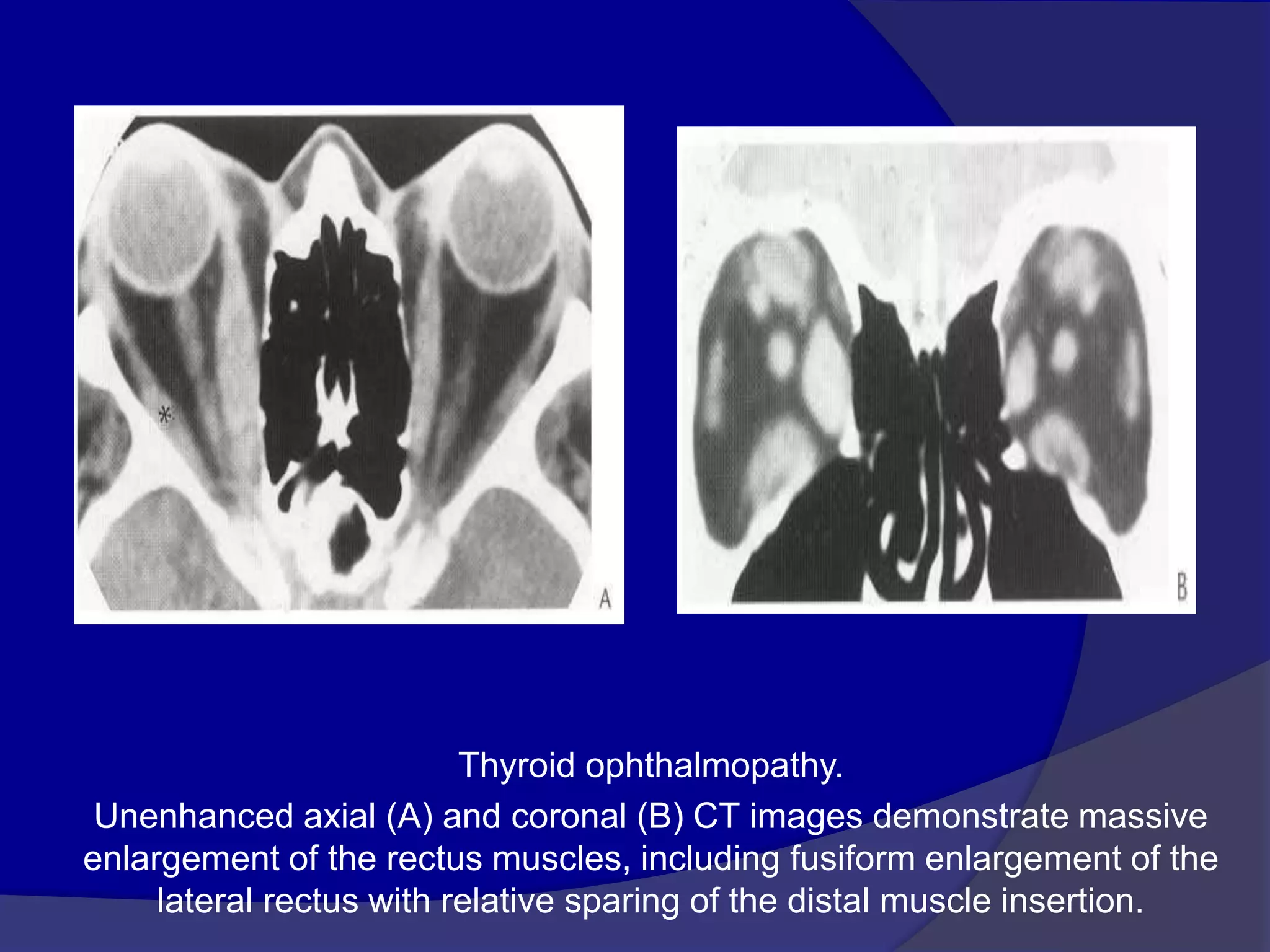
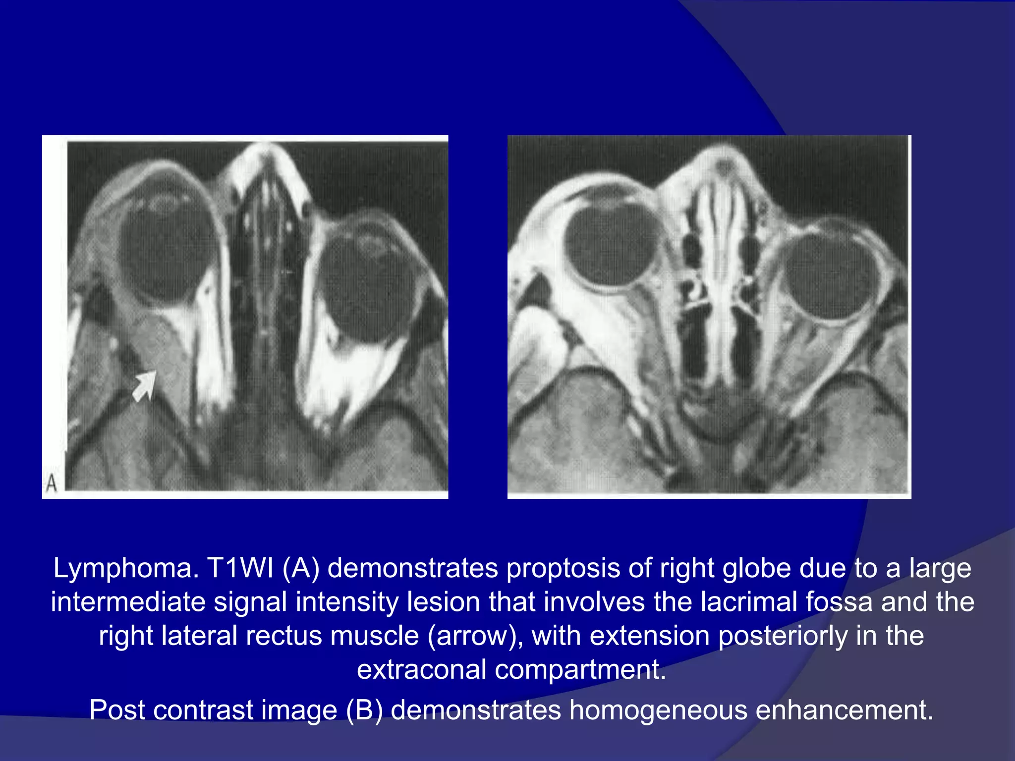
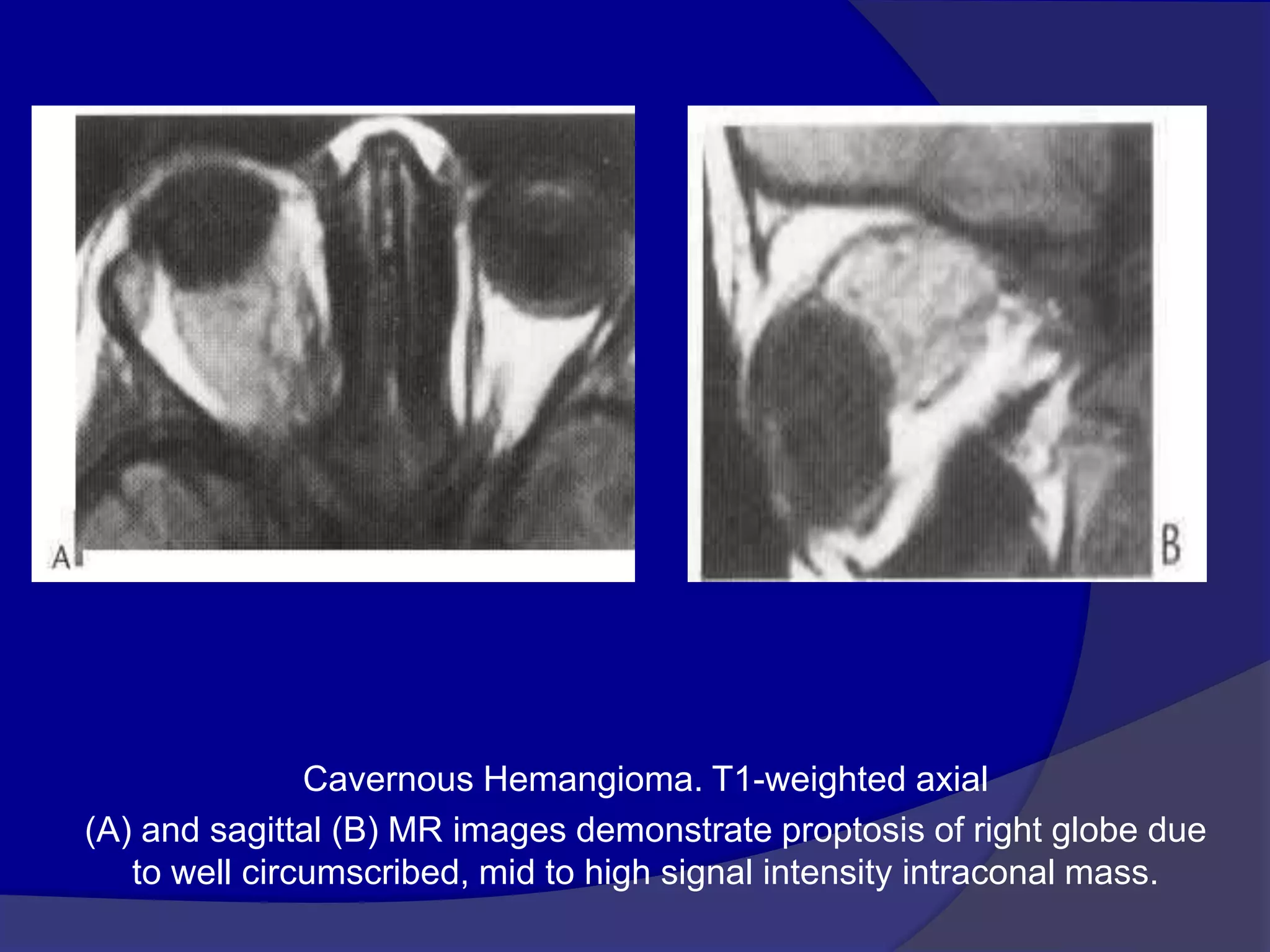
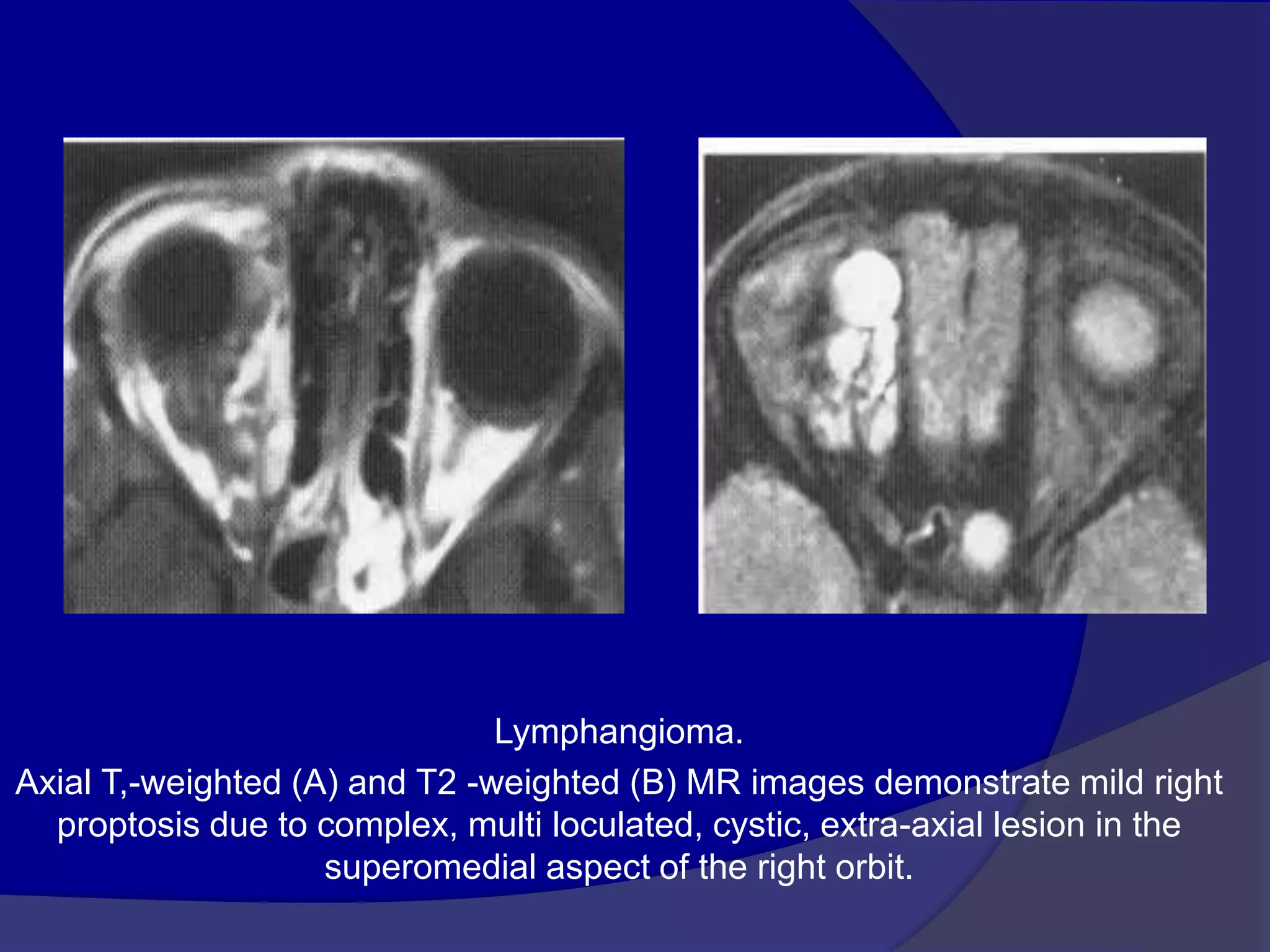
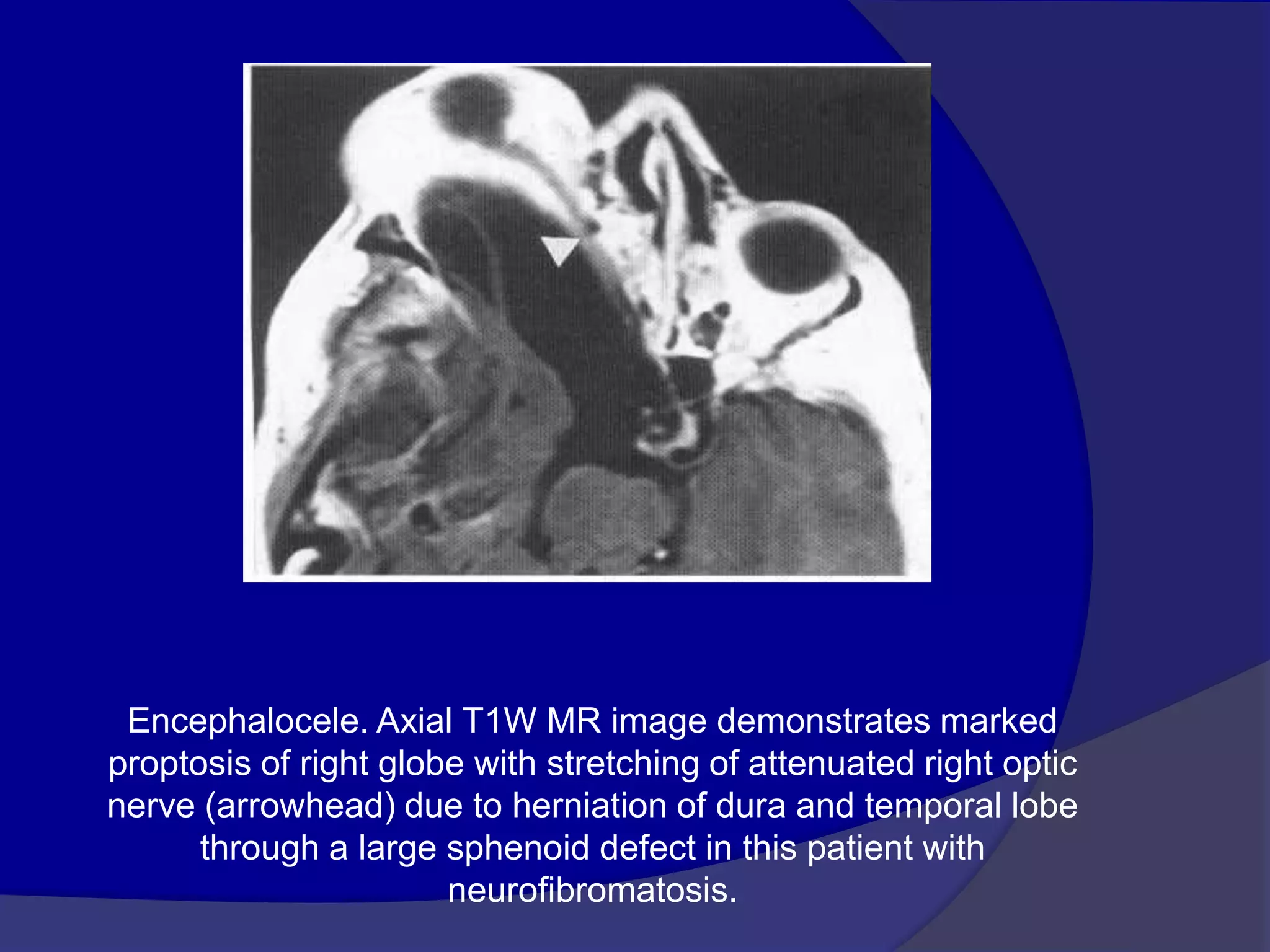
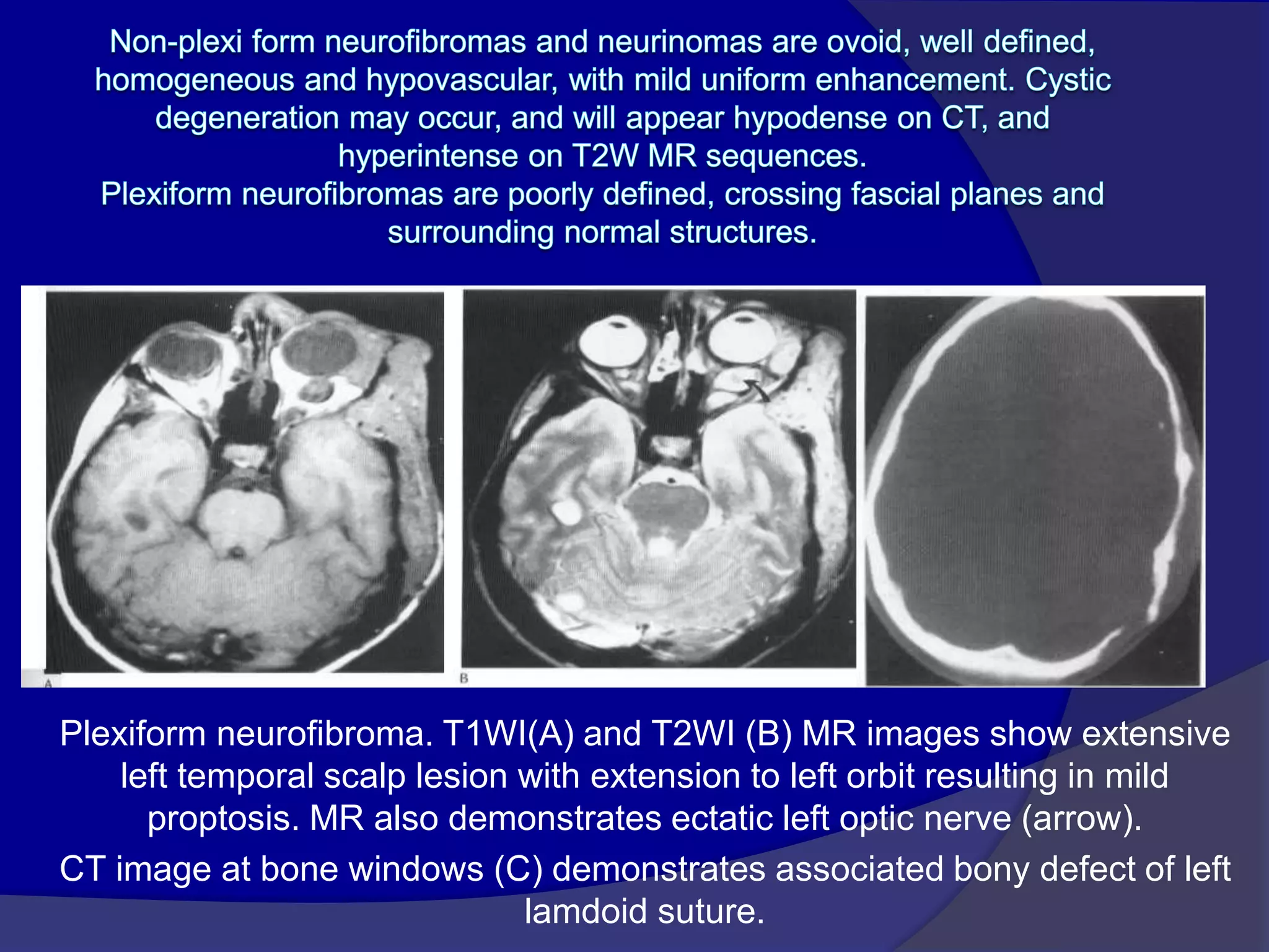
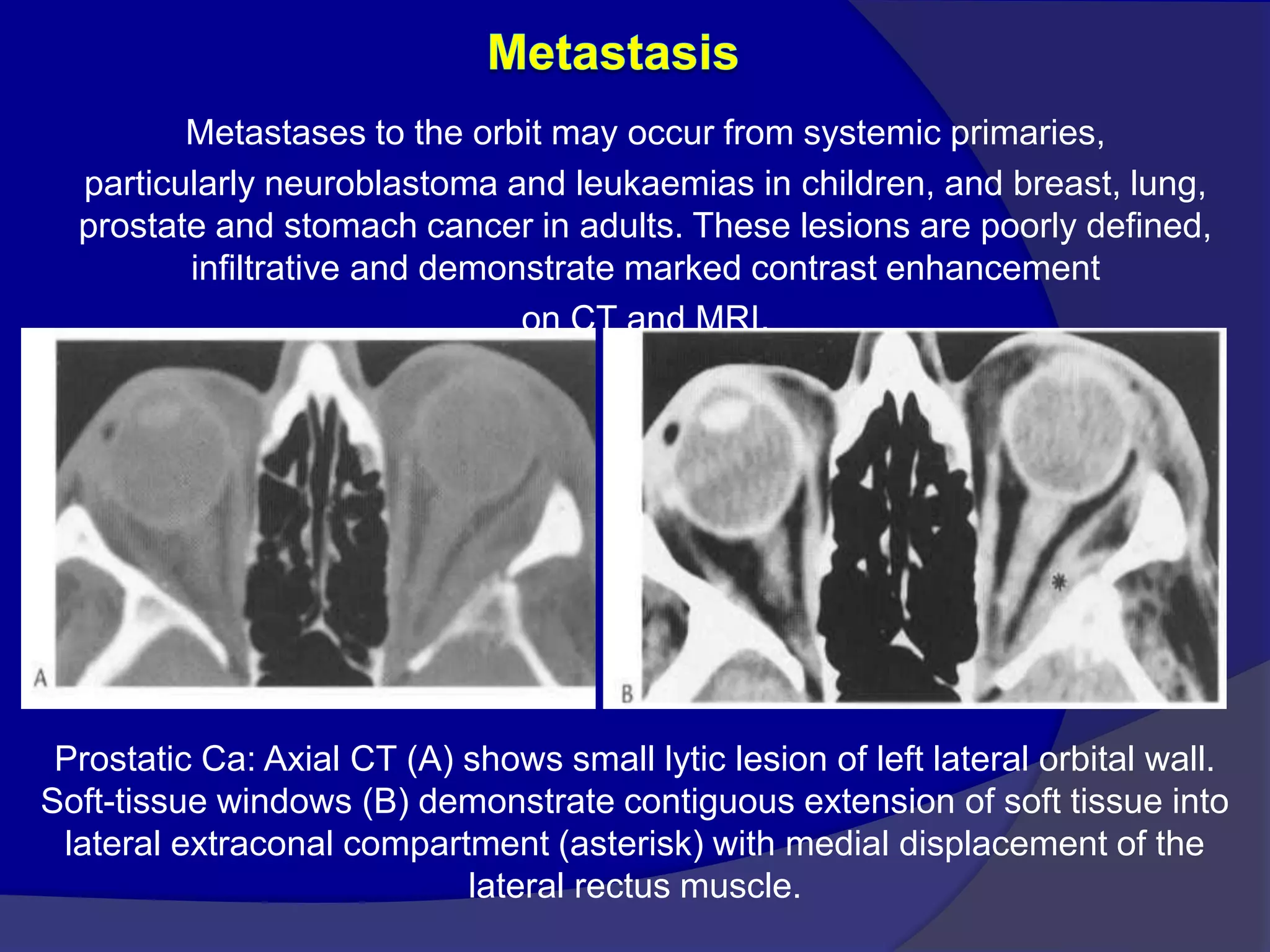
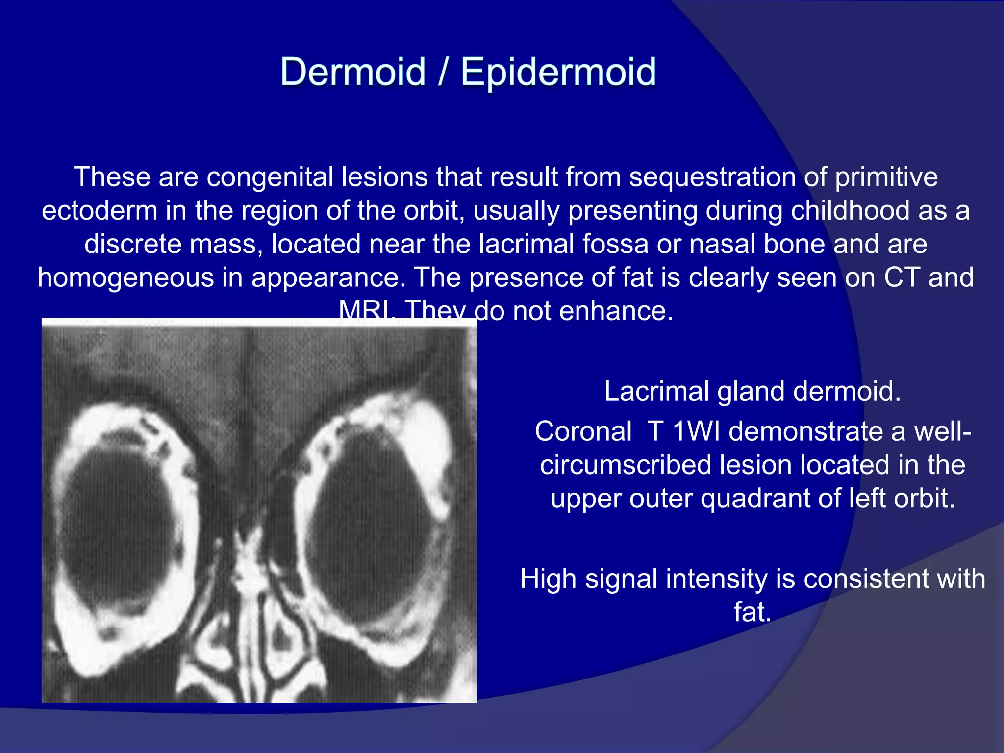
SlideShare a Scribd company logo
Dr. Mohit Goel
JR III
3/07/2014
1. Congenital anomalies
2. Infection / Inflammation
3. Tumors
4. Trauma
5. Neoplasm
Congenital anomalies
MIcropthalmia
Pthisis Bulbi
Coloboma
Colobomatous cyst
Ocular detachments
Retinal detachment.
Inflammatory conditions
Acute inflammation of optic nerve , commonly associated with multiple
sclerosis.
Edema and inflammatory cells infiltrate the nerve resulting in uniform
swelling and focal demyelination.
Imaging : MRI is the modality of choice with hyper intense signal of
T2WI due to fluid and edema. Fat Sat contrast enhanced T1WI will show
areas of demyelination. CT relatively insensitive.
Straightening and thickening
of right optic nerve.
Optic Neuritis. CE Fat Sat T1W axial
(B) MR images demonstrate subtle enlargement and enhancement of the left
optic nerve (curved arrow).
T 2 WI (C) demonstrates corresponding increased signal intensity (straight
arrow).
Retinoblastoma
Retinopathy of Prematurity
Coat’s disease
 Primary retinal telengiectasis
 Vascular anomaly of retina
 Characterized by idiopathic retinal
telengiectatic and aneurysmal retinal vessels
 Progressive deposition of intraretinal and
subretinal exudates
 Leads to massive exudative retinal
detachment
Ocular astrocytic Hamartoma
 Benign yellow-white rare retinal tumor
 Associated with Tuberous Sclerosis or
Neurofibromatosis
 Early on it may look exactly like retinoblastoma
 May involve retina or optic nerve
Juvenile Xanthogranuloma
 Benign cutaneous disorder
 Affects eye and skin
 Affects iris and ciliary body, choroid, retina
and optic nerve
 May present as a solitary orbital mass
Optic nerve head drusen
Tumors
Choroidal osteoma
 Benign tumor
 Unilateral usually
 Affects young white girls
 Patients present with painless progressive
loss of vision
Malignant uveal melanoma
 More common in whites
 May arise from pre-existing nevi
 Metastasizes hematogeneously to liver
Orbital Pseudotumor
• The most common cause of orbital mass in adults.
• Acute form presents with pain, proptosis and
diminished ocular mobility, with histological changes
similar to vasculitis.
• Chronic form may mimic infection or lymphoma both
clinically and histologically. Unilateral presentation is
most common, but findings can be bilateral. All
compartments of the orbit may be affected.
• Imaging -- Heterogeneous poorly marginated
increased CT density and decreased T1 & T2
MRI signal intensity within the intraconal fat
surrounding a thickened sclera or enlarged optic
nerve, sometimes simulating a mass.
• The lacrimal gland may be enlarged.
Enhancement occurs following contrast infusion.
Fig A. Scleral pseudotumour. Marked thickening and irregularity of
the sclera of the right globe involves the adjacent retro-orbital fat.
Fig B. Diffuse pseudotumour. Axial MR T1WI showing a diffuse mass
in the right orbit due to pseudotumour.
Rhabdomyosarcoma
• Rhabdomyosarcoma is the most common primary
orbital malignancy in the pediatric age group. with
most patients presenting below 6 years of age.
• Patients present with rapidly progressive
exophthalmos that may mimic orbital infection.
Spread of the tumor esp. intracranially, portends
poor prognosis.
• Imaging : Both CT and MRI will typically show a
mass involving an extra ocular muscle. Lesions
are isodense on CT and isointense on T1WI
when compared to muscle.
• There may be associated bony destruction and
contiguous extra orbital spread. The tumor
involves the globe less often. Marked
enhancement throughout the mass is seen after
contrast administration.
Rhabdomyosarcoma. CECT image
(A) Orbits demonstrates right proptosis due to large, lobular, intraorbital
mass.
(B) Image at lower level demonstrates invasion of right maxillary
sinus (asterisk) as well as extension through lateral orbital wall (arrow),
consistent with the aggressive nature of this tumor.
• Patients with abnormalities of the optic nerve and
its covering present with proptosis, visual loss and
papilloedema.
• Expansion of the tubular shaped optic nerve and
sheath is well demonstrated on CT and MRI.
• Imaging of the intracranial space is required
because the optic nerve and its coverings are
continuous with the brain and dura mater.
Occur in children
Are low grade astrocytomas.
Associated with NF-1
Imaging:- optic nerve may expand uniformly and diffusely. Plain
films will show asymmetric widening of the optic canal. Post
contrast show uniform enhancement.
Optic nerve glioma.
Enhanced coronal CT image
demonstrates homogeneous
enhancement of enlarged right
optic nerve.
Optic nerve glioma. Enhanced fat-saturated axial T1W image (A)
demonstrates mild enhancement and enlargement of intraorbital
and canalicular segments of left optic nerve.
Coronal image (B) confirms enlargement of nerve and surrounding perioptic
space.
• Meningiomas are dense fibrous tumors. Calcification is
common.
• Plain films may show widening of the optic canal, or
hyperostosis of the sphenoid wing.
• CT generally shows a dense, sharply defined tubular mass
surrounding and paralleling the course of the optic nerve, with
marked enhancement after contrast administration ('tram
track'). Kinking of the nerve may be seen.
• MRI will show a homogeneous mass of decreased T1 and T2
signal intensity, with strong enhancement, especially with the
use of fat suppression sequences.
• Coronal images are preferred, as the encased optic nerve will
be seen in relief against the densely enhanced tumor.
Fig A. Optic nerve Meningiomas. CT -- Enhancement of thickened right
optic nerve with elevation of optic disc (arrowhead).
Fig B. Axial T1-weighted post contrast fat-saturated image (B) demonstrates
peripheral enhancement of the thickened right optic nerve sheath.
Nonenhancing soft tissue within represents the encased optic nerve.
Trauma
Miscellaneous
Thyroid ophthalmopathy.
Unenhanced axial (A) and coronal (B) CT images demonstrate massive
enlargement of the rectus muscles, including fusiform enlargement of the
lateral rectus with relative sparing of the distal muscle insertion.
Lymphoma. T1WI (A) demonstrates proptosis of right globe due to a large
intermediate signal intensity lesion that involves the lacrimal fossa and the
right lateral rectus muscle (arrow), with extension posteriorly in the
extraconal compartment.
Post contrast image (B) demonstrates homogeneous enhancement.
Cavernous Hemangioma. T1-weighted axial
(A) and sagittal (B) MR images demonstrate proptosis of right globe due
to well circumscribed, mid to high signal intensity intraconal mass.
Lymphangioma.
Axial T,-weighted (A) and T2 -weighted (B) MR images demonstrate mild right
proptosis due to complex, multi loculated, cystic, extra-axial lesion in the
superomedial aspect of the right orbit.
Encephalocele. Axial T1W MR image demonstrates marked
proptosis of right globe with stretching of attenuated right optic
nerve (arrowhead) due to herniation of dura and temporal lobe
through a large sphenoid defect in this patient with
neurofibromatosis.
Plexiform neurofibroma. T1WI(A) and T2WI (B) MR images show extensive
left temporal scalp lesion with extension to left orbit resulting in mild
proptosis. MR also demonstrates ectatic left optic nerve (arrow).
CT image at bone windows (C) demonstrates associated bony defect of left
lamdoid suture.
Metastases to the orbit may occur from systemic primaries,
particularly neuroblastoma and leukaemias in children, and breast, lung,
prostate and stomach cancer in adults. These lesions are poorly defined,
infiltrative and demonstrate marked contrast enhancement
on CT and MRI.
Prostatic Ca: Axial CT (A) shows small lytic lesion of left lateral orbital wall.
Soft-tissue windows (B) demonstrate contiguous extension of soft tissue into
lateral extraconal compartment (asterisk) with medial displacement of the
lateral rectus muscle.
These are congenital lesions that result from sequestration of primitive
ectoderm in the region of the orbit, usually presenting during childhood as a
discrete mass, located near the lacrimal fossa or nasal bone and are
homogeneous in appearance. The presence of fat is clearly seen on CT and
MRI. They do not enhance.
Lacrimal gland dermoid.
Coronal T 1WI demonstrate a well-
circumscribed lesion located in the
upper outer quadrant of left orbit.
High signal intensity is consistent with
fat.
Thank You

More Related Content

What's hot

idiopathic orbital inflammatory syndrome
idiopathic orbital inflammatory syndromeidiopathic orbital inflammatory syndrome
idiopathic orbital inflammatory syndrome
NeurologyKota
 
Imaging in Ocular Pathologies
Imaging in Ocular PathologiesImaging in Ocular Pathologies
Imaging in Ocular Pathologies
Sahil Chaudhry
 
Orbital tumours
Orbital tumoursOrbital tumours
Orbital tumours
airwave12
 
Imaging neurology spotters
Imaging   neurology spottersImaging   neurology spotters
Imaging neurology spotters
NeurologyKota
 
Radiology Spotters
Radiology Spotters Radiology Spotters
Radiology Spotters
Anish Choudhary
 
Diagnostic Imaging of Orbital Lesions
Diagnostic Imaging of Orbital LesionsDiagnostic Imaging of Orbital Lesions
Diagnostic Imaging of Orbital Lesions
Mohamed M.A. Zaitoun
 
Imaging in neuro ophthalmology & revisting orbital imaging.2012 (1) (1)
Imaging in neuro ophthalmology & revisting orbital imaging.2012 (1) (1)Imaging in neuro ophthalmology & revisting orbital imaging.2012 (1) (1)
Imaging in neuro ophthalmology & revisting orbital imaging.2012 (1) (1)Dr. Himadri Sikhor Das
 
Ultrasonography of eye
Ultrasonography of eyeUltrasonography of eye
Ultrasonography of eye
Nikita Jaiswal
 
Presentation1.pptx, radiological imaging of skeletal dysplasia
Presentation1.pptx, radiological imaging of skeletal dysplasiaPresentation1.pptx, radiological imaging of skeletal dysplasia
Presentation1.pptx, radiological imaging of skeletal dysplasiaAbdellah Nazeer
 
Orbital pathologies.pptx (part 1)
Orbital pathologies.pptx (part 1)Orbital pathologies.pptx (part 1)
Orbital pathologies.pptx (part 1)
Dr. Mohit Goel
 
Fuchs endothelial dystrophy.
Fuchs endothelial dystrophy.Fuchs endothelial dystrophy.
Fuchs endothelial dystrophy.
DiyarAlzubaidy
 
Keratoconus...
Keratoconus...Keratoconus...
Keratoconus...
Reema Dandavate
 
Imaging of eye and orbit
Imaging of eye and orbitImaging of eye and orbit
Imaging of eye and orbit
Shrikant Nagare
 
USG B Scan
USG B ScanUSG B Scan
Ophthalmic ultrasonography
Ophthalmic ultrasonographyOphthalmic ultrasonography
Ophthalmic ultrasonography
kajal bhagat
 
RADIOLOGY SPOTTERS GIT
RADIOLOGY SPOTTERS GITRADIOLOGY SPOTTERS GIT
RADIOLOGY SPOTTERS GIT
Anish Choudhary
 
Ultrasound of eye - B scan
Ultrasound of eye - B scan Ultrasound of eye - B scan
Ultrasound of eye - B scan
Shruti Laddha
 
Orbital neoplasms & malformations
Orbital neoplasms & malformationsOrbital neoplasms & malformations
Orbital neoplasms & malformations
Bipin Bista
 
Magnetic resonance imaging
Magnetic resonance imagingMagnetic resonance imaging
Magnetic resonance imaging
Mutahir Shah
 

What's hot (20)

idiopathic orbital inflammatory syndrome
idiopathic orbital inflammatory syndromeidiopathic orbital inflammatory syndrome
idiopathic orbital inflammatory syndrome
 
Imaging in Ocular Pathologies
Imaging in Ocular PathologiesImaging in Ocular Pathologies
Imaging in Ocular Pathologies
 
Orbital tumours
Orbital tumoursOrbital tumours
Orbital tumours
 
Imaging neurology spotters
Imaging   neurology spottersImaging   neurology spotters
Imaging neurology spotters
 
Radiology Spotters
Radiology Spotters Radiology Spotters
Radiology Spotters
 
Diagnostic Imaging of Orbital Lesions
Diagnostic Imaging of Orbital LesionsDiagnostic Imaging of Orbital Lesions
Diagnostic Imaging of Orbital Lesions
 
Ct orbit
Ct orbitCt orbit
Ct orbit
 
Imaging in neuro ophthalmology & revisting orbital imaging.2012 (1) (1)
Imaging in neuro ophthalmology & revisting orbital imaging.2012 (1) (1)Imaging in neuro ophthalmology & revisting orbital imaging.2012 (1) (1)
Imaging in neuro ophthalmology & revisting orbital imaging.2012 (1) (1)
 
Ultrasonography of eye
Ultrasonography of eyeUltrasonography of eye
Ultrasonography of eye
 
Presentation1.pptx, radiological imaging of skeletal dysplasia
Presentation1.pptx, radiological imaging of skeletal dysplasiaPresentation1.pptx, radiological imaging of skeletal dysplasia
Presentation1.pptx, radiological imaging of skeletal dysplasia
 
Orbital pathologies.pptx (part 1)
Orbital pathologies.pptx (part 1)Orbital pathologies.pptx (part 1)
Orbital pathologies.pptx (part 1)
 
Fuchs endothelial dystrophy.
Fuchs endothelial dystrophy.Fuchs endothelial dystrophy.
Fuchs endothelial dystrophy.
 
Keratoconus...
Keratoconus...Keratoconus...
Keratoconus...
 
Imaging of eye and orbit
Imaging of eye and orbitImaging of eye and orbit
Imaging of eye and orbit
 
USG B Scan
USG B ScanUSG B Scan
USG B Scan
 
Ophthalmic ultrasonography
Ophthalmic ultrasonographyOphthalmic ultrasonography
Ophthalmic ultrasonography
 
RADIOLOGY SPOTTERS GIT
RADIOLOGY SPOTTERS GITRADIOLOGY SPOTTERS GIT
RADIOLOGY SPOTTERS GIT
 
Ultrasound of eye - B scan
Ultrasound of eye - B scan Ultrasound of eye - B scan
Ultrasound of eye - B scan
 
Orbital neoplasms & malformations
Orbital neoplasms & malformationsOrbital neoplasms & malformations
Orbital neoplasms & malformations
 
Magnetic resonance imaging
Magnetic resonance imagingMagnetic resonance imaging
Magnetic resonance imaging
 

Similar to Orbital pathologies.pptx 1

MRI of Spine and very easy details of sp
MRI of Spine and very easy details of spMRI of Spine and very easy details of sp
MRI of Spine and very easy details of sp
ssuserc66686
 
23204928
2320492823204928
23204928
radgirl
 
RAJ 22.pptx
RAJ 22.pptxRAJ 22.pptx
RAJ 22.pptx
RadiologyReports
 
Presentation1, radiological imaging of intra cranial dermoid tumours.
Presentation1, radiological imaging of intra cranial dermoid tumours.Presentation1, radiological imaging of intra cranial dermoid tumours.
Presentation1, radiological imaging of intra cranial dermoid tumours.
Abdellah Nazeer
 
Imaging in musculoskeletal complications of AIDS
Imaging in musculoskeletal complications of AIDSImaging in musculoskeletal complications of AIDS
Imaging in musculoskeletal complications of AIDS
Dr.Suhas Basavaiah
 
Presentation1.pptx, radiological imaging of spinal cord tumour.
Presentation1.pptx, radiological imaging of spinal cord tumour.Presentation1.pptx, radiological imaging of spinal cord tumour.
Presentation1.pptx, radiological imaging of spinal cord tumour.Abdellah Nazeer
 
Spinal tumors drsant91
Spinal tumors drsant91Spinal tumors drsant91
Spinal tumors drsant91
Santosh Prasad
 
Patterns of Enhancement in the Brain
Patterns of Enhancement in the BrainPatterns of Enhancement in the Brain
Patterns of Enhancement in the Brain
Mohamed M.A. Zaitoun
 
Presentation2, radiological imaging of phakomatosis.
Presentation2, radiological imaging of phakomatosis.Presentation2, radiological imaging of phakomatosis.
Presentation2, radiological imaging of phakomatosis.
Abdellah Nazeer
 
Presentation1, radiological imaging of popliteal fossa masses.
Presentation1, radiological imaging of popliteal fossa masses.Presentation1, radiological imaging of popliteal fossa masses.
Presentation1, radiological imaging of popliteal fossa masses.
Abdellah Nazeer
 
Spinal tuberculosis
Spinal tuberculosisSpinal tuberculosis
Spinal tuberculosis
NeurologyKota
 
Presentation1.pptx, radiological imaging of the nasopharyngeal diseases.
Presentation1.pptx, radiological imaging of the nasopharyngeal diseases.Presentation1.pptx, radiological imaging of the nasopharyngeal diseases.
Presentation1.pptx, radiological imaging of the nasopharyngeal diseases.Abdellah Nazeer
 
29 orbital masses not involving the optic nerve
29 orbital masses not involving the optic nerve29 orbital masses not involving the optic nerve
29 orbital masses not involving the optic nerve
Dr. Muhammad Bin Zulfiqar
 
Imaging cns tb
Imaging   cns tbImaging   cns tb
Imaging cns tb
NeurologyKota
 
Cns infections radiology.
Cns infections radiology.Cns infections radiology.
Cns infections radiology.Raeez Basheer
 
INVESTIGATIONS OF PAROTID GLAND TUMOUR.pptx
INVESTIGATIONS OF PAROTID GLAND TUMOUR.pptxINVESTIGATIONS OF PAROTID GLAND TUMOUR.pptx
INVESTIGATIONS OF PAROTID GLAND TUMOUR.pptx
Pratik Jugnake
 
INVESTIGATIONS OF PAROTID GLAND TUMOUR.pptx
INVESTIGATIONS OF PAROTID GLAND TUMOUR.pptxINVESTIGATIONS OF PAROTID GLAND TUMOUR.pptx
INVESTIGATIONS OF PAROTID GLAND TUMOUR.pptx
Pratik Jugnake
 
CNS Rapid Review of Radiology
CNS Rapid Review of RadiologyCNS Rapid Review of Radiology
CNS Rapid Review of Radiology
Double M
 
Muculoskeletal Pediatic Imaging..pptx
Muculoskeletal Pediatic Imaging..pptxMuculoskeletal Pediatic Imaging..pptx
Muculoskeletal Pediatic Imaging..pptx
Abdellah Nazeer
 

Similar to Orbital pathologies.pptx 1 (20)

MRI of Spine and very easy details of sp
MRI of Spine and very easy details of spMRI of Spine and very easy details of sp
MRI of Spine and very easy details of sp
 
23204928
2320492823204928
23204928
 
RAJ 22.pptx
RAJ 22.pptxRAJ 22.pptx
RAJ 22.pptx
 
Presentation1, radiological imaging of intra cranial dermoid tumours.
Presentation1, radiological imaging of intra cranial dermoid tumours.Presentation1, radiological imaging of intra cranial dermoid tumours.
Presentation1, radiological imaging of intra cranial dermoid tumours.
 
Imaging in musculoskeletal complications of AIDS
Imaging in musculoskeletal complications of AIDSImaging in musculoskeletal complications of AIDS
Imaging in musculoskeletal complications of AIDS
 
Presentation1.pptx, radiological imaging of spinal cord tumour.
Presentation1.pptx, radiological imaging of spinal cord tumour.Presentation1.pptx, radiological imaging of spinal cord tumour.
Presentation1.pptx, radiological imaging of spinal cord tumour.
 
Spinal tumors drsant91
Spinal tumors drsant91Spinal tumors drsant91
Spinal tumors drsant91
 
Patterns of Enhancement in the Brain
Patterns of Enhancement in the BrainPatterns of Enhancement in the Brain
Patterns of Enhancement in the Brain
 
Presentation2, radiological imaging of phakomatosis.
Presentation2, radiological imaging of phakomatosis.Presentation2, radiological imaging of phakomatosis.
Presentation2, radiological imaging of phakomatosis.
 
Presentation1, radiological imaging of popliteal fossa masses.
Presentation1, radiological imaging of popliteal fossa masses.Presentation1, radiological imaging of popliteal fossa masses.
Presentation1, radiological imaging of popliteal fossa masses.
 
Spinal tuberculosis
Spinal tuberculosisSpinal tuberculosis
Spinal tuberculosis
 
Presentation1.pptx, radiological imaging of the nasopharyngeal diseases.
Presentation1.pptx, radiological imaging of the nasopharyngeal diseases.Presentation1.pptx, radiological imaging of the nasopharyngeal diseases.
Presentation1.pptx, radiological imaging of the nasopharyngeal diseases.
 
29 orbital masses not involving the optic nerve
29 orbital masses not involving the optic nerve29 orbital masses not involving the optic nerve
29 orbital masses not involving the optic nerve
 
Imaging cns tb
Imaging   cns tbImaging   cns tb
Imaging cns tb
 
Cns infections radiology.
Cns infections radiology.Cns infections radiology.
Cns infections radiology.
 
INVESTIGATIONS OF PAROTID GLAND TUMOUR.pptx
INVESTIGATIONS OF PAROTID GLAND TUMOUR.pptxINVESTIGATIONS OF PAROTID GLAND TUMOUR.pptx
INVESTIGATIONS OF PAROTID GLAND TUMOUR.pptx
 
INVESTIGATIONS OF PAROTID GLAND TUMOUR.pptx
INVESTIGATIONS OF PAROTID GLAND TUMOUR.pptxINVESTIGATIONS OF PAROTID GLAND TUMOUR.pptx
INVESTIGATIONS OF PAROTID GLAND TUMOUR.pptx
 
CNS Rapid Review of Radiology
CNS Rapid Review of RadiologyCNS Rapid Review of Radiology
CNS Rapid Review of Radiology
 
H&N post ttt
H&N post tttH&N post ttt
H&N post ttt
 
Muculoskeletal Pediatic Imaging..pptx
Muculoskeletal Pediatic Imaging..pptxMuculoskeletal Pediatic Imaging..pptx
Muculoskeletal Pediatic Imaging..pptx
 

More from Anish Choudhary

Pulmonary embolism radiology
Pulmonary embolism radiologyPulmonary embolism radiology
Pulmonary embolism radiology
Anish Choudhary
 
Anatomy of esophgus
Anatomy of esophgusAnatomy of esophgus
Anatomy of esophgus
Anish Choudhary
 
Malignant liver masses
Malignant liver massesMalignant liver masses
Malignant liver masses
Anish Choudhary
 
KEYS OF RADIOLOGY SPOTTERS GIT
KEYS OF RADIOLOGY SPOTTERS GITKEYS OF RADIOLOGY SPOTTERS GIT
KEYS OF RADIOLOGY SPOTTERS GIT
Anish Choudhary
 
radiology Spotters mixed bag
radiology Spotters mixed bagradiology Spotters mixed bag
radiology Spotters mixed bag
Anish Choudhary
 
Usg gb & biliary t
Usg gb & biliary tUsg gb & biliary t
Usg gb & biliary t
Anish Choudhary
 
Ultrasoud hernia
Ultrasoud herniaUltrasoud hernia
Ultrasoud hernia
Anish Choudhary
 
Tvs image gallery
Tvs image galleryTvs image gallery
Tvs image gallery
Anish Choudhary
 
Treat. contrast reaction
Treat. contrast reactionTreat. contrast reaction
Treat. contrast reaction
Anish Choudhary
 
Transitional vertebrae
Transitional vertebraeTransitional vertebrae
Transitional vertebrae
Anish Choudhary
 
Spots with keys
Spots with keysSpots with keys
Spots with keys
Anish Choudhary
 
Spots with keys (2)
Spots with keys (2)Spots with keys (2)
Spots with keys (2)
Anish Choudhary
 
Shoulder ultrasound
Shoulder ultrasoundShoulder ultrasound
Shoulder ultrasound
Anish Choudhary
 
Sectional anatomy of abdomen
Sectional anatomy of abdomenSectional anatomy of abdomen
Sectional anatomy of abdomen
Anish Choudhary
 
Retroperitoneal masses
Retroperitoneal massesRetroperitoneal masses
Retroperitoneal masses
Anish Choudhary
 
Renal doppler
Renal dopplerRenal doppler
Renal doppler
Anish Choudhary
 
Precocious puberty
Precocious pubertyPrecocious puberty
Precocious puberty
Anish Choudhary
 
Pre fess pns ct
Pre fess pns ctPre fess pns ct
Pre fess pns ct
Anish Choudhary
 
Pineal region masses
Pineal region massesPineal region masses
Pineal region masses
Anish Choudhary
 
Peripheral arterial doppler
Peripheral  arterial dopplerPeripheral  arterial doppler
Peripheral arterial doppler
Anish Choudhary
 

More from Anish Choudhary (20)

Pulmonary embolism radiology
Pulmonary embolism radiologyPulmonary embolism radiology
Pulmonary embolism radiology
 
Anatomy of esophgus
Anatomy of esophgusAnatomy of esophgus
Anatomy of esophgus
 
Malignant liver masses
Malignant liver massesMalignant liver masses
Malignant liver masses
 
KEYS OF RADIOLOGY SPOTTERS GIT
KEYS OF RADIOLOGY SPOTTERS GITKEYS OF RADIOLOGY SPOTTERS GIT
KEYS OF RADIOLOGY SPOTTERS GIT
 
radiology Spotters mixed bag
radiology Spotters mixed bagradiology Spotters mixed bag
radiology Spotters mixed bag
 
Usg gb & biliary t
Usg gb & biliary tUsg gb & biliary t
Usg gb & biliary t
 
Ultrasoud hernia
Ultrasoud herniaUltrasoud hernia
Ultrasoud hernia
 
Tvs image gallery
Tvs image galleryTvs image gallery
Tvs image gallery
 
Treat. contrast reaction
Treat. contrast reactionTreat. contrast reaction
Treat. contrast reaction
 
Transitional vertebrae
Transitional vertebraeTransitional vertebrae
Transitional vertebrae
 
Spots with keys
Spots with keysSpots with keys
Spots with keys
 
Spots with keys (2)
Spots with keys (2)Spots with keys (2)
Spots with keys (2)
 
Shoulder ultrasound
Shoulder ultrasoundShoulder ultrasound
Shoulder ultrasound
 
Sectional anatomy of abdomen
Sectional anatomy of abdomenSectional anatomy of abdomen
Sectional anatomy of abdomen
 
Retroperitoneal masses
Retroperitoneal massesRetroperitoneal masses
Retroperitoneal masses
 
Renal doppler
Renal dopplerRenal doppler
Renal doppler
 
Precocious puberty
Precocious pubertyPrecocious puberty
Precocious puberty
 
Pre fess pns ct
Pre fess pns ctPre fess pns ct
Pre fess pns ct
 
Pineal region masses
Pineal region massesPineal region masses
Pineal region masses
 
Peripheral arterial doppler
Peripheral  arterial dopplerPeripheral  arterial doppler
Peripheral arterial doppler
 

Recently uploaded

Pharynx and Clinical Correlations BY Dr.Rabia Inam Gandapore.pptx
Pharynx and Clinical Correlations BY Dr.Rabia Inam Gandapore.pptxPharynx and Clinical Correlations BY Dr.Rabia Inam Gandapore.pptx
Pharynx and Clinical Correlations BY Dr.Rabia Inam Gandapore.pptx
Dr. Rabia Inam Gandapore
 
heat stroke and heat exhaustion in children
heat stroke and heat exhaustion in childrenheat stroke and heat exhaustion in children
heat stroke and heat exhaustion in children
SumeraAhmad5
 
Physiology of Special Chemical Sensation of Taste
Physiology of Special Chemical Sensation of TastePhysiology of Special Chemical Sensation of Taste
Physiology of Special Chemical Sensation of Taste
MedicoseAcademics
 
ACUTE SCROTUM.....pdf. ACUTE SCROTAL CONDITIOND
ACUTE SCROTUM.....pdf. ACUTE SCROTAL CONDITIONDACUTE SCROTUM.....pdf. ACUTE SCROTAL CONDITIOND
ACUTE SCROTUM.....pdf. ACUTE SCROTAL CONDITIOND
DR SETH JOTHAM
 
HOT NEW PRODUCT! BIG SALES FAST SHIPPING NOW FROM CHINA!! EU KU DB BK substit...
HOT NEW PRODUCT! BIG SALES FAST SHIPPING NOW FROM CHINA!! EU KU DB BK substit...HOT NEW PRODUCT! BIG SALES FAST SHIPPING NOW FROM CHINA!! EU KU DB BK substit...
HOT NEW PRODUCT! BIG SALES FAST SHIPPING NOW FROM CHINA!! EU KU DB BK substit...
GL Anaacs
 
New Directions in Targeted Therapeutic Approaches for Older Adults With Mantl...
New Directions in Targeted Therapeutic Approaches for Older Adults With Mantl...New Directions in Targeted Therapeutic Approaches for Older Adults With Mantl...
New Directions in Targeted Therapeutic Approaches for Older Adults With Mantl...
i3 Health
 
Surat @ℂall @Girls ꧁❤8527049040❤꧂@ℂall @Girls Service Vip Top Model Safe
Surat @ℂall @Girls ꧁❤8527049040❤꧂@ℂall @Girls Service Vip Top Model SafeSurat @ℂall @Girls ꧁❤8527049040❤꧂@ℂall @Girls Service Vip Top Model Safe
Surat @ℂall @Girls ꧁❤8527049040❤꧂@ℂall @Girls Service Vip Top Model Safe
Savita Shen $i11
 
Cervical & Brachial Plexus By Dr. RIG.pptx
Cervical & Brachial Plexus By Dr. RIG.pptxCervical & Brachial Plexus By Dr. RIG.pptx
Cervical & Brachial Plexus By Dr. RIG.pptx
Dr. Rabia Inam Gandapore
 
Flu Vaccine Alert in Bangalore Karnataka
Flu Vaccine Alert in Bangalore KarnatakaFlu Vaccine Alert in Bangalore Karnataka
Flu Vaccine Alert in Bangalore Karnataka
addon Scans
 
micro teaching on communication m.sc nursing.pdf
micro teaching on communication m.sc nursing.pdfmicro teaching on communication m.sc nursing.pdf
micro teaching on communication m.sc nursing.pdf
Anurag Sharma
 
Lung Cancer: Artificial Intelligence, Synergetics, Complex System Analysis, S...
Lung Cancer: Artificial Intelligence, Synergetics, Complex System Analysis, S...Lung Cancer: Artificial Intelligence, Synergetics, Complex System Analysis, S...
Lung Cancer: Artificial Intelligence, Synergetics, Complex System Analysis, S...
Oleg Kshivets
 
Superficial & Deep Fascia of the NECK.pptx
Superficial & Deep Fascia of the NECK.pptxSuperficial & Deep Fascia of the NECK.pptx
Superficial & Deep Fascia of the NECK.pptx
Dr. Rabia Inam Gandapore
 
Non-respiratory Functions of the Lungs.pdf
Non-respiratory Functions of the Lungs.pdfNon-respiratory Functions of the Lungs.pdf
Non-respiratory Functions of the Lungs.pdf
MedicoseAcademics
 
POST OPERATIVE OLIGURIA and its management
POST OPERATIVE OLIGURIA and its managementPOST OPERATIVE OLIGURIA and its management
POST OPERATIVE OLIGURIA and its management
touseefaziz1
 
How STIs Influence the Development of Pelvic Inflammatory Disease.pptx
How STIs Influence the Development of Pelvic Inflammatory Disease.pptxHow STIs Influence the Development of Pelvic Inflammatory Disease.pptx
How STIs Influence the Development of Pelvic Inflammatory Disease.pptx
FFragrant
 
BENIGN PROSTATIC HYPERPLASIA.BPH. BPHpdf
BENIGN PROSTATIC HYPERPLASIA.BPH. BPHpdfBENIGN PROSTATIC HYPERPLASIA.BPH. BPHpdf
BENIGN PROSTATIC HYPERPLASIA.BPH. BPHpdf
DR SETH JOTHAM
 
Report Back from SGO 2024: What’s the Latest in Cervical Cancer?
Report Back from SGO 2024: What’s the Latest in Cervical Cancer?Report Back from SGO 2024: What’s the Latest in Cervical Cancer?
Report Back from SGO 2024: What’s the Latest in Cervical Cancer?
bkling
 
Ozempic: Preoperative Management of Patients on GLP-1 Receptor Agonists
Ozempic: Preoperative Management of Patients on GLP-1 Receptor Agonists  Ozempic: Preoperative Management of Patients on GLP-1 Receptor Agonists
Ozempic: Preoperative Management of Patients on GLP-1 Receptor Agonists
Saeid Safari
 
basicmodesofventilation2022-220313203758.pdf
basicmodesofventilation2022-220313203758.pdfbasicmodesofventilation2022-220313203758.pdf
basicmodesofventilation2022-220313203758.pdf
aljamhori teaching hospital
 
Ophthalmology Clinical Tests for OSCE exam
Ophthalmology Clinical Tests for OSCE examOphthalmology Clinical Tests for OSCE exam
Ophthalmology Clinical Tests for OSCE exam
KafrELShiekh University
 

Recently uploaded (20)

Pharynx and Clinical Correlations BY Dr.Rabia Inam Gandapore.pptx
Pharynx and Clinical Correlations BY Dr.Rabia Inam Gandapore.pptxPharynx and Clinical Correlations BY Dr.Rabia Inam Gandapore.pptx
Pharynx and Clinical Correlations BY Dr.Rabia Inam Gandapore.pptx
 
heat stroke and heat exhaustion in children
heat stroke and heat exhaustion in childrenheat stroke and heat exhaustion in children
heat stroke and heat exhaustion in children
 
Physiology of Special Chemical Sensation of Taste
Physiology of Special Chemical Sensation of TastePhysiology of Special Chemical Sensation of Taste
Physiology of Special Chemical Sensation of Taste
 
ACUTE SCROTUM.....pdf. ACUTE SCROTAL CONDITIOND
ACUTE SCROTUM.....pdf. ACUTE SCROTAL CONDITIONDACUTE SCROTUM.....pdf. ACUTE SCROTAL CONDITIOND
ACUTE SCROTUM.....pdf. ACUTE SCROTAL CONDITIOND
 
HOT NEW PRODUCT! BIG SALES FAST SHIPPING NOW FROM CHINA!! EU KU DB BK substit...
HOT NEW PRODUCT! BIG SALES FAST SHIPPING NOW FROM CHINA!! EU KU DB BK substit...HOT NEW PRODUCT! BIG SALES FAST SHIPPING NOW FROM CHINA!! EU KU DB BK substit...
HOT NEW PRODUCT! BIG SALES FAST SHIPPING NOW FROM CHINA!! EU KU DB BK substit...
 
New Directions in Targeted Therapeutic Approaches for Older Adults With Mantl...
New Directions in Targeted Therapeutic Approaches for Older Adults With Mantl...New Directions in Targeted Therapeutic Approaches for Older Adults With Mantl...
New Directions in Targeted Therapeutic Approaches for Older Adults With Mantl...
 
Surat @ℂall @Girls ꧁❤8527049040❤꧂@ℂall @Girls Service Vip Top Model Safe
Surat @ℂall @Girls ꧁❤8527049040❤꧂@ℂall @Girls Service Vip Top Model SafeSurat @ℂall @Girls ꧁❤8527049040❤꧂@ℂall @Girls Service Vip Top Model Safe
Surat @ℂall @Girls ꧁❤8527049040❤꧂@ℂall @Girls Service Vip Top Model Safe
 
Cervical & Brachial Plexus By Dr. RIG.pptx
Cervical & Brachial Plexus By Dr. RIG.pptxCervical & Brachial Plexus By Dr. RIG.pptx
Cervical & Brachial Plexus By Dr. RIG.pptx
 
Flu Vaccine Alert in Bangalore Karnataka
Flu Vaccine Alert in Bangalore KarnatakaFlu Vaccine Alert in Bangalore Karnataka
Flu Vaccine Alert in Bangalore Karnataka
 
micro teaching on communication m.sc nursing.pdf
micro teaching on communication m.sc nursing.pdfmicro teaching on communication m.sc nursing.pdf
micro teaching on communication m.sc nursing.pdf
 
Lung Cancer: Artificial Intelligence, Synergetics, Complex System Analysis, S...
Lung Cancer: Artificial Intelligence, Synergetics, Complex System Analysis, S...Lung Cancer: Artificial Intelligence, Synergetics, Complex System Analysis, S...
Lung Cancer: Artificial Intelligence, Synergetics, Complex System Analysis, S...
 
Superficial & Deep Fascia of the NECK.pptx
Superficial & Deep Fascia of the NECK.pptxSuperficial & Deep Fascia of the NECK.pptx
Superficial & Deep Fascia of the NECK.pptx
 
Non-respiratory Functions of the Lungs.pdf
Non-respiratory Functions of the Lungs.pdfNon-respiratory Functions of the Lungs.pdf
Non-respiratory Functions of the Lungs.pdf
 
POST OPERATIVE OLIGURIA and its management
POST OPERATIVE OLIGURIA and its managementPOST OPERATIVE OLIGURIA and its management
POST OPERATIVE OLIGURIA and its management
 
How STIs Influence the Development of Pelvic Inflammatory Disease.pptx
How STIs Influence the Development of Pelvic Inflammatory Disease.pptxHow STIs Influence the Development of Pelvic Inflammatory Disease.pptx
How STIs Influence the Development of Pelvic Inflammatory Disease.pptx
 
BENIGN PROSTATIC HYPERPLASIA.BPH. BPHpdf
BENIGN PROSTATIC HYPERPLASIA.BPH. BPHpdfBENIGN PROSTATIC HYPERPLASIA.BPH. BPHpdf
BENIGN PROSTATIC HYPERPLASIA.BPH. BPHpdf
 
Report Back from SGO 2024: What’s the Latest in Cervical Cancer?
Report Back from SGO 2024: What’s the Latest in Cervical Cancer?Report Back from SGO 2024: What’s the Latest in Cervical Cancer?
Report Back from SGO 2024: What’s the Latest in Cervical Cancer?
 
Ozempic: Preoperative Management of Patients on GLP-1 Receptor Agonists
Ozempic: Preoperative Management of Patients on GLP-1 Receptor Agonists  Ozempic: Preoperative Management of Patients on GLP-1 Receptor Agonists
Ozempic: Preoperative Management of Patients on GLP-1 Receptor Agonists
 
basicmodesofventilation2022-220313203758.pdf
basicmodesofventilation2022-220313203758.pdfbasicmodesofventilation2022-220313203758.pdf
basicmodesofventilation2022-220313203758.pdf
 
Ophthalmology Clinical Tests for OSCE exam
Ophthalmology Clinical Tests for OSCE examOphthalmology Clinical Tests for OSCE exam
Ophthalmology Clinical Tests for OSCE exam
 

Orbital pathologies.pptx 1

  • 1. Dr. Mohit Goel JR III 3/07/2014
  • 2. 1. Congenital anomalies 2. Infection / Inflammation 3. Tumors 4. Trauma 5. Neoplasm
  • 8.
  • 10.
  • 12.
  • 13.
  • 14.
  • 15.
  • 17.
  • 18.
  • 19.
  • 20.
  • 21.
  • 22. Acute inflammation of optic nerve , commonly associated with multiple sclerosis. Edema and inflammatory cells infiltrate the nerve resulting in uniform swelling and focal demyelination. Imaging : MRI is the modality of choice with hyper intense signal of T2WI due to fluid and edema. Fat Sat contrast enhanced T1WI will show areas of demyelination. CT relatively insensitive. Straightening and thickening of right optic nerve.
  • 23. Optic Neuritis. CE Fat Sat T1W axial (B) MR images demonstrate subtle enlargement and enhancement of the left optic nerve (curved arrow). T 2 WI (C) demonstrates corresponding increased signal intensity (straight arrow).
  • 25.
  • 26.
  • 27.
  • 29.
  • 31.  Primary retinal telengiectasis  Vascular anomaly of retina  Characterized by idiopathic retinal telengiectatic and aneurysmal retinal vessels  Progressive deposition of intraretinal and subretinal exudates  Leads to massive exudative retinal detachment
  • 32.
  • 34.  Benign yellow-white rare retinal tumor  Associated with Tuberous Sclerosis or Neurofibromatosis  Early on it may look exactly like retinoblastoma  May involve retina or optic nerve
  • 35.
  • 37.  Benign cutaneous disorder  Affects eye and skin  Affects iris and ciliary body, choroid, retina and optic nerve  May present as a solitary orbital mass
  • 38.
  • 40.
  • 42. Choroidal osteoma  Benign tumor  Unilateral usually  Affects young white girls  Patients present with painless progressive loss of vision
  • 43.
  • 44.
  • 45.
  • 46. Malignant uveal melanoma  More common in whites  May arise from pre-existing nevi  Metastasizes hematogeneously to liver
  • 47.
  • 48.
  • 49.
  • 50.
  • 51.
  • 52.
  • 53.
  • 54.
  • 55. Orbital Pseudotumor • The most common cause of orbital mass in adults. • Acute form presents with pain, proptosis and diminished ocular mobility, with histological changes similar to vasculitis. • Chronic form may mimic infection or lymphoma both clinically and histologically. Unilateral presentation is most common, but findings can be bilateral. All compartments of the orbit may be affected.
  • 56. • Imaging -- Heterogeneous poorly marginated increased CT density and decreased T1 & T2 MRI signal intensity within the intraconal fat surrounding a thickened sclera or enlarged optic nerve, sometimes simulating a mass. • The lacrimal gland may be enlarged. Enhancement occurs following contrast infusion.
  • 57. Fig A. Scleral pseudotumour. Marked thickening and irregularity of the sclera of the right globe involves the adjacent retro-orbital fat. Fig B. Diffuse pseudotumour. Axial MR T1WI showing a diffuse mass in the right orbit due to pseudotumour.
  • 58. Rhabdomyosarcoma • Rhabdomyosarcoma is the most common primary orbital malignancy in the pediatric age group. with most patients presenting below 6 years of age. • Patients present with rapidly progressive exophthalmos that may mimic orbital infection. Spread of the tumor esp. intracranially, portends poor prognosis.
  • 59. • Imaging : Both CT and MRI will typically show a mass involving an extra ocular muscle. Lesions are isodense on CT and isointense on T1WI when compared to muscle. • There may be associated bony destruction and contiguous extra orbital spread. The tumor involves the globe less often. Marked enhancement throughout the mass is seen after contrast administration.
  • 60. Rhabdomyosarcoma. CECT image (A) Orbits demonstrates right proptosis due to large, lobular, intraorbital mass. (B) Image at lower level demonstrates invasion of right maxillary sinus (asterisk) as well as extension through lateral orbital wall (arrow), consistent with the aggressive nature of this tumor.
  • 61. • Patients with abnormalities of the optic nerve and its covering present with proptosis, visual loss and papilloedema. • Expansion of the tubular shaped optic nerve and sheath is well demonstrated on CT and MRI. • Imaging of the intracranial space is required because the optic nerve and its coverings are continuous with the brain and dura mater.
  • 62. Occur in children Are low grade astrocytomas. Associated with NF-1 Imaging:- optic nerve may expand uniformly and diffusely. Plain films will show asymmetric widening of the optic canal. Post contrast show uniform enhancement. Optic nerve glioma. Enhanced coronal CT image demonstrates homogeneous enhancement of enlarged right optic nerve.
  • 63. Optic nerve glioma. Enhanced fat-saturated axial T1W image (A) demonstrates mild enhancement and enlargement of intraorbital and canalicular segments of left optic nerve. Coronal image (B) confirms enlargement of nerve and surrounding perioptic space.
  • 64. • Meningiomas are dense fibrous tumors. Calcification is common. • Plain films may show widening of the optic canal, or hyperostosis of the sphenoid wing. • CT generally shows a dense, sharply defined tubular mass surrounding and paralleling the course of the optic nerve, with marked enhancement after contrast administration ('tram track'). Kinking of the nerve may be seen. • MRI will show a homogeneous mass of decreased T1 and T2 signal intensity, with strong enhancement, especially with the use of fat suppression sequences. • Coronal images are preferred, as the encased optic nerve will be seen in relief against the densely enhanced tumor.
  • 65. Fig A. Optic nerve Meningiomas. CT -- Enhancement of thickened right optic nerve with elevation of optic disc (arrowhead). Fig B. Axial T1-weighted post contrast fat-saturated image (B) demonstrates peripheral enhancement of the thickened right optic nerve sheath. Nonenhancing soft tissue within represents the encased optic nerve.
  • 67.
  • 68.
  • 69.
  • 70.
  • 71.
  • 72.
  • 73.
  • 74.
  • 75.
  • 76.
  • 78. Thyroid ophthalmopathy. Unenhanced axial (A) and coronal (B) CT images demonstrate massive enlargement of the rectus muscles, including fusiform enlargement of the lateral rectus with relative sparing of the distal muscle insertion.
  • 79. Lymphoma. T1WI (A) demonstrates proptosis of right globe due to a large intermediate signal intensity lesion that involves the lacrimal fossa and the right lateral rectus muscle (arrow), with extension posteriorly in the extraconal compartment. Post contrast image (B) demonstrates homogeneous enhancement.
  • 80. Cavernous Hemangioma. T1-weighted axial (A) and sagittal (B) MR images demonstrate proptosis of right globe due to well circumscribed, mid to high signal intensity intraconal mass.
  • 81. Lymphangioma. Axial T,-weighted (A) and T2 -weighted (B) MR images demonstrate mild right proptosis due to complex, multi loculated, cystic, extra-axial lesion in the superomedial aspect of the right orbit.
  • 82. Encephalocele. Axial T1W MR image demonstrates marked proptosis of right globe with stretching of attenuated right optic nerve (arrowhead) due to herniation of dura and temporal lobe through a large sphenoid defect in this patient with neurofibromatosis.
  • 83. Plexiform neurofibroma. T1WI(A) and T2WI (B) MR images show extensive left temporal scalp lesion with extension to left orbit resulting in mild proptosis. MR also demonstrates ectatic left optic nerve (arrow). CT image at bone windows (C) demonstrates associated bony defect of left lamdoid suture.
  • 84. Metastases to the orbit may occur from systemic primaries, particularly neuroblastoma and leukaemias in children, and breast, lung, prostate and stomach cancer in adults. These lesions are poorly defined, infiltrative and demonstrate marked contrast enhancement on CT and MRI. Prostatic Ca: Axial CT (A) shows small lytic lesion of left lateral orbital wall. Soft-tissue windows (B) demonstrate contiguous extension of soft tissue into lateral extraconal compartment (asterisk) with medial displacement of the lateral rectus muscle.
  • 85. These are congenital lesions that result from sequestration of primitive ectoderm in the region of the orbit, usually presenting during childhood as a discrete mass, located near the lacrimal fossa or nasal bone and are homogeneous in appearance. The presence of fat is clearly seen on CT and MRI. They do not enhance. Lacrimal gland dermoid. Coronal T 1WI demonstrate a well- circumscribed lesion located in the upper outer quadrant of left orbit. High signal intensity is consistent with fat.

Editor's Notes

  1. A, Axial CT scan shows a microphthalmic left eye associated with a small calcification. B, Axial CT scan in another patient shows bilateral microphthalmia with marked calcifications.
  2. Phthisis bulbi: Axial CT scan shows a dense right eye with irregular calcification. This child with acquired immune deficiency syndrome developed cytomegalovirus chorioretinitis, resulting in a disorganized eye with associated dystrophic calcification.
  3. Typical coloboma of the optic disc. Axial CT scan shows a large posterior global defect (arrow) with optic disc excavation on the right side. The defect appears to be surrounded by an enhancing, deformed sclera and seems to have a direct connection with the vitreous body.
  4. Colobomatous cyst. A, Axial CT scan shows bilateral microphthalmia and a large cyst (C) separated from the right globe by a band of enhancement (arrows), which is related to abnormal gliotic tissue. B, Anatomic section of an enucleated right eye. Note the small eye, large colobomatous defect, abnormal white tissues, gliotic tissues (G), and large cyst (C). Note the lens (L) and the optic nerve. C, Histologic section of an eye shows a large retinochoroidal coloboma (arrow), gliotic tissue (G ), cyst (C ), and lens (L).
  5. Colobomatous cyst. Axial CT scan shows microphthalmic eyes with large cysts (arrows).
  6. Posterior hyaloid detachment and retinal detachment in a patient Sag T1 – white arrows is chronic subretinal hemorrhage and black arrows is posterior hyaloid detachment. Sagittal T1-weighted MR image shows two semilunar regions; the posterior region (white arrows) is caused by chronic subretinal hemorrhage, and the anterior region (black arrows) is caused by posterior hyaloid detachment. Surgery confirmed these findings.
  7. Retinal detachment. A - White arrows is retinal detachment B – Black arrows – hyperintense areas - subretinal exudate
  8. Total retinal detachment. Axial T2-weighted MR image shows a detached retina (arrows) with the characteristic V-shaped configuration with the apex at the optic disc. Hypointensity of the left globe is caused by injection of silicone oil into the vitreous, which also has escaped into the subretinal space. The arrowhead points to residual subretinal fluid not replaced by silicone oil.
  9. Retinal detachment. T1-weighted coronal MR image shows the characteristic appearance of retinal folds (arrows) and a hyperintense subretinal exudate (E ).
  10. Serous choroidal detachment. Axial CT scan shows two prominent linear images (solid arrows) in the right eye. Because of the anchoring effect of posterior ciliary arteries and nerves, detached leaves of choroid usually do not appear to converge at the disc, unlike retinal leaves in retinal detachment. The suprachoroidal space (S) is isodense with vitreous, indicating serous choroidal detachment. The enlarged right globe results from known congenital glaucoma. Note the postsurgical changes in the left eye and the scleral-encircling silicone band (curved arrow).
  11. Acute choroidal hemorrhage (detachment). Axial CT scan shows choroidal hematomas (arrows).
  12. A, Endophthalmitis and choroidal abscess. Enhanced axial CT scan shows marked thickening and enhancement of the right globe. Note the focal nodular enhancement (arrow) compatible with choroidal abscess. B, Scleritis: Enhanced axial CT scan shows marked thickening with enhancement of the right globe. C, Endophthalmitis: Enhanced sagittal T1-weighted MR image shows marked irregular thickening of the entire uveal tract associated with distortion of vitreous cavity.
  13. Optic papilledema. Axial CT scan shows bulging of the left optic disc in this patient with bilateral papilledema. There was bilateral optic disc enhancement on enhanced T1-weighted MR images.
  14. Posterior scleritis. Enhanced axial CT scan shows thickening with enhancement of the posterior scleral-uveal coat (arrow- heads).
  15. Granulomatous uveitis. A, Axial T2WI shows hypointense lesions (arrows). B, Axial enhanced, T1WI shows marked enhancement of the entire uveal tract (arrow). C, Unenhanced axial T1-WI shows nodular thickening of the posterior aspect of the right globe (arrow) and thickening of the anterior segment (arrowheads) of the right globe. D, Enhanced axial fat-suppressed, T1WI shows nodular enhancement of the posterior aspect of the right globe (arrowhead and open arrow) related to granulomatous involvement of the choroids.
  16. Granulomatous uveitis. E, Enhanced sagittal T1WI shows granuloma at the optic disc (white arrowhead) as well as involvement of the optic nerve (black arrowhead ). F, Enhanced axial fat-suppressed, T1WI shows enhancement of the markedly thickened uveal tract (arrowheads).
  17. Retinoblastoma. A, Leukokoric left eye (whitish papillary reflex). B, Axial CT scan shows a large calcified intraocular mass (M). Note the noncalcified component (arrowhead). C, Axial T1WI shows a relatively hyperintense infiltrative mass (arrows). D, Axial T2WI shows a hypointense infiltrative mass (M). Note the extension along the temporal aspect of the globe (arrows).
  18. Retinoblastoma with optic nerve involvement. A, Enhanced, fat-suppressed, axial T1-weighted MR image shows marked enhancement of a retinoblastoma (R) with extension into the optic nerve (arrow). B, Photomicrograph of an enucleated eye showing the tumor (T) as well as extension into the optic nerve head (arrow). (Courtesy of D. Ainbinder, MD, Tacoma, WA.)
  19. Tetralateral retinoblastoma. A, Axial T2-weighted MR image shows bilateral retinoblastoma (arrows). B, Enhanced axial T1-weighted MR image shows a markedly enhancing suprasellar mass (m). Note the subarachnoid spread of the tumor, seen as leptomeningeal enhancement along the sylvian fissures (arrows). C, Enhanced axial T1-weighted MR image shows marked enhancement of a pinealoblastoma (arrow). D, Enhanced sagittal T1-weighted MR image obtained a few months later shows diffuse distal spinal cord (C ) and subarachnoid metastases (arrows).
  20. ROP. A, Axial CT scan shows increased density of the globes and left microphthalmos. B, Axial PW MR image shows hyperintensity of both globes, presumably caused by subretinal hemorrhage. Note the retrolental abnormal tissues (arrows) and detached retina (curved arrow). C, Axial T2-weighted MR image shows hyperintensity of the globes and abnormal retrolental soft tissues (arrows). Note the detached retina (curved arrow) and the layered acute hemorrhage in the right subretinal space (arrowhead ).
  21. Coats’ disease. A, Axial CT scan shows generalized increased density of the left globe caused by bullous retinal detachment. The leaves of the detached retina are faintly seen, as shown by the arrows. B, Axial PW MR image shows hyperintensity of the left globe caused by subretinal lipoproteinaceous effusion. Note the leaves of the detached retina (arrows). C, Axial T2-weighted MR image shows the detached retina (arrows).
  22. Astrocytic hamartoma. Axial CT scan shows a mass (arrow) in the posterior aspect of the right eye.
  23. Juvenile xanthogranuloma. Precontrast axial T1-weighted (500/23, TR/TE) (A) and postcontrast T1-weighted (533/23, TR/TE) (B) MR images show an infiltrative enhancing mass involving the left eye (arrow). (Courtesy of A. Hidayat, MD, Washington, DC.)
  24. Optic nerve head drusen. Axial CT scan shows increased density at the optic disc (arrow).
  25. Choroidal osteoma. Axial CT scan shows a peripapillary calcified mass (arrows) compatible with choroidal osteoma.
  26. Malignant melanoma of the choroid. Axial CT scan shows a mushroom-shaped mass with increased density in the temporal quadrant of the left globe (arrowheads).
  27. Malignant melanoma of the choroid. A, Axial CT scan shows a mass (arrow). B, Axial PW MR image (top) and T2-weighted MR image (bottom) show a mass (arrow) and exudative retinal detachment (arrowheads). Retinal detachment is distinguished better on MR imaging than on CT.
  28. Malignant uveal melanoma. A, Macroscopic section showing a mushroom-shaped melanoma (curved arrows) and a detached retina (open arrows). B, Sagittal PW MR image of another patient shows a hyperintense mass (arrows) and retinal detachment (arrow- head)
  29. Malignant uveal melanoma. C, Sagittal T2-weighted MR image shows a mushroom-shaped hypointense melanoma (arrows). The subretinal effusion remains hyperintense.
  30. Uveal metastasis. Axial precontrast (top) and postcon- trast (bottom) T1-weighted MR images show bilateral choroidal metastases (arrows). Mets are usually from the breast or lung and spread is hematogeneous via short posterior ciliary arteries
  31. Choroidal lymphoma. Axial postcontrast T1-weighted MR image shows an irregular, infiltrative, moderately enhancing mass (arrows) involving the left globe. The appearance cannot be differentiated from that of uveal metastasis.
  32. Melanocytoma of the optic disc. Axial T1-weighted (A) and T2-weighted (B) MR images showing a melanocytoma (arrows). C, Axial T2-weighted MR image in another patient shows a melanocytoma (arrow). (Courtesy of M.F. Mafee, MD, FACR, Chicago.)
  33. Choroidal metastasis. A, Axial PW MR image shows a hyperintense lesion (arrows) consistent with an ophthalmoscopic finding of choroidal metastasis. Note the irregularity of the lesion’s surface. B, The lesion remained slightly hyperintense in this axial T2-weighted MR image.
  34. Leiomyoma of the ciliary body. A, Axial PW MR image shows a large, hyperintense mass (arrow). B, Axial T2-weighted MR image shows that the lesion remains hyperintense (arrow). Note the extension into the anterior chamber (arrowhead).
  35. Medulloepithelioma. Axial CT scan shows a hyperdense mass (arrow).
  36. Ocular rupture. Axial CT scan shows deformity of the left eye with uveoscleral infolding due to ocular hypotony related to a rupture.
  37. Ocular trauma and choroidal hematoma. Axial CT scan shows a hyperdense left choroidal hematoma. This can be confused with a choroidal melanoma.
  38. Choroidal hematoma following ocular surgery. Axial CT scan shows multiple choroidal hematomas of various sizes involving the left eye.
  39. Perforation and collapse of the globe. posterior aspect of the globe, Axial CT scans show infolding (arrowheads) of the posterior aspect of the globe, and the lens (arrow) is partially displaced.
  40. A, Axial CT scan shows the intraocular lens (arrow) on the right side. Left globe has lost tone and has partially collapsed, with infolding of the posterior sclera. B, Sagittal reconstruction shows the displacement of the lens (arrowhead) into the posterior aspect of the vitreous compartment.
  41. Acute perforation of the globe on the right side. Phthisis bulbi and calcified lens on the left side. Axial CT scans at narrow (A) and wide (B) window settings. There is inward buckling of the sclera of the right globe after acute trauma. There is calcification along the wall of the globe on the left, with a calcified lens (arrow) from a previous insult.
  42. Perforation of the cornea. Axial CT scan shows perforation of the cornea with hypotony of the aqueous chamber. The fluid space between the cornea and the lens on the right side (arrowhead) is diminished compared to the left. A normal aqueous chamber is seen on the left side (arrow).
  43. Axial CT. Acute perforation of the lens capsule. The abnormal lens (arrowhead) has low density due to the influx of fluid diluting the normally high protein of the lens. Compare with the opposite side.
  44. Dislocated lens, right eye; scleral buckle, left eye. MR imaging. A, Axial T1-weighted image shows the dislocated lens (arrow) posteriorly positioned in the right globe. B, Axial T1-weighted axial image. On the left side, the low-signal areas (arrowheads) on the medial and lateral aspect of the globe represent the scleral buckle. C, Axial T2-weighted image shows the dislocated lens on the right and the scleral buckle (low signal) on the left.
  45. Scleral buckle in retinal detachment. A to D, Axial CT images showing the linear radial density encircling the globe (arrows). Note that if followed on all images, the radiodensity makes a complete ring around the globe.