SlideShare a Scribd company logo
| visit us onfollow us on
European Hospital Verlags GmbH | Theodor-Althoff-Straße 45 | 45133 Essen
www.healthcare-in-europe.com
Trends & innovations in the healthcare market.
25 YEARS
of European
healthcare
communication:
print & online.
Online Portal | Newspaper | Product Guides | Newsletter
Stay
tuned
on all
channels.
g life
medical
ons.
ur customers
es. Original solutions,
ions that meet equally
Shimadzu_RADbook_2013.qxd 07.02.2013
13:33 Uhr Seite 1
RADBOOK2012·TheRadiologyGuidetoTechnology&InformaticsinEurope
W	IT
W	CT
W	MRI
W	Interventional
W	Mammo
W	R / F
W	Nuc
W	Displays / Printers
W	Ultrasound
W	Injectors
W	Testing Devices
The Radiology Guide to Technology & Informatics in Europe
T 19.-
2013
The world’s most advanced dynamic volume CT just got even better. The new Aquilion ONE ViSION Edition provides you and
your patients robust clinical solutions when you need them most. This innovative Dynamic Volume CT enables all patients a
successful examination, with the lowest possible radiation exposure and the highest quality diagnostic outcomes.
For more information please turn to page 12 or visit www.aquilionvision.com.
RADBOOK2014
The Radiology Guide to Technology and Informatics in Europe
T19.-
Shimadzu_RADbo
ok_2013.qxd
07.02.2013
13:33 Uhr Seite 1
RADBOOK2012·TheRadiologyGuidetoTechnology&InformaticsinEurope
W	IT
W	CT
W	MRI
W	Interventional
W	Mammo
W	R / F
W	Nuc
W	Displays / Printers
W	Ultrasound
W	Injectors
W	Testing Devices
The Radiology Guide to Technology & Informatics in Europe
T 19.-
2013
The world’s most advanced dynamic volume CT just got even better. The new Aquilion ONE ViSION Edition provides you and
your patients robust clinical solutions when you need them most. This innovative Dynamic Volume CT enables all patients a
successful examination, with the lowest possible radiation exposure and the highest quality diagnostic outcomes.
For more information please turn to page 12 or visit www.aquilionvision.c
om.
IT
CT
MRI
Interventional
Mammo
R/F
Nuc
Displays/Printers
Ultrasound
Injectors
TestingDevices
Villa Sistemi Medicali’s new general radiographic system, the Moviplan iC, has been conceived for every diagnostic need.
It is available in a wide range of configurations, from basic analog versions up to fully digital and automatized rooms.
THE EUROPEAN FORUM FOR THOSE IN THE BUSINESS OF MAKING HEALTHCARE WORK
CONTENTS
NEWS & MANAGEMENT
1-3
ONCOLOGY
4-5
RADIOLOGY
6-8
MEDICA PRODUCTS
9
INFECTION CONTROL
10
LABORATORY
11-16
www.european-
hospital.com
V O L 2 3 I S S U E 6 / 1 4 • D E C E M B E R 2 0 1 4
Report: Michael Krassnitzer
By definition, an emerging infec-
tious disease is one that has newly
appeared in a population or has
been known for some time but is
rapidly increasing in incidence or
spreading to new geographic areas.
At the International Meeting on
Emerging Diseases and Surveillance
(IMED 2014), in Vienna, this year’s
focus was on one particular emerg-
ing infectious disease: Ebola.
According to the World Health
Organisation (WHO) in the recent
outbreak 5,400 people have died
of this dangerous disease and, as
of 20 November 2014, more than
15,000 cases were reported in eight
countries.
‘This most serious Ebola outbreak
ever was caused by social, geo-
graphic and political factors in the
regions affected,’ says Belgian physi-
cian Hilde De Clerck MD, who works
for the aid organisation Médecins
sans frontières in the crisis regions.
All severely affected countries are
poor, have underdeveloped
health
systems, were involved in wars or
armed conflicts in the past few
years, and the people tend to mis-
trust government agencies and the
countries mistrust each other.
De Clerck: ‘The epidemic start-
ed off slowly and spread quick-
ly, affecting people from all walks
of life. Lack of awareness about
the disease, insufficient protection
measures and the high degree of
mobility of the people between
three of the countries concerned –
be it for business or family purposes
– contributed to Ebola being able
acute Ebola infection cannot travel.
During the severe acute respira-
tory syndrome (SARS) pandemic,
in 2002/2003, 45 million travellers
were screened at airports – with
only a single SARS case being iden-
tified.
It is much more important,
according to the experts, to use the
money for aid programmes right in
the affected regions. ‘It is crucial
to provide help where the epi-
demic broke out, that is in Western
Africa,’ underlines Jack Woodall
MA PhD, an epidemiologist
from
Brazil and vice-editor of ProMED-
mail, the web-based Programme
for Monitoring Emerging Diseases
of the International Society for
Infectious Diseases (ISID). He is
confident: ‘We can stop the disease
from spreading if we manage to
break the chain of infection.’
to spread.’ There is, however, good
news: Oyewale Tomori DVM, PhD,
President of the Nigerian Academy
of Science, describes how Ebola
was contained in his country. On
20 July 2014 a person from Liberia
with acute Ebola symptoms arrived
at Lagos International Airport. The
preliminary diagnosis – Ebola – was
confirmed by a private hospital. This
index case had had contact with 72
people at the airport and in the hos-
pital, who potentially were exposed
to the virus.
The Ministry of Health and the
Nigeria Centre for Disease Control
(NCDC) declared an Ebola emer-
gency. On 23 July the Ministry of
Health, the regional government
of Lagos and international partners
set up an Ebola crisis intervention
centre.
The index case died on 25 July 20.
Subsequent Ebola infections were
reported in Nigeria. All of them
could be traced back to the index
case. Close to 900 identified con-
tact persons were monitored; eight
patients died. On 20 October 2014,
WHO officially declared Nigeria
Ebola-free.
‘It is to a large extent due to
the quickly established crisis inter-
vention centre that we were able
to successfully fight the disease,’
said Tomori. ‘We not only suf-
fer from a real epidemic in West
Africa, we are also suffering from
an epidemic of fear that’s spread-
ing all over the globe,’ said Dr
Pamela Rendi-Wagner, Director of
the Department of Public Health
Services and Medical Affairs at the
Austrian Federal Ministry of Health
and adds, ‘Despite concerted efforts
by all public health authorities it is
difficult to counter the rising public
panic’. In Austria it was reported
that several health professionals quit
their jobs because they feared hav-
ing to care for Ebola patients. ‘It
is our main task to listen to these
fear-driven concerns and to inform
and communicate widely, openly,
early and in a transparent fashion
in order to create trust,’ the health
official emphasised.
Experts agree on one issue: they
consider screening of in-coming
travellers at international airports
for Ebola to be useless, inter alia
because usually people with an
Ebola: Reports of
panic among medics
Experts confirm the disease can be contained by stopping the chain of infection
Médecins Sans Frontières medics: a vital aid organisation in crisis zones
Source:MSF;JulienRey
RADIOLOGY
6-8
• Precision radiotherapy
with 4-D imaging
• MorphMatch technology
for 3-T scans
LABORATORY
11-16
• Breakthrough in hepatitis
C research
• Stain-free 3-D digital pathology
• Recycling blood lost during surgery
Providing Next
Generation
Healthcare in
Georgia
The Aplio
Platinum Edition
“Baby Andrew”;
A Patient Story
Revealing
the Secrets
of the Past
VISIONSMagazine for Medical & Health Professionals I July 2015
36 I MULTI MODALITY
14 I COMPUTED TOMOGRAPHY
41 I ULTRASOUND 60 I HEALTHCARE IT
25V25_Omslag_V2.indd 1 19-06-15 13:31
Imprint
VISIONS magazine is a publication of Toshiba Medical Systems Europe (Toshiba), and is offered free of charge to medical and
health professionals. The magazine is published twice a year but is also available as an online portal. News items and articles
are announced, pre-publication, via social media in dedicated groups. Toshiba stores data of readers and users, as far as known,
of the online VISIONS portal. This data is used to send out the magazine and inform about new developments in the clinical
market. Users can customize their preferences or opt-out in the online VISIONS profile at www.toshiba-medical.eu/visions.
Publisher
TOSHIBA Medical Systems Europe B.V.
Zilverstraat 1
NL-2718 RP Zoetermeer
Tel.: +31 79 368 92 22
Fax: +31 79 368 94 44
Web: www.toshiba-medical.eu
Email: info@tmse.nl
Editor-in-chief
Jack Hoogendoorn (jack.hoogendoorn@toshiba-medical.eu)
Modality coordinators
CT: Roy Irwan
UL: Joerg Schlegel
XR: Jaco Terlouw
Design & Layout
Boerma Reclame (www.boermareclame.com)
Printing
Quadraat Printmedia (www.quadraat.nl)
Text contributions
“Revealing the Secrets of the Past”
by The Creative Practice (www.thecreativepractice.com)
“Continuity, consistency and a lot more clinical power: The Aplio Platinum Edtion”
and“Virtual anatomy teaches real-world skills”by European Hospital Verlags (www.healthcare-in-europe.com)
Subscription Service
www.toshiba-medical.eu/visions
VISIONS magazine is covering Toshiba’s European region and as such reflects products, technologies and services for this
particular area. The mentioned products may not be available in other geographic regions. Please consult your Toshiba
representative sales office in case of any questions.
© 2015 by TOSHIBA Medical Systems Europe
All rights reserved
ISSN 1617-2876
25
A unique Chinese‘Buddha mummy’was scanned on
Toshiba’s Aquilion ONE™ / ViSION Edition CT system
to gather new information and progress scientific
research on the artefact. The scan enabled details
that had previously escaped discovery to be revealed.
Read more on page 14.
Photo: Copyright © 2014 Jan van Esch
V25_Omslag_V2.indd 2 19-06-15 13:31
VISIONS25 | 3
Earlier this year, I celebrated my 12.5 year work anniversary. In the Netherlands, this is considered to be an
important milestone and a sign of loyalty, passion and commitment. In Japan they call this Aisha Seishin – love
of company. Frankly speaking, it was not too difficult to achieve, because Toshiba is simply a great company to
work for, where building and maintaining a strong sense of belonging, gaining group consensus and ensuring
harmonious (work) relationships are part of the daily work ethics.This ensures that loyalty, passion and commit-
ment are mutually beneficial and creates a feeling of pride in being part of the big, worldwide, Toshiba Family.
My personal anniversary, however, is nothing compared to the impressive milestone that Toshiba Medical
Systems now celebrates - its 100th anniversary in the healthcare business1. Toshiba was already involved with
research for manufacturing earlier, but in 1915, Japan’s first domestically-produced X-ray tube was introduced
and was the inspiration for many medical imaging innovations to come; such as Japan’s first X-ray device in
1932, the first linear array ultrasound scanner in 1976, the world’s first commercial MRI system in 1983, and
world’s first continuous rotating CT scanner in 1985.
Now, one hundred years later,Toshiba has become a world leader and great innovator in the healthcare industry.
With a global network of local corporations and representatives in more than 135 countries, the company
provides state-of-the-art products, systems and solutions that significantly improve patient safety and patient
care and provides customers lasting quality with a lifetime of value. In addition, Toshiba offers the best service
experience possible, by offering the most personal attention and contact in the industry. Because of this, many
hospitals and clinics have chosen Toshiba as their main partner to jointly pursue the mutual goal of improving
the healthcare delivery, set new goals and reach new milestones.
Milestones are not an end in themselves, but are a unique opportunity to reflect on. Obviously, it is of great
importance to continue our journey, and I hope, alongside Toshiba, to experience many other interesting
milestones that improve the quality of life for all patients worldwide. Kanpai!
Kind regards,
Dear reader,
EDITORIAL
Jack Hoogendoorn
Sr. Manager Marketing Communications
Toshiba Medical Systems Europe BV
1 Have a look at the 100th anniversary
video at http://tinyurl.com/m7uyoqw
V25_Editorial Jack Hoogendoorn.indd 3 19-06-15 13:32
©2015 TOSHIBA MEDICAL SYSTEMS4 | VISIONS25
9	 Celebrating 100 years in
	 the healthcare business
14	Revealing the Secrets of the Past
20	First Clinical Results of
	 Coronary CT Subtraction
23	Elastography in the Diagnosis of the Structure
of Atherosclerotic Plaques in Carotid Arteries
31	 Getting it right!
36	 Providing Next Generation
	 Healthcare in Georgia
38	Clinical Experience with
4D Ortho Application
23
36
38
Reliable methods to diagnose possible
complications in primary stroke prevention.
Lancet Medical Center works with a
full range of Toshiba’s medical diagnostic
imaging equipment.
Wide area-detector CT is suited to dynamic
study of joints, allowing volumetric study
of bone and intra-articular ligaments
during physiologic motion or under stress
maneuvers.
CONTENT
COMPUTED TOMOGRAPHY
MULTI MODALITY
COMPUTED TOMOGRAPHY
ULTRASOUND
COMPUTED TOMOGRAPHY
COMPUTED TOMOGRAPHY
HISTORY HIGHLIGHTS
3D reconstruction, mummy
Coronary Subtraction, CT Angiography, Coronary Artery Disease
Elastography, atherosclerotic plaques, carotid artieries
CT Pulmonary Angiography
Musculoskeletal System, Post Processing
V25_Contentpages.indd 4 19-06-15 14:29
VISIONS25 | 5
41
58
54
The Aplio architecture enabled so many
significant changes that it arrives with a
new name, the Aplio Platinum Edition
Manually selecting the kV setting is time
consuming; therefore Toshiba has developed
SUREkV to improve workflow.
The University of Heidelberg
brought a CT scanner directly
into the anatomy lab.
COMPUTED TOMOGRAPHY
ULTRASOUND
41 Continuity, consistency and
a lot more clinical power
The Aplio Platinum Edition
52 Dual Energy
54 Virtual anatomy teaches
real-world skills
56 PUREViSION in Clinical Practice
58 SUREkV - total patient protection
60 Vital patient story - Baby Andrew
63 Coronary Artery Adaptive
Motion Correction Software
CAD, CTA, Adaptive Motion Correction (AMC)
03 Editorial
06 News
12 Message from the President
COMPUTED TOMOGRAPHY
Volumetric Dual Energy, liver, Hepato-Callular Carcinoma
COMPUTED TOMOGRAPHY
COMPUTED TOMOGRAPHY
ADVANCED VISUALISATION
COMPUTED TOMOGRAPHY
Detector, radiaton dose reduction, iodine dose reduction
Low tube-voltage scanning, dose reduction, contrast reduction
Anatomy
VISIONS
SPECIAL
V25_Contentpages.indd 5 19-06-15 14:29
©2015 TOSHIBA MEDICAL SYSTEMS6 | VISIONS25
NEWS
Long lasting quality
After 22 years of hard work and loyal services the Toshiba SSH-140A
Ultrasound system of Dr Stefan Hanggi finally stopped working due
to a severe defect.
Bridging time by a demo unit, a brand-new Aplio 300 could already
be delivered to a him within three weeks by Mr Fritz Aeppli leaving
behind a very satisfied customer for many years to come.
Dr Stefan Hanggi (left) and Mr Fritz Aeppli, Account Manager Toshiba
Medical Systems Switzerland (right)
Toshiba held an event in Mitsukoshi, the department store
in Japan, to encourage customers to enjoy the leading
innovation technology. The video is a report of the event,
featuring a communication android, a vegetable tasting
booth of the plant factory, a digital signage that brings back
the scenery of the Japanese Edo period (AD 1600-1868) and
some other exhibitions.
Watch the video at: http://tinyurl.com/plsfebc (English subs)
Toshiba was National Sponsor of the Final Six
Water polo Champions League held (Barcelona,
Spain) showing its support to Sports in general,
Water polo in particular, participants in the ‘Final
Six’ and - last but not least, the Olympic City
of Barcelona. Participants were: Szolnoki VSK
(Hungary), ZF Eger (Hungary), VK Jug Dubrovnik
(Croatia), VK Primorje Erste Bank (Croatia), Pro
Recco Nuoto e Pallanuoto (Italy) and Club Natació
Atlètic-Barceloneta (Spain).
Pro Recco achieved their eighth Champions title
in history by beating Primorje in an exciting final
that honored the equality that had been seen
throughout the competition. Igor Milanovic´s men prevailed by
7-8 in a tight, tension-filled final and brought the Final Six to a
close in Barcelona.
Toshiba also became official sponsor of Club Natació Atlètic-
Barceloneta, the organizer of the tournament, by providing a
portable Ultrasound Diagnostic System.
Toshiba sponsors Water Polo League
V25_News and Photopage.indd 6 19-06-15 14:09
Kawasaki Tsurumi Rinko Bus Co., Ltd. recently has begun to take
Toshiba’s first commercial electric bus in use on its Kawasaki
Municipal Hospital route (Japan). Toshiba’s SCiB™ lithium-ion
rechargeable battery cells, which excel in quick charging and
long life, will help the electric bus reduce to CO2 emissions by
approximately 40%1
, compared to diesel buses.
1 Result of tests conducted by Toshiba
VISIONS25 | 7
Follow us
for the latest news:
https://www.youtube.com/user/ToshibaMedicalEurope
https://twitter.com/toshiba_med
http://www.linkedin.com/company/
toshiba-medical-systems-europe
https://plus.google.com/116571489645577418994
https://www.facebook.com/ToshibaMedicalSystemsEurope
http://www.slideshare.net/toshibamedical
Next page is part of the VISIONS Photo Page Series reflecting an eye for
the beauty of our planet, the environment and the direct surroundings
where Toshiba’s systems are installed by Toshiba and its customers. Not
the actual imaging products but photos of sceneries, cities, countries or
other cultural aspects are highlighted on this photo page.
The Photo Page is based upon an idea of Prof. Edwin van Beek.
Every reader of VISIONS can participate and get their picture published.
The submitted content should include: high resolution (300dpi) image,
photo of the hospital and a brief text, name of photographer and Toshiba
system(s) installed. The complete result is shown on the opposite page.
Send your pictures and texts to: jhoogendoorn@tmse.nl, Subject: Photo Page
►
IBA (Ion Beam Applications S.A.) and
Toshiba Corporation signed a global
collaboration to expand access to
advanced particle therapy worldwide.
Toshiba Medical Systems Corporation
will become the distributor in Japan
for Proteus®ONE, IBA’s compact single-
room proton therapy solution, and IBA will become the agent for
Toshiba’s Carbon Therapy Solutions outside Japan.
IBA and Toshiba will collaborate on activities such as customer
education for Proteus®ONE and Toshiba’s carbon therapy solutions.
IBA and Toshiba Sign Strategic Partnership
in Particle Therapy
Next page is part of the VISIONS Photo Page Series reflecting an eye for
►
V25_News and Photopage.indd 7 19-06-15 14:09
©2015 TOSHIBA MEDICAL SYSTEMS8 | VISIONS25
Tobata Kyoritsu Hospital (Japan) was founded in 1912
and, as a trusted hospital, serves the Japanese commu-
nity for already over 100 years. Interacting with society
and with a strong emphasizing on good teamwork it
continuously transforms itself to meet the ever-chang-
ing expectations of patients. Starting out with 18 bed, it
now holds 218 beds and has over 125 Toshiba products
and systems in use ranging from Hospital Information
Systems and Bio Chemistry Analysers to CT, MR, X-ray
and Ultrasound imaging systems.
Text source and photography: www.kyoaikai.com
Tokyo SkytreeSM
is a free-standing broadcasting tower with a restaurant
inside the observation deck located in Sumida, Tokyo, Japan. It
became the tallest structure in Japan in 2010 and reached its full
height of 634.0 metres in March 2011, making it the tallest tower in
the world. The two illumination patterns Iki (chic, stylish) sky blue and
Miyabi (elegance, refinement) purple are used, alternating daily.
Text Source: Wikipedia – Photography: Reiko Aoki
V25_News and Photopage.indd 8 19-06-15 14:09
VISIONS25 | 9
V25_News and Photopage.indd 9 19-06-15 14:09
©2015 TOSHIBA MEDICAL SYSTEMS10 | VISIONS25
Starting with Japan’s first domestically produced X-ray
tube in 1915, Toshiba celebrates its 100th anniversary
in the healthcare business. Here you will find an
overview of our rich company history and related
innovations that clearly demonstrates that curiosity
and passion are part of our DNA.
1914
Research on
manufacturing
X-ray tubes is
started.
1932
The GIBA 75 X-ray
device is introduced. 1946
KXO-8, the first post-war
X-ray system is completed.
1969
Toshiba delivers the first gamma camera to
National Kohnodai Hospital. It can visualize
scintigrams instantaneously without the
need for scanning the organs.
1978
The first cassette-less
X-ray device is launched.
1955
The development of
an X-ray angiography
system proceeds.
1955
Japan’s first
image intensifier
is introduced to
the market.
1966
Toshiba enters
the ultrasound
market.
1967
A high-energy
therapeutic system
is developed.
1960
Japan’s first X-ray
TV devices are
launched (image-
scope 506 and 507).
1976
The first linear array
ultrasound scanner
is introduced.
1983
The world’s first
commercial MRI
system is introduced.
1985
Toshiba introduces the
world’s first continuously
rotating CT scanner with
slip-ring technology.
1978
Japan’s first
whole-body
CT scanner is
released.
KXC-180-6 STX-20
TCT-60A TCT-900S
MRT-15A
1915
GIBA, Japan’s first
domestically
produced X-ray
tube is introduced.
V25_News and Photopage.indd 10 19-06-15 14:09
VISIONS25 | 11
1990
The triple-head digital
gamma camera GCA-
9300 A wins the Image
of the Year award at the
annual meeting of the
U.S. Society of Nuclear
Medicine.
2003
Toshiba’s first digital
X-ray system with flat
panel detector Ultimax
is launched.
2012
Iterative dose reduction
becomes standard on all
Toshiba CT scanners.
Safer imaging, clearer outcomes
Toshiba’s revolutionary PUREViSION
dtector makes CT imaging safer
for all patients. Delivering up to
40% increased efficiency it enables
superior imaging with significantly
reduced radiation dose and iodine.
Making the unseen visible
Toshiba’s innovative SMI ultrasound
technology on the Aplio Platinum
Series provides a safe and contrast-free
way to examine the microvasculature,
advancing diagnostic accuracy and
patient outcome.
Innovating clinical pathways
Toshiba introduces Infinix 4D CT, a
powerful hybrid imaging system
combining the world’s most flexible
angio suite with the world’s most
advanced dynamic volume CT.
Comprehensive dose management
Toshiba’s DoseRite technology is a
comprehensive dose management
solution. The dose Tracking System
provides continuous real-time
information on skin dose deliverd to
the patient.
Adaptive diagnostics
To make complex exams easier,
to minimize patient dose, and
to improve diagnostic accuracy
and reproducibility, Toshiba has
introduced Adaptive Diagnostics
technology to its CT systems.
Minimizing patient risk
To minimize the potential risks
associated with gadolinium-based
contrast agents Toshiba provides
clinicians with unique, non-contrast
MRA sequences that minimize
risk to patients while producing
exceptional images.
2007
The world’s first
wide-area detector
CT Aquilion ONE is
launched.
1990
The world’s first
helical scan CT system is
developed.
1999
Toshiba’s noise
reduction technology
Pianissimo revolution-
izes the MRI market.
Ultimax
V25_News and Photopage.indd 11 19-06-15 14:10
©2015 TOSHIBA MEDICAL SYSTEMS12 | VISIONS25
PRESIDENT’S
MESSAGE
“We are committed to
developing solutions to
improve clinical work-
flow, advance patient
outcomes and patients’
quality of life.”
Throughout its 100-year history in the medical device
business, Toshiba has responded to customers’ needs by
providing a wide range of high-value solutions. These
efforts have made us the top company in our business
segment in Japan, and have supported expansion of our
global presence into over 135 countries around the globe.
Delivering many Japanese- and world’s firsts, we have
always been committed to developing solutions that help
improve clinical workflow and advance patient outcomes
and patients’ quality of life. From corporate programs to
the wide range of product features that protect patients
and healthcare providers, we have made safety a top
priority in everything we do.
During the second half of 2014, we introduced the
following systems and technology to the market:
In X-Ray, as most of you know, we introduced the Infinix
4D CT at The Radiological Society of North America’s
Annual Meeting, RSNA2014. This powerful hybrid
imaging system combines the world’s most flexible
angio suite with the world’s most advanced dynamic
volume CT. This outstanding image-guided solution for
interventions also easily accommodates a wide range
of interventional procedures from TACE (Transarterial
Chemo Embolization) to Trauma, and from Neuro to
Cardiac. Introducing new technology, we promoted
the new XR Dose Right DTS (Dose Tracking System), a
real time, peak skin dose visualization tool, which color-
maps the exposure of the patient during endovascular
treatment, and enables the interventionalist to better
manage the dose delivered to the patients to help avoid
serious, undesirable effects on the skin.
In Nuclear Medicine, we introduced the Large-Bore TOF
PET-CT Celesteion.
The versatile system combines high performance PET
and CT for all radiation and oncology imaging needs,
including tumor-detection, treatment-evaluation and
CT-simulation. It features the industry’s largest bore, state-
of-the-art PET- and CT technology and dose reduction.
20150326_V25_Message from the President_DEF_V2.indd 12 19-06-15 14:39
VISIONS25 | 13
In ComputedTomography, we introduced a new detector
for the Aquilion ONETM ViSION Edition - PUREViSION.
Toshiba’s commitment to ALARA, while delivering 4D
functional dynamic studies at conventional dose levels,
was a key driver in the development of PUREViSION.
Having achieved a further 40% increase in light output
and a 28% decrease in electronic noise with this
development, PUREViSION provides the latest generation
in detector performance for clinical- and diagnostic
excellence, while ensuring patient safety at all times.
In MRI, we introduced our new Saturn Gradient, software,
and 16ch Flex SPEEDER coils for 3T systems to boost image
quality, speed, and flexibility. This new combination has
been in operation at the VU University Medical Center
(VUMC) Amsterdam, the Netherlands, since January 2015.
Toshiba’s Vantage Elan was welcomed by the market,
with its excellent image quality, ease-of-use, compact
footprint, and total lifetime value. In addition, the results of
REACT(REnalArteryContrast-freeTrial)Multi-CenterStudy,
which validated the usefulness of non-contrast enhanced
MRA as a diagnostic tool for renal artery stenosis, were
published in The American Journal of Roentgenology
(AJR) in January 2015. An increasing awareness of the
potential risks associated with Gadolinium-based contrast
agents has revealed the need for alternative, contrast-
free imaging techniques. We provide clinicians with
unique technologies that minimize risk to patients, while
producing exceptional images.
In Ultrasound, we launched the Aplio Platinum Series, a
system that provides powerful clinical imaging tools for
advanced visualization, quantification and intervention.
We also brought new tools to clinical practice, such as
BEAM(anewtechniquethatimprovesneedlevisualization
during ultrasound-guided procedures) and an advanced
implementation of ShearWave Elastography (that quickly
and non-invasively measures tissue stiffness of the liver to
potentially reduce requirements for expensive biopsies).
Improvements to current technologies, such as Superb
Micro-Vascular Imaging (SMI), have further enhanced
diagnostic capabilities for day-to-day examinations, as
well as for Interventionists or Specialized Radiologists.
In Vital, we introduced Version 6.7 of our Vitrea®
software, with new features and enhancements to
its Cardiovascular-, Neurovascular- and Oncology
applications, as well as service, integration and
efficiency enhancements. General and integration
enhancements include improvements to Vitrea’s STL
export functionality, providing flexibility to export
from Vitrea for use in 3D printing capabilities, such as
CAD-modeling, computational-modeling and surgical-
simulation. Version 6.7 also includes enhancements
to partner technologies with Olea Sphere®, Visia™ CT
Lung CAD, Mirada RTX and Cedars-Sinai Medical Center
Cardiac Suite.
Last, but not least, let me mention the developments that
have occurred within Toshiba.
In 2013, Mr. Tanaka, Toshiba’s CEO and President,
reshaped Toshiba’s focus onto three core areas of
innovation and business development – Toshiba’s three
pillars. Across Toshiba’s many business lines, our people
develop solutions, products and technologies in different
ways. From lifestyle-sensing devices, designed by our
Semi-Conductor Division, through to world-leading
cancer treatment systems from our Power Division,
these Toshiba innovations are now being brought
together to better meet the needs of the total healthcare
environment.
In the field of ‘Big Data’, Toshiba is partnering with
leading institutions around the world. Together with
The Johns Hopkins University in the United States (US),
Toshiba is participating in work being carried out to
analyze treatment pathways and outcomes in head- and
neck cancer patients, towards improving outcomes for
individuals in the future. Another ‘Big Data’ project is
underway in Japan, in which Toshiba has joined forces
with Tohoku University and Nihon Kohden to study life
factors of more than 150,000 people from the Tohoku
area in Japan. This comprehensive project, sponsored
under the Japanese Government’s ‘Center of Innovation’
(COI) Program, has already led to clinical benefits for the
community and tangible business benefits for Toshiba. In
December 2014, Toshiba commenced with the offer of
genome testing to support the research requirements of
Japanese Universities through rapid service and greatly
reduced costs, thanks to knowledge developed as part
of the COI Project.
While always strictly adhering to our company’s well-
knowntraditionofexcellenceandquality,wewillcontinue
to respond to changes in the industry and the needs of
our customers, and pursue our mutual goal of improving
healthcare delivery. Our products and company are
dedicated to our ‘Made for Life’ philosophy. We deeply
appreciate your continuing support and cooperation in
improving human health through continually improving
healthcare for all patients. Our company is the product
of our employees and I am proud of our ‘Toshiba Family’.
I will always strive to protect our brand name and image
now and in the future.
Toshio Takiguchi
President and Chief Executive Officer
Toshiba Medical Systems Corporation
20150326_V25_Message from the President_DEF_V2.indd 13 19-06-15 14:39
©2015 TOSHIBA MEDICAL SYSTEMS14 | VISIONS25
V25_CTEU140096 Revealing the Secrets of the Past.indd 14 19-06-15 14:17
VISIONS25 | 15
The artefact appeared to be a large, highly ornate, gilded
statue of a Buddhist saint, in half-lotus position from the
outside, but as work began on restoration, the mummi-
fied body of a monk, who lived around the year 1100,
was discovered inside the plaster casing. The mummy is
the only one of its kind known and available for scientific
research outside of China. It is a truly unique exemplar
and has been the star exhibit at expositions organized
by the Drents Museum in Assen, the Netherlands and the
Hungarian Natural History Museum in Budapest. 
Initial investigations provided some information, but
also raised even more questions. In the quest to find out
more about this intriguing, culturally and historically sig-
nificant artefact, and with the potential offered by con-
tinually advancing research technology, unravelling the
mystery of the mummy has become an ongoing story.
Toshiba heard about the project through Erik Bruijn,
an independent researcher, Buddhism expert and Guest
Curator of the Tibet and Japan Departments of the
Wereldmuseum in Rotterdam, the Netherlands, who
inspired his neighbour, Dr. Ben Heggelman, Radiologist
at the Meander Medical Center in Amersfoort, the
Netherlands, to also become involved in the project.
Toshiba offered use of its state-of-the-art Aquilion ONE
to assist with the research.
“We are delighted to support scientific research that
could provide fresh insights into this remarkable artefact,”
said Jos Ruis - Vice President Toshiba Medical Systems
Europe. “Investigations like this are extremely rare. For
museums, it offers the possibility to perform research
that is normally beyond their means. For Toshiba, such
projects are important, because we can help the scien-
tist to better investigate and understand the subject of
their research and thus demonstrate the usefulness of
our imaging technology beyond regular clinical practice.
New technological advances are allowing investigators
to non-invasively‘look’into the subjects of their research.
We can, so to speak, ‘peel off’ layer-after-layer, and pro-
gressively learn more and more about historically-signif-
icant specimens. By using advanced 3D reconstruction
CTEU140096
technology, we can visualize otherwise hidden features
and even enable the creation of‘life-like’reconstructions
in combination with 3D printing technology.”
A SURPRISING DISCOVERY
In 1996, a Dutch-Asian art collector bought the statue
from another Dutch collector. One year later, a skilled
craftsman removed the statue’s bottom wooden plate
to assess the statue for restoration. He discovered a linen
scroll annotated with Chinese script dating from the
1300s, along with a number of dead beetles, and realized
that the statue was far more than just a piece of religious
artwork. In 2013, a CT scan was made at the Mannheim
Hospital in Germany with the statue-owners’ approval.
The result clearly revealed the skeletal remains of the
mummified person inside. Subsequent research indi-
cates that the artefact was restored in the 14th Century.
Translation of the Chinese scroll text found inside the
mummy, which dates from that period, suggests that the
mummified remains were of a Buddhist monk, whose
name is still unknown. Extensive examination of the outer
casing and carbon dating confirmed that the mummy
dates from the 11th or 12th Century and that the monk
probably died between 1022 and 1155, as a relatively
young man of around 35 to 40 years old.
The Buddhist monk would have been alive during
the Song Dynasty – a time when China experienced
turbulent and violent development, and Buddhism
was opposed by Confucianism and Taoism, steeped as
it was in corruption and decay. Its wealthy monasteries
and dazzling shrines had become a heavy burden on
the nation’s economy. In the wars that ensued and, in
particular, a revolution against mainstream Buddhism,
many religious artefacts were destroyed or plundered,
and as a consequence, are extremely rare finds today.
Revealing the Secrets of the Past
Toshiba’smottois‘MadeforLife’,butarecentcollaborationwithaleadingDutchmuseum
has demonstrated that its technology extends beyond this! Toshiba’s Aquilion ONE™ /
ViSION Edition CT system was used to scan a unique Chinese ‘Buddha mummy’ to
gather new information and progress scientific research on the artefact. The advanced
technological features of the state-of-the-art Toshiba system enabled details that had
previously escaped discovery to be revealed. And the new data collected will be used
for‘life-like’three-dimensional (3D) reconstruction.
HISTORY COMPUTED TOMOGRAPHY
3D reconstruction, mummy
Copyright©2014JanvanEsch
gather new information and progress scientific research on the artefact. The advanced
technological features of the state-of-the-art Toshiba system enabled details that had
previously escaped discovery to be revealed. And the new data collected will be used
technology, we can visualize otherwise hidden features
and even enable the creation of‘life-like’reconstructions
in combination with 3D printing technology.”
In 1996, a Dutch-Asian art collector bought the statue
from another Dutch collector. One year later, a skilled
craftsman removed the statue’s bottom wooden plate
to assess the statue for restoration. He discovered a linen
scroll annotated with Chinese script dating from the
1300s, along with a number of dead beetles, and realized
that the statue was far more than just a piece of religious
The Drents Museum (www.drentsmuseum.nl) encourages
visitors to discover, learn and interpret history. They do this,
amongst other things, by organizing spectacular, international
exhibitions that offer ‘once-in-a-lifetime’ experiences.
V25_CTEU140096 Revealing the Secrets of the Past.indd 15 19-06-15 14:17
©2015 TOSHIBA MEDICAL SYSTEMS16 | VISIONS25
Erik Bruijn is the independent researcher and expert in
Buddhism, who is leading the investigation into the mum-
my’s significance. He explained more about the artefact:
“The monk would appear to have belonged to the Ch’an
Sect of Buddhism, although we cannot implicitly verify
the truth of the scroll inserted during restoration in the
1300s. Ch’an philosophy steered Buddhism back towards
the values of simplicity, intuition and nature. It embraced
all sorts of elements from other Buddhist traditions, and
in this way appealed to many people. Ch’an Buddhism
incorporated many sub sects, many of which survived
the decline of mainstream Buddhism in this period. We
can deduce from study of the many details on the plas-
ter casing of the mummy that the monk was a master
– for example, the positioning of the hands incorporates
secret symbolism. The statue would have been a focus
of worship for pilgrims in a Buddhist temple. The plaster
casing was made with holes in the ears and nose so that
the holy monk could‘listen’to the prayers of the pilgrims
and ’smell’the incense and flowers offered by them.
UNANSWERED QUESTIONS
While the research revealed some basic information,
many unanswered questions about the mummy remain.
It is not clear how the Buddhist monk died, for exam-
ple. The initial CT scan revealed a molar abscess in the
lower, left jaw, which might have progressed to sepsis
and proved fatal in a time before antibiotics existed.
Or, this could be an example of ‘self-mummification’,
which was achieved through dehydration and starva-
tion during the end of the monk’s life, as practiced by
some Buddhist monks in the past. Mummification was
achieved by different methods in China than in Egypt.
The brain was not removed through the nose, but left
intact. In the initial- and latest CT scans, the remains of
former brain meninges can be seen. No other remains
of internal organs exist. They could have been removed
during mummification, decomposed or destroyed by
insects later. Metal- and wooden rods support the spine
and chin respectively, so that the mummy could assume
an upright crossed-legged position.
Of particular interest, were two previously uniden-
tified structures found within the body cavity: what
appears to be a scrolled piece of cloth inside the center
of the abdominal area, and a highly layered structure in
the right side of the thoracic region within the skeleton.
The scroll could have been placed there at the time of
mummification or during restoration in the 14th Century.
The thoracic structure did not appear to be comprised of
previously living tissue and could be an insect-created
nest. These questions and many others prompted inves-
tigation with the advanced Aquilion ONE. In addition to
Figure 1: Maximum Intensity Projection showing
a scrolled piece of cloth or paper
The examination was preceded by a short ceremony.
Copyright©2014JanvanEsch
Copyright©2014ErikBruijn
V25_CTEU140096 Revealing the Secrets of the Past.indd 16 19-06-15 14:17
VISIONS25 | 17
the CT, further scientific research was carried out with an
endoscope at Meander Hospital, Amersfoort and further
laboratory tests at The Rijksmuseum, in Amsterdam, the
Netherlands. The endoscopic examination did confirm
the presence of an as yet unidentified material in the
thoracic cavity, which appeared to include scraps of rice
paper covered with Chinese characters written in black
ink. The endoscopic examination did confirm the pres-
ence of corpora aliena in the thoracic cavity.
HIGH EXPECTATIONS
Erik’s neighbour, Ben Heggelman, is providing radio-
logical and medical expertise in the current phase of
research. Working in Radiology since CT was first intro-
duced; he has seen the evolution of this modality from its
very beginning and harbored high expectations for the
results of the scan on the Buddha mummy.
“When I first qualified in radiology at Erasmus
University, Rotterdam, in the Netherlands, CT had just
been introduced. While its immediate potential was
evident, technology has developed so much since
then. Today’s clinical practice relies on CT techniques
and technology that we couldn’t have imagined might
even be possible then,” said Ben. “The Aquilion ONE™ /
ViSION Edition is considered the top of the range in the
healthcare industry and we are delighted to have access
to it to further our research on this unusual project. We
anticipate that its advanced capabilities will provide use-
ful additional information. The possibilities with CT now
are extensive and – they are not science fiction, they are
simply science fact! ”
“The combination of Toshiba’s innovative technol-
ogy and the expertise, inventiveness and stamina of our
staff can yield surprising results and insights. We dem-
onstrated this already in our collaboration with Caspar
Berger, an award-winning Dutch artist and sculptor,
who created an exact copy of his own skeleton using a
Toshiba Aquilion CT scanner and 3D-print technology
and then incorporated this into a wide variety of creative
interpretations entitled‘Bone’1. Caspar’s innovative work,
which combines artistic expertise with modern tech-
niques to creatively interpret and reflect the true qualities
and beauty of the human body, was recognized with the
prestigious Dutch Singer Prize in 2013,” added Jos Ruis.
“We hope that the same combination of technology and
people can help the researchers obtain greater under-
standing of this intriguing story and unravel the mystery
of this unknown Buddhist monk.”
Figure 2: 3D Image displaying the layered structure
in the apex of the lung
Copyright © 2014 Erik Bruijn
CTEU140096
Skeleton / Self-portrait 20, 2012 - by Caspar Berger
A cast of the artists right upper arm bone (the humerus)
in three kilogramme of gold.
V25_CTEU140096 Revealing the Secrets of the Past.indd 17 19-06-15 14:17
©2015 TOSHIBA MEDICAL SYSTEMS18 | VISIONS25
MEETING THE NEEDS OF
THE MOST UNUSUAL ‘PATIENTS’
After intricate planning in agreement with the Drents
museumandownersoftheartefact,Toshiba’sHeadquarters
in Zoetermeer welcomed its latest and most unusual
‘patient’, who was carefully transported from the Drents
Museum in a custom-built, shock-protected, carrying case.
A short Buddhist ceremony was held to observe religious
customs.The Buddha mummy was carefully positioned for
its CT by Erik. Because the statue consists of a rigid, but
obviously fragile construction, very careful adjustment
was required to secure and support it. The table of the
Aquilion ONE features Tech Assist Lateral Slide, a unique
Toshiba innovation designed to make positioning patients
easy and strain-free. The artefact could be fitted safely into
the scanner, thanks to its large open bore. Aquilion ONE™
/ ViSION Edition has a 78cm open bore – a 10 cm wider
aperture than the previous scanner used at Mannheim,
providing ideal access.
Roy Verlaan, Clinical Application Specialist at Toshiba’s
Computed Tomography Business Group carried out the
scan. Within a matter of minutes, the statue had been
scanned and data collected within a single scan.
“Using Toshiba’s Aquilion ONE, we anticipated that the
image quality would be far superior than any CT investiga-
tion previously made of the Buddha mummy and com-
binedwitheasyaccess,artifactreductionformetalartifacts
and iterative reconstruction, we were able to contribute to
further insights,”he said.
“The scan indeed provided a much higher resolution;
the information that we had before was better presented
with far more detail, and enabled us to derive even more
information,” remarked Ben. “In addition, we were even
able to find things that weren’t apparent in the initial
investigation, and through 3D reconstruction, were
able to gather information on their exact shape, size
and density of these ‘newly discovered’ structures.
Extracting such information from CT is particularly
important in this case, because it is unlikely that intact
DNA still exists, since the mummy is around 950 years
old and for a large part has been kept in a temple with
temperatures of around 30°C. We may possibly be able
to extract information from dental DNA in subsequent
research, if this is permitted by the owner of the exhibit,
but even this could prove unsuccessful.”
Copyright©2014JanvanEsch
Careful and exact positioning of the mummy
in the Aquilion ONE scanner gantry
Copyright©2014JanvanEsch
V25_CTEU140096 Revealing the Secrets of the Past.indd 18 19-06-15 14:17
VISIONS25 | 19
References:
1.	”Bone as Art – From CT scan to solid gold”, VISIONS Magazine 20,
p36-p39 | http://tinyurl.com/mkpnoy3
2. 	Vilsteren, V. van (ed.), 2014: Mummies, overleven na de dood.
Zwolle. (191 pages, ISBN9789462580046)
Note: at the end of last year, a Toshiba CT scanner was used to study
the mummy, as described in this article. The scans provided new and
surprising insights that were previously unknown. Toshiba is proud to
have been able to contribute to these advances in cultural and historical
knowledge. Since then, and entirely unknown at the time of scanning, the
ownership of the mummy has become the subject of media controversy.
An illustrated book2 with many
beautiful photographs edited by
Vincent van Vilsteren, the Curator of
the Drents Museum, expands upon
thedifferentcausesofnaturalmum-
mification and provides further
information on scientific research,
restoration of mummies and on the
medieval trade in Mumia (currently
only available in Dutch).
Ben will assess the results of the CT scan fully, and
together with those from further research at the Meander
Medical Center and Rijksmuseum, Erik will compile and
publish a book, which he hopes to publish in 2016.
“It is incredible that we can gather so much new infor-
mation from an image made really within a matter of
seconds,” said Erik. “We are confident that the latest
phase of research, including today’s scan, will deliver a
large amount of new data, helping us to solve many of
the current puzzles around the Buddha mummy.”
“One word describes this day - Amazing!” concluded
Roy. “This has been an absolutely unique experience.
The story behind the Buddha mummy is intriguing. The
artefact itself is breathtaking and the images and data
we have been able to obtain with the Aquilion ONE™ /
ViSION Edition have exceeded our expectations.”
FUTURE ROLE
Advanced imaging has a clear role in helping to progress
scientific research into museum artefacts and enhancing
our knowledge of the past.
“New technological developments in medical imaging
are, of course, implemented for the benefit of the living,
however,thebenefitsofourdiscoveriescouldalsobeused
for immense progress in the domain of scientific research
into artefacts, like the study of this mummy,”remarked Jos.
“Delivering this benefit in a wider context in the future
would rely on easing the access to this type of imaging
technology for museums and researchers. Mobile CT and
MRI scanners could play an important role in this.”
Sagittal reconstruction (l) and 3D reconstruction (r) being displayed inside the scan room
with the Chinese mummy in the background
CTEU140096
Copyright©2014JanvanEsch
V25_CTEU140096 Revealing the Secrets of the Past.indd 19 19-06-15 14:17
stented arteries. Coronary Subtraction can be performed
in two breath-hold or in a single breath-hold (Figure 1).
The non-contrast CTA volume is then subtracted
from the contrast-enhanced CTA volume, removing
the calcium/metallic content of coronary arteries. To
obtain an accurate registration of two volumes and
minimize misregistration artefacts, the single breath-
hold methodology was chosen. After pre-oxygenation
with low-flow oxygen and immediately after contrast
injection, a non-contrast scan and a contrast scan with
The introduction of Aquilion ONE™ 320-row CT scanner,
and later the Aquilion ONE™/ ViSION Edition with a
maximum fastest rotation time of 0.275s, have it made
possibletoobtainanon-invasivecoronaryangiographyin
asingleheartbeatandseveralstudieshavedemonstrated
its high diagnostic accuracy in the depiction of epicardial
coronary stenotic lesions7,8. Despite technological
advances in CT, improving both spatial and temporal
resolution, there are still several limitations due to
increased heart rate, presence of small stents (less than
3 mm diameter), or extensive coronary artery
calcification. Recently, Toshiba has developed
a new application (Coronary Subtraction) to
allow the subtraction of calcium and metal
stents in the coronary arteries to improve
the diagnostic performance in these clinical
scenarios.
Coronary subtraction CT requires two
scans,onepre-contrastandonepost-contrast.
The subtraction of three-dimensional (3D)
CTA volumes is significantly more challenging
than the two-dimensional (2D) subtraction
performed in X-Ray digital subtraction
angiography (DSA). Subtraction in two
dimensions only requires shifting of the mask
image in two axes: x and y. In 3D volumes,
not only manual shifting of the mask in three
axes (x,y and z), but also rotational movement
is required. The fact that the two studies
have to be performed consecutively implies
that respiratory, pulsatile and spatial motion
potential changes can lead to volumes of data
with somewhat different spatial relationships
between bony structures and calcified or
Precontrast
CCTA
Contrast
CCTA
Precontrast
CCTA
Contrast
CCTA
Voltage: 120 kV
0.5mm slice thickness
Voltage: 120 kV
0.5mm slice thickness
First breath-hold
Single breath-hold
Two separate breath-holds
Second breath-hold
Single breath-hold
Contrast injection
0s 30s
Contrast injection
SURE
Start
SURE
Start
Precontrast
CCTA
Contrast
CCTA
Precontrast
CCTA
Contrast
CCTA
Voltage: 120 kV
0.5mm slice thickness
Voltage: 120 kV
0.5mm slice thickness
First breath-hold
Single breath-hold
Two separate breath-holds
Second breath-hold
Single breath-hold
Contrast injection
0s 30s
Contrast injection
SURE
Start
SURE
Start
First Clinical Results of
Coronary CT Subtraction
Computed tomography angiography (CTA) of the coronary arteries is a useful non-
invasive tool to rule out significant coronary artery disease (CAD) in many clinical
situations. Recent guidelines of stable CAD1 and non-ST segment elevation myocardial
infarction2 endorse the use of CTA in symptomatic patients with low to intermediate
likelihood of the disease, given the particularly high negative predictive value of the
technique. However, in patients with high pre-test likelihood of CAD, the technique
is not recommended, and one of the reasons is the high probability of coronary
calcification in these patients, which interferes with the analysis of the images and
reduces the specificity and negative predictive value of CTA3-6.
D. Viladés-Medel MD 1,2), R. Leta MD1,2), A. Hidalgo MD PhD 1,2), F. Carreras MD PhD 1,2), G. Pons-Lladó MD PhD 1,2),
X. Alomar MD 1)
CARDIOLOGY COMPUTED TOMOGRAPHY
Coronary Subtraction, CT Angiography, Coronary Artery Disease
1) Clinica Creu Blanca,
Barcelona
2) Unitat d’Imatge
Cardíaca Hospital de
Sant Creu i Sant Pau
Spain Figure 1: Image acquisition methodologies for CTA subtraction.
D. Viladés-Medel
©2014 TOSHIBA MEDICAL SYSTEMS20 | VISIONS25
V25_CTEU140095_First clinical results of coronary CT Subtraction_v4.indd 20 19-06-15 14:15
CASE 2
69 year-old male, past smoker with hypertension and
moderate chronic renal failure with a glomerular filtration
rate of 40 mL/kg/min. No previous cardiac history. During
the last weeks, he presented with atypical chest pain and
an inconclusive exercise ECG (subtle changes in cardiac
repolarization without associated angina).
Cardiac computed tomography angiography (CTA)
showed a high degree of calcification in the left anterior
descending artery that impeded the assessment of the
wall lumen in these segments (Agatston score 845)
(Figure A). After coronary subtraction, a vessel without
any significant lesion could be observed (Figure B), as
confirmed in a further invasive coronary angiography
(Figure C).
CASE 3
64 year-old male, past smoker with hypertension
and hypercholesterolemia. Chronic ischemic heart
disease with previous inferior myocardial infarction and
stenting of right coronary artery (RCA). After this he
presented with unstable angina in the context of a total
chronic occlusion at the site of the stent. Again, he was
revascularized with bioabsorbable vascular scaffold. Six
months later, the patient was referred for a CTA to check
this last revascularization procedure.
Cardiac computed tomography angiography (CTA)
showed a patent stent in the proximal RCA and focal
calcification in the distal segment of the RCA, making it
impossible to rule out significant stenosis in this segment
(Agaston score 530) (Figure A). After coronary subtraction,
an RCA without significant stenosis was observed (Figure
B), subsequently confirmed with an invasive coronary
angiography (Figure C).
a z-coverage of up to 16cm was performed in the same
breath-hold. Prospective ECG-triggering was used with
a variable exposure window depending on the heart
rate. Coronary CTA analysis was performed on an off-line
workstation (Vitrea Fx; Vital Images).
Three clinical cases from daily practice are presented
to illustrate how the Coronary Subtraction application
works and its correlation with the invasive coronary
anatomy seen by an invasive catheterization.
CASE 1
51 year-old male, active smoker with hypertension, type
II diabetes mellitus and hypercholesterolemia. Chronic
ischemic heart disease previously revascularized with
stents in left main and proximal to mid left anterior
descending artery (2008). During the last month,
he presented with atypical chest pain unrelated to
physical effort. Baseline ECG did not show repolarization
abnormalities.
Cardiac computed tomography angiography (CTA)
showed previous implanted stents, with extensive
metal artefact, making an assessment of neointimal
hyperplasia or de novo atherosclerosis within the stents
difficult (Figure A). After coronary subtraction, a vessel
without any significant lesion could be observed
(Figure B), as confirmed in a further invasive coronary
angiography (Figure C).
CTEU140095
Case 1
Case 2
Case 3
VISIONS25 | 21
V25_CTEU140095_First clinical results of coronary CT Subtraction_v4.indd 21 19-06-15 14:15
CONCLUSIONS
The Toshiba Coronary Subtraction application is a
promising tool to minimize calcium and metallic
artefacts, as these three clinical cases have exemplified.
Coronary calcium subtraction may improve the
diagnostic accuracy of CTA, especially in those patients
with extensive calcifications or small stents. More studies
are needed to validate this diagnostic tool and find out
what role this could have in clinical practice.
Toshiba’s Aquilion ONE / ViSION Edition
offers all patients a successful examination,
with the lowest possible radiation exposure
and the highest quality diagnostic
outcomes - www.aquilionvision.com
Refences
- Subtraction Coronary CT Angiography for the Evaluation of Severely
Calcified Lesions Using a 320-Detector Row Scanner, Yoshioka K 
Tanaka R, Current Cardiovascular Imaging Reports, 2011; 4(6):437-446
- Subtraction Coronary CT Angiography for Calcified Lesions, Yoshioka
K, Tanaka R, Muranaka K.Y, Cardiol Clinics, 2012; 30(1):93-102
- Improved evaluation of calcified segments on coronary CT
angiography: a feasibility of coronary calcium subtraction, Tanaka R,
Yoshioka K, Muranaka K, Chiba T, Ueda T, Sasaki T, Fusazaki T, Ehara S,
International Journal of Cardiovascular Imaging, 2013, Epub.
- Accurate Registration of Coronary Arteries for volumetric CT Digital
Subtraction Angiography, M. Razeto, J. Matthews, S. Masood, J. Steel,
K. Arakita, Proc. SPIE Vol. 8768, 2013.
Bibliography
1. Members TF, Montalescot G, Sechtem U, Achenbach S, Andreotti F, Arden C, et al.
2013 ESC guidelines on the management of stable coronary artery disease: The
Task Force on the management of stable coronary artery disease of the European
Society of Cardiology . Eur Hear J 2013;34 (38):2949–3003.
2. Hamm CW, Bassand J-P, Agewall S, Bax J, Boersma E, Bueno H, et al. ESC Guidelines
for the management of acute coronary syndromes in patients presenting
without persistent ST-segment elevation: The Task Force for the management
of acute coronary syndromes (ACS) in patients presenting without persistent
ST-segment elevation of the European Society of Cardiology (ESC). Eur Hear J
2011;32 (23):2999–3054.
3. Stolzmann P, Scheffel H, Leschka S, Plass A, Baumüller S, Marincek B, et al.
Influence of Calcifications on Diagnostic Accuracy of Coronary CT Angiography
Using Prospective ECG Triggering. Am J Roentgenol. 2008;191(6):1684–9.
4. Vavere AL, Arbab-Zadeh A, Rochitte CE, Dewey M, Niinuma H, Gottlieb I, et al.
Coronary Artery Stenoses: Accuracy of 64–Detector Row CT Angiography in
Segments with Mild, Moderate, or Severe Calcification—A Subanalysis of the
CORE-64 Trial. Radiology 2011;261(1):100–8.
5. Abdulla J, Pedersen KS, Budoff M, Kofoed KF. Influence of coronary calcification
on the diagnostic accuracy of 64-slice computed tomography coronary
angiography: a systematic review and meta-analysis. Int J Cardiovasc Imaging.
2012;28(4):943–53.
6. Meijboom WB, van Mieghem C a G, Mollet NR, Pugliese F, Weustink AC, van Pelt
N, et al. 64-Slice ComputedTomography Coronary Angiography in Patients With
High, Intermediate, or Low Pretest Probability of Significant Coronary Artery
Disease. J Am Coll Cardiol. 2007;50(15):1469–75.
7. Dewey M, Zimmermann E, Deissenrieder F, Laule M, Dübel H-P, Schlattmann P, et
al. Noninvasive Coronary Angiography by 320-Row ComputedTomographyWith
Lower Radiation Exposure and Maintained Diagnostic Accuracy: Comparison
of Results With Cardiac Catheterization in a Head-to-Head Pilot Investigation .
Circulation 2009;120 (10):867–75.
8. De Graaf FR, Schuijf JD, van Velzen JE, Kroft LJ, de Roos A, Reiber JHC, et al.
Diagnostic accuracy of 320-row multidetector computed tomography coronary
angiography in the non-invasive evaluation of significant coronary artery disease.
Eur Hear J 2010;31 (15):1908–15.
©2014 TOSHIBA MEDICAL SYSTEMS22 | VISIONS25
V25_CTEU140095_First clinical results of coronary CT Subtraction_v4.indd 22 19-06-15 14:15
VISIONS25 | 23
Today there are two major mechanisms for pathogen-
esis of ischemic cerebrovascular disorders (CVD): hemo-
dynamic (narrowing of the artery with insufficiency
of cerebral blood flow) and embolic (destruction by
atherosclerotic plaque - ASP - with ulceration of the
tectum). Developed over 50 years ago, carotid endar-
terectomy (CEA) is still the most efficient intervention
to prevent stroke in patients with carotid artery steno-
sis. According to A. V. Pokrovsky, the indications for CEA
are determined by the presence or absence of clinical
manifestations of cerebrovascular insufficiency, as well
ULEU150044
CASE REPORTS ULTRASOUND
Elastography, atherosclerotic plaques, carotid artieries
as by the extent and nature of changes in the carotid
arteries: the degree of the internal carotid artery (ICA)
stenosis (expressed as a percentage); structural charac-
terization of atherosclerotic plaque; and the condition
of the ASP tectum3.
Ultrasound (US) today is the gold standard in carotid
artery examination and risk assessment for CVD devel-
opment in patients with carotid atherosclerosis. Being
inexpensive, non-invasive, and precise, it allows the phy-
sician to explore the patency of the vessel, its diameter,
vascular wall and perivascular tissue condition4.
To characterize the echogenic properties of plaques, in
most cases the A. Gray-Weale classification is used, supple-
mented by C. M. Steffen, G. Geroulacos, and by the Russian
scientific centre of surgery (RSSS) named after acad. B. V.
Petrovsky of RAMS. [Guided / Ed. by A. V. Pokrovsky]:
• type I - completely hypoechoic ASP with thin echo-
genic tectum (homogeneous);
• type II - predominantly hypoechoic ASP with 50% of
hyperechoic areas (heterogeneous);
• type III - predominantly hyperechoic ASP with 50% of
hypoechoic areas (heterogeneous);
• type IV - completely hyperechoic ASP (homogeneous);
• type V - ASP can not be identified due to pronounced
calcification causing acoustic shadow.
• type VI - isoehoic ASP with echo-structure with the
intensity of the reflected US signal similar to that of the
vascular wall.
US enables detection of cerebrovascular embolic event
risk factors, evaluation of ASP structural features and of
stenosis configuration. The sources of microemboli are
known5 to be homogeneous hypoechoic ASP, heteroge-
neous ASP with a prevalence of hypoechoic zones, plaque
A. R. Zubarev 1), I. V. Rychkova 1), M. B. Saratov 2), A. K. Demidova 1), E. L. Tumanova,
V. N. Fedorova, V. Y. Panko, N. V. Krivosheeva.
Capabilities of Ultrasound
Elastography in the Diagnosis of
the Structure of Atherosclerotic
Plaques in Carotid Arteries
The proportion of acute ischemic cerebrovascular accidents associated with carotid
atherosclerosis is 68%1. This disease is associated with high mortality and disability
rates, therefore reliable methods to diagnose possible complications in primary stroke
prevention are necessary, as about 70% of strokes develop without any precursors2.
1) Department of ultra-
sonography. Russian
National Research
Medical University
named after N.I. Pirogov.
Moscow. Russia
2) Department of vascular
surgery. State-owned
Federal State Institution
“Main Clinical Military
Hospital of Federal
Security Service of the
Russian Federation”.
Moscow. Russia
V25_ULEU150044 Elastography Athesclerosis.indd 23 19-06-15 14:50
©2015 TOSHIBA MEDICAL SYSTEMS24 | VISIONS25
with irregular contours and with “silent” areas on color
Doppler mapping (CDM), as well as complicated plaques
(with bleeding and / or ulceration). New US modes with
enhanced quality of US images do not, unfortunately, pro-
vide any additional US criteria for interpretation of hypo-
and anechoic zones forming a heterogeneous plaque6.
The possibilities of US in assessing the ASP structure in
case of pronounced calcification in the examined area
are also limited due to the fact that the acoustic shadow
makes it impossible to reliably estimate the structure of
ASP, creating the illusion of a“hypoechoic component.”
To date, the most accurate non-invasive technique
in the diagnosis of atherosclerotic plaque instability is
MRI16-18. However, the procedure is quite expensive and
time consuming. Ultrasonic elastography (USEG) is an
inexpensive technique, implemented in many modern
devices, allowing determination of the mechanical prop-
erties of tissue that can be used in addition to a standard
US exam13-15. The method is based on an objective
assessment of the degree of displacement of the tissue,
depending on its stiffness in response to outside pres-
sure. Given that the mechanical properties of tissues
are directly dependent on their structural organization,
we assumed that the various components of ASP being
coded in B-mode as a hypoechoic component of ASP
may differ on elastograms7. Thus, the aim of our study
was to assess the ability of USEG to diagnose various
components of ASP and its instability.The results of USEG
were compared with morphological and histological
data obtained after separation of ASP by CEA.
MATERIALS AND METHODS
To achieve the goal of the study, to evaluate the ability
of USEG to determine the components and stability
of atherosclerotic plaque, and given the novelty of the
methodology and the lack of sufficient data on its use in
clinical practice at the time of beginning the study, we
had to perform several preliminary stages.
At the beginning of the study the methodology was
tested on a specially designed gel phantom. At this stage
the best machine settings were revealed, and repeatable
results were achieved by the doctors involved in the
project.
In the second stage the methodology was tested
on healthy volunteers. At this stage, two variants of the
methodology showed similar results: performed with
own tissue pulsation and during compression; several
machine settings were adjusted for optimal visualization
against the background of own pulsation.
At the next stage a study was conducted on volunteers
with carotid atherosclerosis. To confirm the safety of the
procedure we carried out an emboli detection procedure
in the medial cerebral arteries (MCA) on both sides simul-
taneously with the US and elastography. First the patients
signed informed consent notices, which described the
essence of the procedure and possible risks.
At the fourth stage, all patients with indications for CEA
received a standard US in combination with USEG and
transcranial microemboli detection. The visualization
results obtained were detailed for the operating surgeon
and sketched on the scheme developed. The obtained
results of complex US and elastography exams were
compared with intraoperative results and the histopatho-
logical examination data. Subsequently, all the data were
recorded in the database and processed. All the methods
were compared with each other, and also differences and
possible reasons for the discrepancies were assessed.
The standard US and elastographic exams were con-
ducted on a Toshiba Aplio XG, supplied with a high
frequency linear transducer, using the dynamic flow
technique.
The study was conducted with the patient in the
normal position. At the stage of standard US in longitu-
dinal and transverse scanning of arteries the following
were evaluated: patency, diameter, and geometry of the
vessel, vascular wall along its entire length, surface and
internal structure of the atherosclerotic plaque (ASP).
Using Doppler technology, the blood flow was quanti-
tatively determined. Using CDI, power Doppler mapping,
and dynamic-flow techniques allowed us to identify and
assess the nature of hypoechoic areas, and to assess the
surface of the plaque.
Then the targeted elastography of the ASP identified
was conducted. Given the need to compress the study
area to obtain an elastogram and the high risk of compli-
cations developing during compression in patients with
severe carotid atherosclerosis, the study was conducted
with extreme caution, in the presence of a treating phy-
sician. Use of the own pulsation of arteries to develop
elastograms, as in the case of normal unaffected arteries,
appearedimpossible;whichislikelytobeduetothesevere
rigidity of the walls of the affected vessels. After several
cycles of compression-decompression the device started
the qualitative and quantitative elastography mode, and
data processing was performed in off-line mode.
RESULTS AND DISCUSSION
We obtained data from five patients.
V25_ULEU150044 Elastography Athesclerosis.indd 24 19-06-15 14:50
VISIONS25 | 25ULEU150044
An US examination of the carotid arteries revealed circular
prolonged heterogeneous ASP, calcified with a predomi-
nance of hypoechoic component, of the left CCA with
transition to the ICA. Maximum stenosis on the cross-
sectional area of the vessel reached 65-70% (Fig. 1a, 1b).
In the USEG mode the ASP region is colored het-
erogeneously. In the distal segment of plaque an area
of marked soft-elastic consistency was found, followed
by a tight area and a small soft-elastic component of the
plaque with green staining of the tectum zone in the
proximal segment (Fig. 1c).
It should be noted that in this case USEG identified
differences between these ASP areas, while in B-mode
they appeared as areas of lowered echogenicity.
Examination of ASP macropreparation, obtained by
endarterectomy, showed a dense fragment of the tissue
tube wall. In cross-section the tissue deep in the frag-
ment is heterogeneous - with yellow, grey, and brown
areas (Fig. 1d).
Figure 1: Patient 1
1a and 1b - grey-scale image of the circular prolonged heterogeneous calcified atherosclerotic plaque with a predominance of
hypoechoic component.
1c - elastogram of atherosclerotic plaque in the lumen of the left CCA. Heterogeneous staining of ASP is noted. Left to right:
area of pronounced soft consistency, colored predominantly red, area of more dense consistency, colored blue, and a small
area of moderately reduced elasticity, colored green. Noteworthy green staining along the ASP tectum that probably reflects
active inflammation with macrophagal infiltration of ASP tissue.
1d - photo of macropreparations obtained by endarterectomy. ASP is dissected along the back wall. In the interior of the
plaque a cavity with gelatinous contents is noted. In the distal region an area raising suspicions of haemorrhage is found.
1e - micropreparation of tectum area, accumulation of macrophages in fibrous tissue adjacent to the centre of the plaque.
Thus the data obtained in the B-mode exam did not allow us to accurately determine the structure of the plaque examined.
The hypoechoic component in this case could be a lipid core, as well as a manifestation of hemorrhage into the depth of the
atherosclerotic plaque. Elastography data confirmed by the histopathological study, enable differentiation of the differences
in the structure of atherosclerotic plaque. An active inflammatory component, presented as active tissue infiltration by leuko-
cytes, was also reflected on the elastogram.
Pathohistological study revealed three zones, similar
to the elastography data. In the distal fragment of the
ASP the thinning of the fibrous tectum was revealed in
conjunction with the accumulation of foam cells and
structureless masses containing erythrocytes (zone of
hemorrhage), white blood cells, and macrophages, which
corresponded to the soft component of the plaque on
the elastogram (red color). The medium fragment rep-
resented a necrotic core, consisting of detritus, deposits
of lipids and cholesterol, with a small amount of foam
cells, and contained a dissection zone in the middle. No
data for active inflammatory process were obtained. An
elastogram of this area corresponded to the blue staining,
i.e. to the relatively dense consistency area. The proximal
zone of the ASP had a similar composition, but on the
periphery of the core a significant accumulation of foam
cells and fibrous tissue infiltration of adjacent cells and
macrophages were noted (Fig. 1e), which is consistent
with green staining on the elastogram.
Patient 1
V25_ULEU150044 Elastography Athesclerosis.indd 25 19-06-15 14:50
©2015 TOSHIBA MEDICAL SYSTEMS26 | VISIONS25
An US examination of the carotid arteries revealed a
heterogeneous prolonged plaque from CCA bifurca-
tion to the proximal third of the ICA, with a hypoechoic
component and irregular contour. A detailed study of
the tectum area in the hypoechoic inclusion area (fig.
2c - marked with an asterisk) gives the impression of its
thinning.
According to the USEG, the plaque area is relatively
homogeneously stained, excluding the area in the ASP
tectum projection, colored green and detectable in
transverse scan, indicating a local increase in the elasticity
of the tissue in this area (fig. 2d, 2e).
The ASP macropreparation, obtained by endarter-
ectomy, consists of two fragments, one of which is the
dense inner contents of the plaque, with greyish-brown
color on the outside and brown on the cross section, and
the second is the dense tubular vascular cast (fig. 2f, 2g).
On the surface of the plaque facing the inside of the ves-
sel the damaged ASP tectum is visible.
During pathological examination of the greyish-
brown fragment corresponding to the internal content of
plaque was shown to be decaying detritus with foam cells
and an accumulation of red blood cells. ASP cavity walls
are formed by fibrous tissue with fibrous areas of infiltra-
tion by lymphocytes and macrophages in small quantities,
and with edema and pulping fibrous tissue area.
Figure 2: Patient 2
2a (longitudinal scan), 2b, and 2c (transverse scan) - a heterogeneous prolonged atherosclerotic plaque from the CCA bifurca-
tion to the proximal third of the ICA area, with hypoechoic component and irregular contour. Arrows highlight areas, where the
structure should be clarified. An asterisk denotes a zone of expressed thinning / interruption of the ASP tectum.
2d, 2e - elastography image of atherosclerotic plaque in the lumen of the ICA (magnified). 2d - cycle before compression,
2e - cycle of maximum compression. Homogeneous blue staining area of the plaque, which indicates a relatively hard
consistency of the structure, except for the area in the ASP tectum projection, which is colored green and is detectable on
transverse scan (indicated by asterisk) and corresponds to the hypoechoic area in the B-mode exam.
2f, 2g - ASP fragments, obtained by endarterectomy, one of which represents dense inner contents of the plaque of
greyish-brown color on the outside and brown on the cross section, and the second is the dense tubular vascular cast.
Thus, the standard US data do not provide a clear answer about the structure of the hypoechoic inclusion, but they were
supplemented by the USEG results, which provided additional information on the damage of the ASP tectum and pres-
ence of an active inflammatory process.
Patient 2
V25_ULEU150044 Elastography Athesclerosis.indd 26 19-06-15 14:50
VISIONS25 | 27
Standard US revealed a heterogeneous circular pro-
longed ASP in the right ICA, with a predominant hyper-
echoic component, calcium inclusion, and irregular
contour. Transverse scan showed a hypoechoic area,
communicating with the vessel lumen, which may
be a manifestation of atherosclerotic plaque contour
irregularities, and we cannot rule out the presence of
complicated ASP (Fig. 3a, 3b, 3c).
USEG showed homogeneous ASP staining in longitu-
dinal scan, and a heterogeneous one with predominantly
blue staining of the ASP area in transverse scan, which
may indicate the hard consistency of the ASP. (fig. 3d, 3e).
In the projection of hypoechoic zone, visually communi-
cating with the lumen of the vessel, which was detected
in B-mode (indicated by an asterisk), no relative difference
with the surrounding tissues in elasticity was noted.
The study of ASP macropreparations, obtained by endar-
terectomy, showed a dense-elastic fragment in the form
of a tissue tube wall portion, with non-uniform tissue of
yellow-grey color (fig. 3f, 3g).
The pathohistological pattern is typical for atheroscle-
rosis in the atherocalcification stage: a sufficiently dense
fibrous tectum covers the necrotizing centre (atheroma)
of the plaque, presented by a large accumulation of lipids
and cholesterol, moderate deposits of fine particles of
lime, a small amount of foam cells, and cell-free masses. In
the adjacent fibrous tissue one notes deposits of calcium
salts, focal lympho-macrophagal infiltration. Elements of
plaque are presented predominantly as fibrous tissue and
fibrous matrix with sufficiently large lime deposits in the
interior. The fibrous tectum contains small cluster of foam
cells and some scattered lymphocytes (fig. 3h).
Patient 3
ULEU150044
Figure 3: Patient 3
3a and 3b (longitudinal scan), 3c (transverse scan) - US exam of the right ICA. Spotted circular prolonged heterogeneous
ASP is noted with predominant hyperechoic component, inclusion of calcium, and irregular contour. Transverse scan shows
hypoechoic area, communicated with the lumen of the vessel (arrow).
3d - transverse, and 3e - longitudinal scans in ultrasound elastography mode. Heterogeneous, predominantly blue colored
ASP, showing the hard consistency of the structure under study. In the projection of the hypoechoic zone, visually commu-
nicating with the lumen of the vessel that was detected in B-mode (fig. 3d, indicated by an asterisk) no relative difference in
elasticity with the surrounding tissues was noted.
3f, 3g - nonhomogeneous dense fragment as a portion of the tissue tube wall, of yellow-grey color, obtained by endarterectomy.
3h - microscopic image of the plaque fragment: under the dense fibrous tectum is noted the atheroma of the plaque, presented
as an accumulation of lipids and cholesterol, moderate deposits of fine particles of lime, a small amount of foam cells, and
cell-free masses.
Thus the suspicious hypoechoic area, identified in the B-mode during transverse scanning, is a reflection of the inner contour
of the ASP. Elastography in this case helped us to identify atherosclerotic plaque as relatively stable, without threatened
complications in the near future.
V25_ULEU150044 Elastography Athesclerosis.indd 27 19-06-15 14:50
©2015 TOSHIBA MEDICAL SYSTEMS28 | VISIONS25
Standard US revealed a heterogeneous circular pro-
longed ASP in the right ICA, with a hypoechoic com-
ponent. This hypoechoic component may in part be an
illusion, due to the presence of the acoustic shadow of
hyperechoic inclusions. It should be noted that these
standard US data in this case do not provide a clear pic-
ture of the ASP structure. The hypoechoic area revealed
could be a manifestation of an acoustic shadow artefact
as well as a true hypoechoic component of the ASP, and
therefore the ASP can be assumed to be unstable.
Cross-sectional USEG showed green color in the
projection area of the studied ASP tectum. No acous-
tic shadow, detectable in B-mode, was found on the
elastograms.
The study of ASP macropreparations revealed heteroge-
neous tissue with areas of bone density, with crumbling
grey-yellow masses, and yellow area.
According to the histological study, the structure of
the plaque at the level of ultrasound and elastography
studies is non-uniform. In the centre it contains a cavity
containing detritus. The walls of the cavity contain focal
accumulations of foam cells. A significant portion of the
plaque is filled with fibrous tissue with a marked fibrillary
component. There are small deposits of brown pigment
(most likely due to recent small hemorrhage).
Figure 4: Patient 4
4a, 4b - US of the right ICA. Prolonged heterogeneous ASP of the right ICA with a hypoechoic component is found.
4c and 4d - elastogram and the corresponding B-mode image (4c). Green color is identified above the ASP tectum projection,
which could indicate the softer consistency of the area.
4e - photo of macropreparation, obtained by endarterectomy. Dashed line indicates the approximate direction of the scan
during USEG.
4f - microscopic image of a dense fibrous plaque.
Thus, in this case the elastogram reflected dense consistency of ASP core and walls. However, the reason for the clear green
staining in the projection of plaque tectum is not completely clear. While assessing the location of the green stained area its
shift toward the vessel lumen is noteworthy. It is possible that this pattern may be a manifestation of slow blood flow and
turbulence near the wall.
Patient 4
V25_ULEU150044 Elastography Athesclerosis.indd 28 19-06-15 14:50
VISIONS25 | 29
US of the carotid arteries revealed circular heterogene-
ous ASP in the left CCA, with a predominant hypoechoic
component. Maximum stenosis reaches 65-70% (fig. 5).
B-mode revealed in this case a predominance of hypo-
echoic component of the ASP, which may be related to
the peculiarities of the internal ASP structure, as well as to
poor visualization due to acoustic shadowing.
USEG identified two areas of ASP, differing in elasticity
(fig. 5b).
The study of macropreparation revealed an area rais-
ing suspicions of local necrosis (fig. 5c).
The data obtained were confirmed by histopatho-
logical study, which evaluated the structure of the
two ASP fragments. The first fragment (on elastogram
corresponds to the blue staining) consists of predomi-
nantly fibrillary fibrous tissue with areas of calcified detri-
tus. The second segment, having a green color on the
elastogram, is represented by fibrillary fibrous tissue with
lipoidosis portions, accumulation of cholesterol crystals,
and accumulation of macrophages.
Figure 5: Patient 5
5a - US of ASP in the left CCA. The prevalence of hypoechoic component in ASP is noteworthy.
5b - US elastography mode. Heterogeneous staining is revealed: the two ASP areas differ in elasticity, with the left which is
harder, and the right one having a soft component.
5c - the study of macropreparation revealed an area raising suspicions of local necrosis (arrow).
Thus, the USEG data allowed us to allocate a site of active inflammation in the studied ASP.
Patient 5
DISCUSSION
So, as mentioned earlier, the classification of different
types of atherosclerotic plaques is based on the assess-
ment of echo-structural ASP features, and a reliable
judgement on the ASP structure is not always possible
based on the standard US scanning using Doppler and
other advanced technologies. Our study showed the
possibility of USEG to differentiate the biomechanical
properties of the various ASP components. All of our
patients had heterogeneous plaques with a marked
hypoechoic component, which could be a reflection of
the lipid component of the plaque8, but also of recent
hemorrhage in the interior of the plaque, or of thrombotic
surface depositions9. Usage of USEG enabled us to obtain
additional data on the structure of the atherosclerotic
plaques studied. The fibrillary component and necrotic
core of plaques consisted of cholesterol, cellular debris
and lime, and naturally had a more dense structure. A soft
consistency is noted in structures that are a reflection of
plaque destabilization: pronounced leukocyte infiltration,
haemorrhage in the interior of the plaque, and a region
of tectum rupture (Table 1).These data are common with
earlier studies, which showed that the fibrous acellular
component of ASP has a greater stiffness as compared
with the components of plaque, containing a large num-
ber of cellular elements, as well as higher stiffness values
of calcified and fibrous plaque components, as compared
with non-fibrous components20,21.
ULEU150044
V25_ULEU150044 Elastography Athesclerosis.indd 29 19-06-15 14:50
©2015 TOSHIBA MEDICAL SYSTEMS30 | VISIONS25
Bibliography
1. Z. A. Suslina, N. V. Vereshchagin, M. A. Piradov. Subtypes of Ischemic
Cerebrovascular Events: Diagnosis andTreatment. Cons. Med. 2001; 3 (5).
2. American Heart Association. Heart Disease and Stroke Statistics-2004
Update. Dallas, Tex: American Heart Association; 2003.
3. Clinical Angiology: Guided / Ed. by A. V. Pokrovsky. In two volumes.
“Medicine”Publishers, JSC, 2004. V.1. p. 758.
4. DaviesM.J.,RichardsonP.,WoolfN.,KatzD.,MannJ..Riskofthrombosisin
human atherosclerotic plaque: role of extracellular lipid, macrophages
and smooth muscle cell content. Br Heart J 1993;69:377–381.
5. Orlandi G., Parenti G., Bertolucci A. Carotid plaque fetures on
angiography and asymtomatic cerebral microembolism. Acta Neurol
Scand 1997; 96: 183–6
6. I. E. Thymina, E. A. Burceva, N. D. Scuba, A. V. Pokrovsky, G. I. Kuntcevich.
A comparison of the structure of atherosclerotic plaques in the carotid
artery by complex ultrasound and histological studies. Ultrasound and
functional diagnostics. No. 3, 200. p. 81-87. Ultrasound and Functional
Diagnostics. Number 3, 200.81-87.
7. A. R. Zubarev, V. N. Fedorova, A. K. Demidova, etc. Ultrasound
elastography as a new step in the differential diagnosis of thyroid
nodules: Literature review and preliminary clinical data. //Medical
Imaging 2010. v.1. p. 11.
8. Mazzone A. M., Urbani M. P., Picano E. et al. In vivo ultrasonic parametric
imaging of carotid atherosclerotic plaque by videodensitometric
technique // Angiology. 1995. V. 46. № 8. P. 663–672.
9. Schulte_Altedorneburg G., Droste D. W., Haas N. et al. Preoperative
B_mode ultrasound plaque appearance compared with carotid
endarterectomy specimen histology // Acta Neurol. Scand. 2000. V.
101. № 3. P. 188–194.
10. Оphir, J., Alam, S. K., Garra, B. S., Kallel, F., Konofagou, E. E., Krouskop, T.,
Merritt, C. R. B., Righetti, R., Souchon, R., Srinivasan, S. and Varghese,
T.: Elastography: Imaging the Elastic Properties of Soft Tissues with
Ultrasound. Journal of Med Ultrasound, Vol. 29, Winter, pp. 155-171,
2002. Invited Review Article.
11. Fung Y. C.: Biomechanical properties of living tissues, Ch. 7, Springer
Verlag, NY, 1981
12. Biomechanics: Mechanical Properties of Living Tissues (2nd)
(Hardcover) Author: Y. C. Fung, 2nd (1993-06-18), Publisher: Springer,
568 pages
13. de Korte C. L., van der Steen A. F., Cépedes E. I., Pasterkamp G., Carlier
S. G., Mastik F., Schoneveld A. H., Serruys P. W., Bom N. Characterization
of plaque components and vulnerability with intravascular ultrasound
elastography. Phys Med Biol. 2000 Jun;45(6):1465-75.
14. Carlier S. G., de Korte C. L., Brusseau E., Schaar J. A., Serruys P. W., van der
Steen A. F. Imaging of atherosclerosis. Elastography. J Cardiovasc Risk.
2002 Oct;9(5):237-45.
15. Reza Karimi, Ting Zhu, Brett E. Bouma, and Mohammad R. Kaazempur
Mofrad. Estimation of Nonlinear Mechanical Properties of Vascular
Tissues via Elastography Cardiovasc Eng. 2008 December; 8(4): 191–202.
16. Cai J., Hatsukami T. S., Ferguson M. S. et al (2005) In vivo quantitative
measurement of intact fibrous cap and lipid-rich necrotic core size in
atherosclerotic carotid plaque: comparison of highresolution, contrast-
enhanced magnetic resonance imaging and histology. Circulation
112:3437–3444
17. Cappendijk V. C., Cleutjens K. B. J. M., Kessels A. G. H. et al (2005)
Assessment of human atherosclerotic carotid plaque components
with multisequence MR imaging: initial experience. Radiology
234:487–492
18. Takaya N., Yuan C., Chu B. et al (2006) Association between carotid
plaque characteristics and subsequent ischemic cerebrovascular
events: a prospective assessment with MRI—initial results. Stroke
37:818–823
19. Maurice R. L., Soulez G., Giroux M. F., Cloutier G. (2008) Noninvasive
vascular elastography for carotid artery characterization on subjects
without previous history of atherosclerosis. Med Phys 35:3436–3443
20. H. M. Loree, B. J. Tobias, L. J. Gibson, R. D. Kamm, D M Small and R T
Lee. Mechanical properties of model atherosclerotic lesion lipid pools.
Arteriosclerosis, Thrombosis, and Vascular Biology.1994; 14: 230-234
21. Lee R. T., Grodzinsky A. J., Frank E. H., Kamm R. D., Schoen F. J. Structure-
dependent dynamic mechanical behavior of fibrous caps from human
atherosclerotic plaques. Circulation. 1991;83:1764-1770.
CONCLUSION
Based on our data it is too early to discuss the identifica-
tion of criteria for plaque destabilization. More in-depth
research is undoubtedly required, but now it can be
assumed that the use of a combination of standard ultra-
sound and USEG will improve the quality of noninvasive
diagnosis of atherosclerotic lesion of carotid arteries, and
perhaps in the future give reasons for the revision of cur-
rent indications for carotid endarterectomy. Perhaps in
the future, based on the data on the rigidity of the various
ASP components, the course of atherosclerosis can be
monitored and the degree of the ASP structural instability
can be established.
Morphology Standardultrasound Ultrasonicelastography
Necrosis Hypoechoic component Blue color - tight-elastic component
Detritus Hypoechoic component Blue color - tight-elastic component
Lipid core Hypoechoic component Blue color - tight-elastic component
Thinning of the plaque tectum
Hypoechoic component, interruption
of the structure
Green color - soft-elastic component
Calcium deposition Hyperechoic component Blue color - tight-elastic component
Hemorrhage Hypoechoic component Red and green color - soft-elastic component
Inflammation Hypoechoic component Green color - soft-elastic component
Fibrotic component Hyperechoic component Blue color - tight-elastic component
Standardultrasound
Hypoechoic component
Hypoechoic component, interruption
of the structure
Hyperechoic component
Hypoechoic component
Hyperechoic component
Ultrasonicelastography
Blue color - tight-elastic component
Green color - soft-elastic component
Blue color - tight-elastic component
Red and green color - soft-elastic component
Green color - soft-elastic component
Blue color - tight-elastic component
V25_ULEU150044 Elastography Athesclerosis.indd 30 19-06-15 14:50
VISIONS25 | 31
Blood in the veins is more likely to clot, because of the
slower flow and lower pressure compared to arteries.
Usually a blood clot is formed in a deep vein in the legs,
called DVT or Deep Venous Thrombosis. When a clot
breaks free, it may enter the Inferior Vena Cava through
the right heart and lodge in a pulmonary artery. There it
can block the flow of blood to the lung tissue causing an
infarction.The symptoms of PE are sudden chest pain and
breathlessness. Large emboli may be lethal, particularly if
left untreated. Risk factors include smoking, pregnancy,
recent surgery, long bed rest, and long haul flights.
DIAGNOSTIC TESTS
Originally invasive catheter pulmonary angiography was
the gold standard to diagnose PE. In search of noninva-
sive methods to evaluate patients with suspected PE,
the results of the PIOPED (Prospective Investigation of
Pulmonary Embolism Diagnosis) were published in 1990.
The conclusion was that the Ventilation Q lung scan was
an effective, noninvasive way to diagnose or exclude the
1) Royal Bournemouth
Hospital, UK
2) TMSE, Zoetermeer,
Netherlands
CTEU150103
REPORT COMPUTED TOMOGRAPHY
CT Pulmonary Angiography
Review: ≤ 4 points – PE unlikely;
 4 points – PE likely (Wells, 2000)
presence of acute PE. In 2006 the PIOPED II study was
published, a prospective, multicenter study to investigate
the diagnostic accuracy of CT Angiography for diagnos-
ing acute PE. This study showed that CTPA had a high
sensitivity and specificity making it the preferred diagnos-
tic test for acute PE. The development of the multislice in
general and in particular over 64 row helical scanners has
increased this high sensitivity and specificity even more.
The Wells Score is a series of clinical tests that clinicians
can use at the bedside to suggest the increased likeli-
hood of symptoms being as a result of a PE.
Also, there is a blood test to diagnose thrombosis which
is based on finding the D-dimer concentration. D-dimer
is a fibrin degradation product (FDP), a protein in the
blood when a blood clot is degraded by fibrinolysis. The
D-dimer test can have false positive and false negative
readings and the final diagnosis or exclusion of PE can
therefore not be based on this test alone. Thus, patients
presenting with suspected PE, the clinical probability
of PE is first assessed using the Wells clinical prediction
rule. If based on this prediction rule the presence of PE is
unlikely, the D-dimer test is used for final rule out of PE. In
case of a higher probability of PE (or a positive D-dimer
test), a CTPA scan is typically performed.
CT TECHNIQUE
Once it has been decided that the patient should have
a CT scan as a test to detect PE, it is essential that all the
parameters are chosen in such a way that the results are
optimal and at the lowest possible dose. The patient is
placed supine, feet first on the couch. An 18 gauge can-
nula is placed in the ante-cubital vein, preferably on the
right side because of the shorter route.The scan direction
is cranio-caudal.The majority of emboli are located in the
lower lobes and because of this phenomenon it is often
recommended to scan in the caudo-cranial direction.
Getting it right!
Matthew Benbow
Cees Verlooij
After myocardial infarction and cerebral stroke, pulmonary embolism (PE) is the third
most common acute cardiovascular disease. In the United States 650,000 cases are
reported per year and pulmonary embolism is responsible for 200,000 deaths per year.
Blood can clot in vessels and form a thrombus. When a thrombus breaks off and travels
with the bloodstream, it is called a thromboembolism.
Symptons Points
Clinical signs and symptoms of DVT
(min. swelling and pain when palpated)
3
PE more likely than alternative diagnosis 3
Heart rate  100 bpm 1.5
Immobilization (at least 3 days) or
surgery in previous 4 weeks
1.5
DTV or PE in history 1.5
Hemoptysis 1
Malignancy (till 6 months after
last treatments or palliative)
1
Points
3
3
1.5
1.5
1.5
1
1
Optimizing scan protocol, contrast administration
and patient instruction for CT pulmonary angiography
Matthew Benbow 1), Cees Verlooij 2)
V25_CTEU150103 CT Pulmonary Angiography.indd 31 19-06-15 14:37
©2015 TOSHIBA MEDICAL SYSTEMS32 | VISIONS25
This was also done to prevent image quality issues when
the patient started breathing at the end of the scan. The
very fast scan times and an optimal contrast timing justify
the cranio-caudal scan direction. The images are recon-
structed with three different WW/WL defaults: 400/50
for mediastinum; 1300/-500 for lung parenchyma and
700/150 for detecting emboli. The Aquilion ONE allows
scanning in a helical mode and a Wide Volume mode.
The Wide Volume mode does not give direct benefits
unless dual or triple rule out imaging is desired.Therefore
helical scan using 80 rows of 0.5 mm is the best option in
terms of scan time and patient dose. Routinely patients
are scanned with 100 kVp and patients with a low BMI
can be scanned with 80 kVp. Iterative reconstruction
(AIDR 3D) is integrated in the automatic exposure control
(SUREExposure) and used for all patients. The scan is per-
formed with 0.5 s rotation speed and a fast pitch (1:1.39)
resulting in a total scan time of app. 3 seconds.
DOSE CONSIDERATIONS
The use of 80 kVp and 100 kVp results in lower CTDIvol
values and lower patient doses. SUREExposure is optimized
to obtain sufficient image quality to evaluate the CTPA
examination. A target SD value of 20 for 5 mm reconstruc-
tions is a good choice. Based on the attenuation values
of the two scanograms, the target SD and the fact that
the system knows iterative reconstruction is used, the mA
will drop to low values. Minimizing the scan range is an
efficient way of reducing the total DLP. According to Dr.
N.S. Paul limiting the range from the aortic arch to the
base of the right ventricle‘results in a significant saving in
radiation dose and does not miss any significant findings’.
(Paul, 2012)
The benefit of this technique is that the areas of the chest
not exposed are also those that would require the highest
doses, i.e. through the shoulders to image the lung apices,
or through the upper abdomen to image the lung bases.
CONTRAST TIMING
The ideal examination will have optimal filling of the pul-
monary circulation with not too much enhancement in
the superior vena cava, because of high density streak
artefacts. The left atrium and the left ventricle should be
slightly enhanced to know for certain that the contrast
has passed the entire pulmonary system. The nephrotox-
icity should also be taken into account so the amount of
contrast should be as low as possible. SUREStart isToshiba’s
bolus triggering software that will measure the HU until
the set threshold is reached, starting the helical scan. To
save dose the measurement can be set to intermittent,
choosing the delay. For CTPA intermittent 1 second inter-
vals are selected. Because of this method the uncertain
factor of cardiac output of the patient is accounted for.
A ROI is set in the pulmonary trunk and a target of 150
HU, absolute value. Accurate timing like this can generate
problems. Since we are dealing with the small circulation
here the following scenario is a realistic possibility: the
contrast arrives, the threshold is reached, the breathing
command is given (4 seconds), the table moves and the
scan is done. In the meanwhile the contrast is in the aorta
and the pulmonary circulation is empty. To prevent this
scenario it is not uncommon that hospitals prolong the
injection duration by administration of 100 ml contrast
agent or even more. Since the goal is to decrease the
amount of contrast another approach is clearly needed.
A part of SUREStart is the voice delay. This is set to 5 sec-
onds resulting in the breathing command already given 5
second after injection start and during SUREStart. Once the
threshold is reached the scan will start within 1 second
while the patient is already holding his breath. The voice
delay is also helping to avoid Valsalva which is covered
elsewhere in this article.
WHAT CAN GO WRONG?
Valsalva is one of the main reasons of a failed CT Angio
examination. It is named after the Italian physician
and anatomist Antonio Maria Valsalva (1666-1723).
He described the method to test the patency of the
Eustachian tube or to expel pus from the middle ear. The
maneuver is to force expiration while the nose is pinched
resulting in a blocked airway. This is the same method
performed in an airplane to clear the ears when the pres-
sure changes. Patients will do this when they take a (too)
deep breath. In an attempt to endure intra-thoracic pres-
sure will build up preventing the contrast to flow into the
right atrium. There will be enough contrast in the pulmo-
nary trunk to let the bolus triggering start the actual scan,
but the rest of the contrast will be stuck in the Superior
Vena Cava and the pulmonary system will be empty.
Another pitfall can be the so-called ‘transient inter-
ruption of contrast’. When the patient breathes in as
deep as possible, the diaphragm will cause a decreased
intra-thoracic pressure and simultaneously an increased
abdominal pressure. This pressure drags the blood
mA
Axillae
Chest
Heart
Liver
Z Position
Axillae
Liver
Most dose, but least‘interesting bit’
Least dose, but most‘interesting bit’
V25_CTEU150103 CT Pulmonary Angiography.indd 32 19-06-15 14:37
Toshiba's VISIONS Magazine - issue 25

More Related Content

Similar to Toshiba's VISIONS Magazine - issue 25

European-Hospital Magazine - Israel Focus
European-Hospital Magazine - Israel FocusEuropean-Hospital Magazine - Israel Focus
European-Hospital Magazine - Israel Focus
Talor Sax
 
Covid reference01 book Dr. Freddy Flores Malpartida
Covid reference01 book Dr. Freddy Flores MalpartidaCovid reference01 book Dr. Freddy Flores Malpartida
Covid reference01 book Dr. Freddy Flores Malpartida
Freddy Flores Malpartida
 
Covid reference01 book
Covid reference01 bookCovid reference01 book
Covid reference01 book
Freddy Flores Malpartida
 
Telehealth in Urology: A Systematic Review of the Literature.
Telehealth in Urology: A Systematic Review of the Literature.Telehealth in Urology: A Systematic Review of the Literature.
Telehealth in Urology: A Systematic Review of the Literature.
Valentina Corona
 
120130 the sars experience
120130   the sars experience120130   the sars experience
120130 the sars experienceAnnita Mau
 
International Hospital Federation's journal 50-2
International Hospital Federation's journal 50-2International Hospital Federation's journal 50-2
International Hospital Federation's journal 50-2
Battur Lk
 
European directory of health apps 2013
European directory of health apps 2013European directory of health apps 2013
European directory of health apps 2013
Alfonso Gadea
 
The future of the emergency room | Jean-Louis Vincent at TBS23
The future of the emergency room | Jean-Louis Vincent at TBS23The future of the emergency room | Jean-Louis Vincent at TBS23
The future of the emergency room | Jean-Louis Vincent at TBS23
scanFOAM
 
Sensors in Medicine 2015 Post-Conference Summary
Sensors in Medicine 2015 Post-Conference SummarySensors in Medicine 2015 Post-Conference Summary
Sensors in Medicine 2015 Post-Conference Summary
Captum Capital Limited
 
Глобальний звіт з туберкульозу ВООЗ 2016
Глобальний звіт з туберкульозу ВООЗ 2016Глобальний звіт з туберкульозу ВООЗ 2016
Глобальний звіт з туберкульозу ВООЗ 2016
Центр громадського здоров'я МОЗ України
 
Who 2016 global tuberculosis report
Who 2016 global tuberculosis reportWho 2016 global tuberculosis report
Who 2016 global tuberculosis report
romeo pomari juarez
 
2016 tbc reporte global
2016 tbc reporte global2016 tbc reporte global
2016 tbc reporte global
Omar Zapata
 
Dr Marc Sprenger
Dr Marc SprengerDr Marc Sprenger
Carl koppeschaar: Disease Radar: Measuring and Forecasting the Spread of Infe...
Carl koppeschaar: Disease Radar: Measuring and Forecasting the Spread of Infe...Carl koppeschaar: Disease Radar: Measuring and Forecasting the Spread of Infe...
Carl koppeschaar: Disease Radar: Measuring and Forecasting the Spread of Infe...
Flávio Codeço Coelho
 
Detlev Ganten Skolkovo biomedical technologies opportunities and challenges
Detlev Ganten  Skolkovo biomedical technologies   opportunities and challengesDetlev Ganten  Skolkovo biomedical technologies   opportunities and challenges
Detlev Ganten Skolkovo biomedical technologies opportunities and challengesigorod
 
Μάκης Παπαταξιάρχης, 2nd Health Innovation Conference
Μάκης Παπαταξιάρχης, 2nd Health Innovation ConferenceΜάκης Παπαταξιάρχης, 2nd Health Innovation Conference
Μάκης Παπαταξιάρχης, 2nd Health Innovation Conference
Starttech Ventures
 
Germany-Health statistics
Germany-Health statisticsGermany-Health statistics
Germany-Health statistics
Bharathi Dhasan
 
PPP Case Studies to combat COVID-19
PPP Case Studies to combat COVID-19PPP Case Studies to combat COVID-19
PPP Case Studies to combat COVID-19
Natalia Korchakova-Heeb
 
Impact of health facilities and low death rate of COVID-19-in Germany compare...
Impact of health facilities and low death rate of COVID-19-in Germany compare...Impact of health facilities and low death rate of COVID-19-in Germany compare...
Impact of health facilities and low death rate of COVID-19-in Germany compare...
SubmissionResearchpa
 
Journal of-general-medicine
Journal of-general-medicine Journal of-general-medicine
Journal of-general-medicine
kalpita Raut
 

Similar to Toshiba's VISIONS Magazine - issue 25 (20)

European-Hospital Magazine - Israel Focus
European-Hospital Magazine - Israel FocusEuropean-Hospital Magazine - Israel Focus
European-Hospital Magazine - Israel Focus
 
Covid reference01 book Dr. Freddy Flores Malpartida
Covid reference01 book Dr. Freddy Flores MalpartidaCovid reference01 book Dr. Freddy Flores Malpartida
Covid reference01 book Dr. Freddy Flores Malpartida
 
Covid reference01 book
Covid reference01 bookCovid reference01 book
Covid reference01 book
 
Telehealth in Urology: A Systematic Review of the Literature.
Telehealth in Urology: A Systematic Review of the Literature.Telehealth in Urology: A Systematic Review of the Literature.
Telehealth in Urology: A Systematic Review of the Literature.
 
120130 the sars experience
120130   the sars experience120130   the sars experience
120130 the sars experience
 
International Hospital Federation's journal 50-2
International Hospital Federation's journal 50-2International Hospital Federation's journal 50-2
International Hospital Federation's journal 50-2
 
European directory of health apps 2013
European directory of health apps 2013European directory of health apps 2013
European directory of health apps 2013
 
The future of the emergency room | Jean-Louis Vincent at TBS23
The future of the emergency room | Jean-Louis Vincent at TBS23The future of the emergency room | Jean-Louis Vincent at TBS23
The future of the emergency room | Jean-Louis Vincent at TBS23
 
Sensors in Medicine 2015 Post-Conference Summary
Sensors in Medicine 2015 Post-Conference SummarySensors in Medicine 2015 Post-Conference Summary
Sensors in Medicine 2015 Post-Conference Summary
 
Глобальний звіт з туберкульозу ВООЗ 2016
Глобальний звіт з туберкульозу ВООЗ 2016Глобальний звіт з туберкульозу ВООЗ 2016
Глобальний звіт з туберкульозу ВООЗ 2016
 
Who 2016 global tuberculosis report
Who 2016 global tuberculosis reportWho 2016 global tuberculosis report
Who 2016 global tuberculosis report
 
2016 tbc reporte global
2016 tbc reporte global2016 tbc reporte global
2016 tbc reporte global
 
Dr Marc Sprenger
Dr Marc SprengerDr Marc Sprenger
Dr Marc Sprenger
 
Carl koppeschaar: Disease Radar: Measuring and Forecasting the Spread of Infe...
Carl koppeschaar: Disease Radar: Measuring and Forecasting the Spread of Infe...Carl koppeschaar: Disease Radar: Measuring and Forecasting the Spread of Infe...
Carl koppeschaar: Disease Radar: Measuring and Forecasting the Spread of Infe...
 
Detlev Ganten Skolkovo biomedical technologies opportunities and challenges
Detlev Ganten  Skolkovo biomedical technologies   opportunities and challengesDetlev Ganten  Skolkovo biomedical technologies   opportunities and challenges
Detlev Ganten Skolkovo biomedical technologies opportunities and challenges
 
Μάκης Παπαταξιάρχης, 2nd Health Innovation Conference
Μάκης Παπαταξιάρχης, 2nd Health Innovation ConferenceΜάκης Παπαταξιάρχης, 2nd Health Innovation Conference
Μάκης Παπαταξιάρχης, 2nd Health Innovation Conference
 
Germany-Health statistics
Germany-Health statisticsGermany-Health statistics
Germany-Health statistics
 
PPP Case Studies to combat COVID-19
PPP Case Studies to combat COVID-19PPP Case Studies to combat COVID-19
PPP Case Studies to combat COVID-19
 
Impact of health facilities and low death rate of COVID-19-in Germany compare...
Impact of health facilities and low death rate of COVID-19-in Germany compare...Impact of health facilities and low death rate of COVID-19-in Germany compare...
Impact of health facilities and low death rate of COVID-19-in Germany compare...
 
Journal of-general-medicine
Journal of-general-medicine Journal of-general-medicine
Journal of-general-medicine
 

More from Canon Medical Systems Europe

Canon Medical Systems VISIONS Magazine - issue 37
Canon Medical Systems VISIONS Magazine - issue 37Canon Medical Systems VISIONS Magazine - issue 37
Canon Medical Systems VISIONS Magazine - issue 37
Canon Medical Systems Europe
 
VISIONS Veterinary Special
VISIONS Veterinary SpecialVISIONS Veterinary Special
VISIONS Veterinary Special
Canon Medical Systems Europe
 
Canon Medical Systems VISIONS Magazine - issue 35
Canon Medical Systems VISIONS Magazine - issue 35Canon Medical Systems VISIONS Magazine - issue 35
Canon Medical Systems VISIONS Magazine - issue 35
Canon Medical Systems Europe
 
VISIONS MR Special
VISIONS MR SpecialVISIONS MR Special
VISIONS MR Special
Canon Medical Systems Europe
 
Canon Medical Systems VISIONS Magazine - issue 34
Canon Medical Systems VISIONS Magazine - issue 34Canon Medical Systems VISIONS Magazine - issue 34
Canon Medical Systems VISIONS Magazine - issue 34
Canon Medical Systems Europe
 
Canon Medical Systems VISIONS Magazine - issue 33
Canon Medical Systems VISIONS Magazine - issue 33Canon Medical Systems VISIONS Magazine - issue 33
Canon Medical Systems VISIONS Magazine - issue 33
Canon Medical Systems Europe
 
Canon Medical Systems VISIONS Magazine - issue 32
Canon Medical Systems VISIONS Magazine - issue 32Canon Medical Systems VISIONS Magazine - issue 32
Canon Medical Systems VISIONS Magazine - issue 32
Canon Medical Systems Europe
 
Canon Medical Systems VISIONS Magazine - issue 31
Canon Medical Systems VISIONS Magazine - issue 31Canon Medical Systems VISIONS Magazine - issue 31
Canon Medical Systems VISIONS Magazine - issue 31
Canon Medical Systems Europe
 
Canon Medical Systems VISIONS Magazine - issue 30
Canon Medical Systems VISIONS Magazine - issue 30Canon Medical Systems VISIONS Magazine - issue 30
Canon Medical Systems VISIONS Magazine - issue 30
Canon Medical Systems Europe
 
Toshiba Medical's VISIONS Magazine - issue 29
Toshiba Medical's VISIONS Magazine - issue 29Toshiba Medical's VISIONS Magazine - issue 29
Toshiba Medical's VISIONS Magazine - issue 29
Canon Medical Systems Europe
 
Toshiba's VISIONS Magazine - issue 28
Toshiba's VISIONS Magazine - issue 28Toshiba's VISIONS Magazine - issue 28
Toshiba's VISIONS Magazine - issue 28
Canon Medical Systems Europe
 
Toshiba's VISIONS Magazine - issue 27
Toshiba's VISIONS Magazine - issue 27Toshiba's VISIONS Magazine - issue 27
Toshiba's VISIONS Magazine - issue 27
Canon Medical Systems Europe
 
Exercise Myocardial Performance - Dr. Guido E. Pieles
Exercise Myocardial Performance - Dr. Guido E. PielesExercise Myocardial Performance - Dr. Guido E. Pieles
Exercise Myocardial Performance - Dr. Guido E. Pieles
Canon Medical Systems Europe
 
Lung SURESubtraction in Everyday Practice - Dr R. Bull
Lung SURESubtraction in Everyday Practice - Dr R. BullLung SURESubtraction in Everyday Practice - Dr R. Bull
Lung SURESubtraction in Everyday Practice - Dr R. Bull
Canon Medical Systems Europe
 
Toshiba's VISIONS Magazine - issue 23
Toshiba's VISIONS Magazine - issue 23Toshiba's VISIONS Magazine - issue 23
Toshiba's VISIONS Magazine - issue 23
Canon Medical Systems Europe
 
Dual energy case study
Dual energy case studyDual energy case study
Dual energy case study
Canon Medical Systems Europe
 
First Clinical Results of Coronary CT Subtraction
First Clinical Results of Coronary CT SubtractionFirst Clinical Results of Coronary CT Subtraction
First Clinical Results of Coronary CT Subtraction
Canon Medical Systems Europe
 
White paper "Advances in ultrasound - smart fusion technology"
White paper "Advances in ultrasound - smart fusion technology"White paper "Advances in ultrasound - smart fusion technology"
White paper "Advances in ultrasound - smart fusion technology"
Canon Medical Systems Europe
 
White paper "coronary ct angiography with reduced radiation dose using the aq...
White paper "coronary ct angiography with reduced radiation dose using the aq...White paper "coronary ct angiography with reduced radiation dose using the aq...
White paper "coronary ct angiography with reduced radiation dose using the aq...
Canon Medical Systems Europe
 
White paper "the clinical utility of smi"
White paper "the clinical utility of smi"White paper "the clinical utility of smi"
White paper "the clinical utility of smi"
Canon Medical Systems Europe
 

More from Canon Medical Systems Europe (20)

Canon Medical Systems VISIONS Magazine - issue 37
Canon Medical Systems VISIONS Magazine - issue 37Canon Medical Systems VISIONS Magazine - issue 37
Canon Medical Systems VISIONS Magazine - issue 37
 
VISIONS Veterinary Special
VISIONS Veterinary SpecialVISIONS Veterinary Special
VISIONS Veterinary Special
 
Canon Medical Systems VISIONS Magazine - issue 35
Canon Medical Systems VISIONS Magazine - issue 35Canon Medical Systems VISIONS Magazine - issue 35
Canon Medical Systems VISIONS Magazine - issue 35
 
VISIONS MR Special
VISIONS MR SpecialVISIONS MR Special
VISIONS MR Special
 
Canon Medical Systems VISIONS Magazine - issue 34
Canon Medical Systems VISIONS Magazine - issue 34Canon Medical Systems VISIONS Magazine - issue 34
Canon Medical Systems VISIONS Magazine - issue 34
 
Canon Medical Systems VISIONS Magazine - issue 33
Canon Medical Systems VISIONS Magazine - issue 33Canon Medical Systems VISIONS Magazine - issue 33
Canon Medical Systems VISIONS Magazine - issue 33
 
Canon Medical Systems VISIONS Magazine - issue 32
Canon Medical Systems VISIONS Magazine - issue 32Canon Medical Systems VISIONS Magazine - issue 32
Canon Medical Systems VISIONS Magazine - issue 32
 
Canon Medical Systems VISIONS Magazine - issue 31
Canon Medical Systems VISIONS Magazine - issue 31Canon Medical Systems VISIONS Magazine - issue 31
Canon Medical Systems VISIONS Magazine - issue 31
 
Canon Medical Systems VISIONS Magazine - issue 30
Canon Medical Systems VISIONS Magazine - issue 30Canon Medical Systems VISIONS Magazine - issue 30
Canon Medical Systems VISIONS Magazine - issue 30
 
Toshiba Medical's VISIONS Magazine - issue 29
Toshiba Medical's VISIONS Magazine - issue 29Toshiba Medical's VISIONS Magazine - issue 29
Toshiba Medical's VISIONS Magazine - issue 29
 
Toshiba's VISIONS Magazine - issue 28
Toshiba's VISIONS Magazine - issue 28Toshiba's VISIONS Magazine - issue 28
Toshiba's VISIONS Magazine - issue 28
 
Toshiba's VISIONS Magazine - issue 27
Toshiba's VISIONS Magazine - issue 27Toshiba's VISIONS Magazine - issue 27
Toshiba's VISIONS Magazine - issue 27
 
Exercise Myocardial Performance - Dr. Guido E. Pieles
Exercise Myocardial Performance - Dr. Guido E. PielesExercise Myocardial Performance - Dr. Guido E. Pieles
Exercise Myocardial Performance - Dr. Guido E. Pieles
 
Lung SURESubtraction in Everyday Practice - Dr R. Bull
Lung SURESubtraction in Everyday Practice - Dr R. BullLung SURESubtraction in Everyday Practice - Dr R. Bull
Lung SURESubtraction in Everyday Practice - Dr R. Bull
 
Toshiba's VISIONS Magazine - issue 23
Toshiba's VISIONS Magazine - issue 23Toshiba's VISIONS Magazine - issue 23
Toshiba's VISIONS Magazine - issue 23
 
Dual energy case study
Dual energy case studyDual energy case study
Dual energy case study
 
First Clinical Results of Coronary CT Subtraction
First Clinical Results of Coronary CT SubtractionFirst Clinical Results of Coronary CT Subtraction
First Clinical Results of Coronary CT Subtraction
 
White paper "Advances in ultrasound - smart fusion technology"
White paper "Advances in ultrasound - smart fusion technology"White paper "Advances in ultrasound - smart fusion technology"
White paper "Advances in ultrasound - smart fusion technology"
 
White paper "coronary ct angiography with reduced radiation dose using the aq...
White paper "coronary ct angiography with reduced radiation dose using the aq...White paper "coronary ct angiography with reduced radiation dose using the aq...
White paper "coronary ct angiography with reduced radiation dose using the aq...
 
White paper "the clinical utility of smi"
White paper "the clinical utility of smi"White paper "the clinical utility of smi"
White paper "the clinical utility of smi"
 

Recently uploaded

The Impact of Meeting: How It Can Change Your Life
The Impact of Meeting: How It Can Change Your LifeThe Impact of Meeting: How It Can Change Your Life
The Impact of Meeting: How It Can Change Your Life
ranishasharma67
 
Navigating Healthcare with Telemedicine
Navigating Healthcare with  TelemedicineNavigating Healthcare with  Telemedicine
Navigating Healthcare with Telemedicine
Iris Thiele Isip-Tan
 
Myopia Management & Control Strategies.pptx
Myopia Management & Control Strategies.pptxMyopia Management & Control Strategies.pptx
Myopia Management & Control Strategies.pptx
RitonDeb1
 
GLOBAL WARMING BY PRIYA BHOJWANI @..pptx
GLOBAL WARMING BY PRIYA BHOJWANI @..pptxGLOBAL WARMING BY PRIYA BHOJWANI @..pptx
GLOBAL WARMING BY PRIYA BHOJWANI @..pptx
priyabhojwani1200
 
GURGAON Call Girls ❤8901183002❤ #ℂALL# #gIRLS# In GURGAON ₹,2500 Cash Payment...
GURGAON Call Girls ❤8901183002❤ #ℂALL# #gIRLS# In GURGAON ₹,2500 Cash Payment...GURGAON Call Girls ❤8901183002❤ #ℂALL# #gIRLS# In GURGAON ₹,2500 Cash Payment...
GURGAON Call Girls ❤8901183002❤ #ℂALL# #gIRLS# In GURGAON ₹,2500 Cash Payment...
ranishasharma67
 
Essential Metrics for Palliative Care Management
Essential Metrics for Palliative Care ManagementEssential Metrics for Palliative Care Management
Essential Metrics for Palliative Care Management
Care Coordinations
 
Demystifying-Gene-Editing-The-Promise-and-Peril-of-CRISPR.pdf
Demystifying-Gene-Editing-The-Promise-and-Peril-of-CRISPR.pdfDemystifying-Gene-Editing-The-Promise-and-Peril-of-CRISPR.pdf
Demystifying-Gene-Editing-The-Promise-and-Peril-of-CRISPR.pdf
SasikiranMarri
 
Introduction to Forensic Pathology course
Introduction to Forensic Pathology courseIntroduction to Forensic Pathology course
Introduction to Forensic Pathology course
fprxsqvnz5
 
Artificial Intelligence to Optimize Cardiovascular Therapy
Artificial Intelligence to Optimize Cardiovascular TherapyArtificial Intelligence to Optimize Cardiovascular Therapy
Artificial Intelligence to Optimize Cardiovascular Therapy
Iris Thiele Isip-Tan
 
Telehealth Psychology Building Trust with Clients.pptx
Telehealth Psychology Building Trust with Clients.pptxTelehealth Psychology Building Trust with Clients.pptx
Telehealth Psychology Building Trust with Clients.pptx
The Harvest Clinic
 
Empowering ACOs: Leveraging Quality Management Tools for MIPS and Beyond
Empowering ACOs: Leveraging Quality Management Tools for MIPS and BeyondEmpowering ACOs: Leveraging Quality Management Tools for MIPS and Beyond
Empowering ACOs: Leveraging Quality Management Tools for MIPS and Beyond
Health Catalyst
 
10 Ideas for Enhancing Your Meeting Experience
10 Ideas for Enhancing Your Meeting Experience10 Ideas for Enhancing Your Meeting Experience
10 Ideas for Enhancing Your Meeting Experience
ranishasharma67
 
How many patients does case series should have In comparison to case reports.pdf
How many patients does case series should have In comparison to case reports.pdfHow many patients does case series should have In comparison to case reports.pdf
How many patients does case series should have In comparison to case reports.pdf
pubrica101
 
POLYCYSTIC OVARIAN SYNDROME (PCOS)......
POLYCYSTIC OVARIAN SYNDROME (PCOS)......POLYCYSTIC OVARIAN SYNDROME (PCOS)......
POLYCYSTIC OVARIAN SYNDROME (PCOS)......
Ameena Kadar
 
ICH Guidelines for Pharmacovigilance.pdf
ICH Guidelines for Pharmacovigilance.pdfICH Guidelines for Pharmacovigilance.pdf
ICH Guidelines for Pharmacovigilance.pdf
NEHA GUPTA
 
HEAT WAVE presented by priya bhojwani..pptx
HEAT WAVE presented by priya bhojwani..pptxHEAT WAVE presented by priya bhojwani..pptx
HEAT WAVE presented by priya bhojwani..pptx
priyabhojwani1200
 
India Clinical Trials Market: Industry Size and Growth Trends [2030] Analyzed...
India Clinical Trials Market: Industry Size and Growth Trends [2030] Analyzed...India Clinical Trials Market: Industry Size and Growth Trends [2030] Analyzed...
India Clinical Trials Market: Industry Size and Growth Trends [2030] Analyzed...
Kumar Satyam
 
A Community health , health for prisoners
A Community health  , health for prisonersA Community health  , health for prisoners
A Community health , health for prisoners
Ahmed Elmi
 
CHAPTER 1 SEMESTER V - ROLE OF PEADIATRIC NURSE.pdf
CHAPTER 1 SEMESTER V - ROLE OF PEADIATRIC NURSE.pdfCHAPTER 1 SEMESTER V - ROLE OF PEADIATRIC NURSE.pdf
CHAPTER 1 SEMESTER V - ROLE OF PEADIATRIC NURSE.pdf
Sachin Sharma
 
R3 Stem Cells and Kidney Repair A New Horizon in Nephrology.pptx
R3 Stem Cells and Kidney Repair A New Horizon in Nephrology.pptxR3 Stem Cells and Kidney Repair A New Horizon in Nephrology.pptx
R3 Stem Cells and Kidney Repair A New Horizon in Nephrology.pptx
R3 Stem Cell
 

Recently uploaded (20)

The Impact of Meeting: How It Can Change Your Life
The Impact of Meeting: How It Can Change Your LifeThe Impact of Meeting: How It Can Change Your Life
The Impact of Meeting: How It Can Change Your Life
 
Navigating Healthcare with Telemedicine
Navigating Healthcare with  TelemedicineNavigating Healthcare with  Telemedicine
Navigating Healthcare with Telemedicine
 
Myopia Management & Control Strategies.pptx
Myopia Management & Control Strategies.pptxMyopia Management & Control Strategies.pptx
Myopia Management & Control Strategies.pptx
 
GLOBAL WARMING BY PRIYA BHOJWANI @..pptx
GLOBAL WARMING BY PRIYA BHOJWANI @..pptxGLOBAL WARMING BY PRIYA BHOJWANI @..pptx
GLOBAL WARMING BY PRIYA BHOJWANI @..pptx
 
GURGAON Call Girls ❤8901183002❤ #ℂALL# #gIRLS# In GURGAON ₹,2500 Cash Payment...
GURGAON Call Girls ❤8901183002❤ #ℂALL# #gIRLS# In GURGAON ₹,2500 Cash Payment...GURGAON Call Girls ❤8901183002❤ #ℂALL# #gIRLS# In GURGAON ₹,2500 Cash Payment...
GURGAON Call Girls ❤8901183002❤ #ℂALL# #gIRLS# In GURGAON ₹,2500 Cash Payment...
 
Essential Metrics for Palliative Care Management
Essential Metrics for Palliative Care ManagementEssential Metrics for Palliative Care Management
Essential Metrics for Palliative Care Management
 
Demystifying-Gene-Editing-The-Promise-and-Peril-of-CRISPR.pdf
Demystifying-Gene-Editing-The-Promise-and-Peril-of-CRISPR.pdfDemystifying-Gene-Editing-The-Promise-and-Peril-of-CRISPR.pdf
Demystifying-Gene-Editing-The-Promise-and-Peril-of-CRISPR.pdf
 
Introduction to Forensic Pathology course
Introduction to Forensic Pathology courseIntroduction to Forensic Pathology course
Introduction to Forensic Pathology course
 
Artificial Intelligence to Optimize Cardiovascular Therapy
Artificial Intelligence to Optimize Cardiovascular TherapyArtificial Intelligence to Optimize Cardiovascular Therapy
Artificial Intelligence to Optimize Cardiovascular Therapy
 
Telehealth Psychology Building Trust with Clients.pptx
Telehealth Psychology Building Trust with Clients.pptxTelehealth Psychology Building Trust with Clients.pptx
Telehealth Psychology Building Trust with Clients.pptx
 
Empowering ACOs: Leveraging Quality Management Tools for MIPS and Beyond
Empowering ACOs: Leveraging Quality Management Tools for MIPS and BeyondEmpowering ACOs: Leveraging Quality Management Tools for MIPS and Beyond
Empowering ACOs: Leveraging Quality Management Tools for MIPS and Beyond
 
10 Ideas for Enhancing Your Meeting Experience
10 Ideas for Enhancing Your Meeting Experience10 Ideas for Enhancing Your Meeting Experience
10 Ideas for Enhancing Your Meeting Experience
 
How many patients does case series should have In comparison to case reports.pdf
How many patients does case series should have In comparison to case reports.pdfHow many patients does case series should have In comparison to case reports.pdf
How many patients does case series should have In comparison to case reports.pdf
 
POLYCYSTIC OVARIAN SYNDROME (PCOS)......
POLYCYSTIC OVARIAN SYNDROME (PCOS)......POLYCYSTIC OVARIAN SYNDROME (PCOS)......
POLYCYSTIC OVARIAN SYNDROME (PCOS)......
 
ICH Guidelines for Pharmacovigilance.pdf
ICH Guidelines for Pharmacovigilance.pdfICH Guidelines for Pharmacovigilance.pdf
ICH Guidelines for Pharmacovigilance.pdf
 
HEAT WAVE presented by priya bhojwani..pptx
HEAT WAVE presented by priya bhojwani..pptxHEAT WAVE presented by priya bhojwani..pptx
HEAT WAVE presented by priya bhojwani..pptx
 
India Clinical Trials Market: Industry Size and Growth Trends [2030] Analyzed...
India Clinical Trials Market: Industry Size and Growth Trends [2030] Analyzed...India Clinical Trials Market: Industry Size and Growth Trends [2030] Analyzed...
India Clinical Trials Market: Industry Size and Growth Trends [2030] Analyzed...
 
A Community health , health for prisoners
A Community health  , health for prisonersA Community health  , health for prisoners
A Community health , health for prisoners
 
CHAPTER 1 SEMESTER V - ROLE OF PEADIATRIC NURSE.pdf
CHAPTER 1 SEMESTER V - ROLE OF PEADIATRIC NURSE.pdfCHAPTER 1 SEMESTER V - ROLE OF PEADIATRIC NURSE.pdf
CHAPTER 1 SEMESTER V - ROLE OF PEADIATRIC NURSE.pdf
 
R3 Stem Cells and Kidney Repair A New Horizon in Nephrology.pptx
R3 Stem Cells and Kidney Repair A New Horizon in Nephrology.pptxR3 Stem Cells and Kidney Repair A New Horizon in Nephrology.pptx
R3 Stem Cells and Kidney Repair A New Horizon in Nephrology.pptx
 

Toshiba's VISIONS Magazine - issue 25

  • 1. | visit us onfollow us on European Hospital Verlags GmbH | Theodor-Althoff-Straße 45 | 45133 Essen www.healthcare-in-europe.com Trends & innovations in the healthcare market. 25 YEARS of European healthcare communication: print & online. Online Portal | Newspaper | Product Guides | Newsletter Stay tuned on all channels. g life medical ons. ur customers es. Original solutions, ions that meet equally Shimadzu_RADbook_2013.qxd 07.02.2013 13:33 Uhr Seite 1 RADBOOK2012·TheRadiologyGuidetoTechnology&InformaticsinEurope W IT W CT W MRI W Interventional W Mammo W R / F W Nuc W Displays / Printers W Ultrasound W Injectors W Testing Devices The Radiology Guide to Technology & Informatics in Europe T 19.- 2013 The world’s most advanced dynamic volume CT just got even better. The new Aquilion ONE ViSION Edition provides you and your patients robust clinical solutions when you need them most. This innovative Dynamic Volume CT enables all patients a successful examination, with the lowest possible radiation exposure and the highest quality diagnostic outcomes. For more information please turn to page 12 or visit www.aquilionvision.com. RADBOOK2014 The Radiology Guide to Technology and Informatics in Europe T19.- Shimadzu_RADbo ok_2013.qxd 07.02.2013 13:33 Uhr Seite 1 RADBOOK2012·TheRadiologyGuidetoTechnology&InformaticsinEurope W IT W CT W MRI W Interventional W Mammo W R / F W Nuc W Displays / Printers W Ultrasound W Injectors W Testing Devices The Radiology Guide to Technology & Informatics in Europe T 19.- 2013 The world’s most advanced dynamic volume CT just got even better. The new Aquilion ONE ViSION Edition provides you and your patients robust clinical solutions when you need them most. This innovative Dynamic Volume CT enables all patients a successful examination, with the lowest possible radiation exposure and the highest quality diagnostic outcomes. For more information please turn to page 12 or visit www.aquilionvision.c om. IT CT MRI Interventional Mammo R/F Nuc Displays/Printers Ultrasound Injectors TestingDevices Villa Sistemi Medicali’s new general radiographic system, the Moviplan iC, has been conceived for every diagnostic need. It is available in a wide range of configurations, from basic analog versions up to fully digital and automatized rooms. THE EUROPEAN FORUM FOR THOSE IN THE BUSINESS OF MAKING HEALTHCARE WORK CONTENTS NEWS & MANAGEMENT 1-3 ONCOLOGY 4-5 RADIOLOGY 6-8 MEDICA PRODUCTS 9 INFECTION CONTROL 10 LABORATORY 11-16 www.european- hospital.com V O L 2 3 I S S U E 6 / 1 4 • D E C E M B E R 2 0 1 4 Report: Michael Krassnitzer By definition, an emerging infec- tious disease is one that has newly appeared in a population or has been known for some time but is rapidly increasing in incidence or spreading to new geographic areas. At the International Meeting on Emerging Diseases and Surveillance (IMED 2014), in Vienna, this year’s focus was on one particular emerg- ing infectious disease: Ebola. According to the World Health Organisation (WHO) in the recent outbreak 5,400 people have died of this dangerous disease and, as of 20 November 2014, more than 15,000 cases were reported in eight countries. ‘This most serious Ebola outbreak ever was caused by social, geo- graphic and political factors in the regions affected,’ says Belgian physi- cian Hilde De Clerck MD, who works for the aid organisation Médecins sans frontières in the crisis regions. All severely affected countries are poor, have underdeveloped health systems, were involved in wars or armed conflicts in the past few years, and the people tend to mis- trust government agencies and the countries mistrust each other. De Clerck: ‘The epidemic start- ed off slowly and spread quick- ly, affecting people from all walks of life. Lack of awareness about the disease, insufficient protection measures and the high degree of mobility of the people between three of the countries concerned – be it for business or family purposes – contributed to Ebola being able acute Ebola infection cannot travel. During the severe acute respira- tory syndrome (SARS) pandemic, in 2002/2003, 45 million travellers were screened at airports – with only a single SARS case being iden- tified. It is much more important, according to the experts, to use the money for aid programmes right in the affected regions. ‘It is crucial to provide help where the epi- demic broke out, that is in Western Africa,’ underlines Jack Woodall MA PhD, an epidemiologist from Brazil and vice-editor of ProMED- mail, the web-based Programme for Monitoring Emerging Diseases of the International Society for Infectious Diseases (ISID). He is confident: ‘We can stop the disease from spreading if we manage to break the chain of infection.’ to spread.’ There is, however, good news: Oyewale Tomori DVM, PhD, President of the Nigerian Academy of Science, describes how Ebola was contained in his country. On 20 July 2014 a person from Liberia with acute Ebola symptoms arrived at Lagos International Airport. The preliminary diagnosis – Ebola – was confirmed by a private hospital. This index case had had contact with 72 people at the airport and in the hos- pital, who potentially were exposed to the virus. The Ministry of Health and the Nigeria Centre for Disease Control (NCDC) declared an Ebola emer- gency. On 23 July the Ministry of Health, the regional government of Lagos and international partners set up an Ebola crisis intervention centre. The index case died on 25 July 20. Subsequent Ebola infections were reported in Nigeria. All of them could be traced back to the index case. Close to 900 identified con- tact persons were monitored; eight patients died. On 20 October 2014, WHO officially declared Nigeria Ebola-free. ‘It is to a large extent due to the quickly established crisis inter- vention centre that we were able to successfully fight the disease,’ said Tomori. ‘We not only suf- fer from a real epidemic in West Africa, we are also suffering from an epidemic of fear that’s spread- ing all over the globe,’ said Dr Pamela Rendi-Wagner, Director of the Department of Public Health Services and Medical Affairs at the Austrian Federal Ministry of Health and adds, ‘Despite concerted efforts by all public health authorities it is difficult to counter the rising public panic’. In Austria it was reported that several health professionals quit their jobs because they feared hav- ing to care for Ebola patients. ‘It is our main task to listen to these fear-driven concerns and to inform and communicate widely, openly, early and in a transparent fashion in order to create trust,’ the health official emphasised. Experts agree on one issue: they consider screening of in-coming travellers at international airports for Ebola to be useless, inter alia because usually people with an Ebola: Reports of panic among medics Experts confirm the disease can be contained by stopping the chain of infection Médecins Sans Frontières medics: a vital aid organisation in crisis zones Source:MSF;JulienRey RADIOLOGY 6-8 • Precision radiotherapy with 4-D imaging • MorphMatch technology for 3-T scans LABORATORY 11-16 • Breakthrough in hepatitis C research • Stain-free 3-D digital pathology • Recycling blood lost during surgery Providing Next Generation Healthcare in Georgia The Aplio Platinum Edition “Baby Andrew”; A Patient Story Revealing the Secrets of the Past VISIONSMagazine for Medical & Health Professionals I July 2015 36 I MULTI MODALITY 14 I COMPUTED TOMOGRAPHY 41 I ULTRASOUND 60 I HEALTHCARE IT 25V25_Omslag_V2.indd 1 19-06-15 13:31
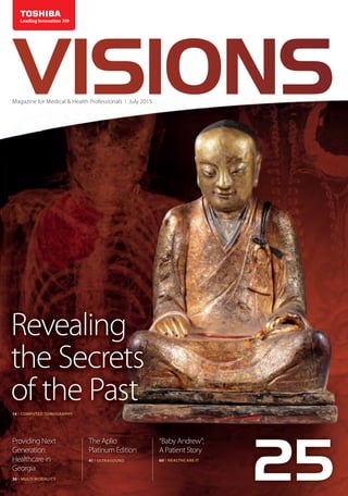
  • 2. Imprint VISIONS magazine is a publication of Toshiba Medical Systems Europe (Toshiba), and is offered free of charge to medical and health professionals. The magazine is published twice a year but is also available as an online portal. News items and articles are announced, pre-publication, via social media in dedicated groups. Toshiba stores data of readers and users, as far as known, of the online VISIONS portal. This data is used to send out the magazine and inform about new developments in the clinical market. Users can customize their preferences or opt-out in the online VISIONS profile at www.toshiba-medical.eu/visions. Publisher TOSHIBA Medical Systems Europe B.V. Zilverstraat 1 NL-2718 RP Zoetermeer Tel.: +31 79 368 92 22 Fax: +31 79 368 94 44 Web: www.toshiba-medical.eu Email: info@tmse.nl Editor-in-chief Jack Hoogendoorn (jack.hoogendoorn@toshiba-medical.eu) Modality coordinators CT: Roy Irwan UL: Joerg Schlegel XR: Jaco Terlouw Design & Layout Boerma Reclame (www.boermareclame.com) Printing Quadraat Printmedia (www.quadraat.nl) Text contributions “Revealing the Secrets of the Past” by The Creative Practice (www.thecreativepractice.com) “Continuity, consistency and a lot more clinical power: The Aplio Platinum Edtion” and“Virtual anatomy teaches real-world skills”by European Hospital Verlags (www.healthcare-in-europe.com) Subscription Service www.toshiba-medical.eu/visions VISIONS magazine is covering Toshiba’s European region and as such reflects products, technologies and services for this particular area. The mentioned products may not be available in other geographic regions. Please consult your Toshiba representative sales office in case of any questions. © 2015 by TOSHIBA Medical Systems Europe All rights reserved ISSN 1617-2876 25 A unique Chinese‘Buddha mummy’was scanned on Toshiba’s Aquilion ONE™ / ViSION Edition CT system to gather new information and progress scientific research on the artefact. The scan enabled details that had previously escaped discovery to be revealed. Read more on page 14. Photo: Copyright © 2014 Jan van Esch V25_Omslag_V2.indd 2 19-06-15 13:31
  • 3. VISIONS25 | 3 Earlier this year, I celebrated my 12.5 year work anniversary. In the Netherlands, this is considered to be an important milestone and a sign of loyalty, passion and commitment. In Japan they call this Aisha Seishin – love of company. Frankly speaking, it was not too difficult to achieve, because Toshiba is simply a great company to work for, where building and maintaining a strong sense of belonging, gaining group consensus and ensuring harmonious (work) relationships are part of the daily work ethics.This ensures that loyalty, passion and commit- ment are mutually beneficial and creates a feeling of pride in being part of the big, worldwide, Toshiba Family. My personal anniversary, however, is nothing compared to the impressive milestone that Toshiba Medical Systems now celebrates - its 100th anniversary in the healthcare business1. Toshiba was already involved with research for manufacturing earlier, but in 1915, Japan’s first domestically-produced X-ray tube was introduced and was the inspiration for many medical imaging innovations to come; such as Japan’s first X-ray device in 1932, the first linear array ultrasound scanner in 1976, the world’s first commercial MRI system in 1983, and world’s first continuous rotating CT scanner in 1985. Now, one hundred years later,Toshiba has become a world leader and great innovator in the healthcare industry. With a global network of local corporations and representatives in more than 135 countries, the company provides state-of-the-art products, systems and solutions that significantly improve patient safety and patient care and provides customers lasting quality with a lifetime of value. In addition, Toshiba offers the best service experience possible, by offering the most personal attention and contact in the industry. Because of this, many hospitals and clinics have chosen Toshiba as their main partner to jointly pursue the mutual goal of improving the healthcare delivery, set new goals and reach new milestones. Milestones are not an end in themselves, but are a unique opportunity to reflect on. Obviously, it is of great importance to continue our journey, and I hope, alongside Toshiba, to experience many other interesting milestones that improve the quality of life for all patients worldwide. Kanpai! Kind regards, Dear reader, EDITORIAL Jack Hoogendoorn Sr. Manager Marketing Communications Toshiba Medical Systems Europe BV 1 Have a look at the 100th anniversary video at http://tinyurl.com/m7uyoqw V25_Editorial Jack Hoogendoorn.indd 3 19-06-15 13:32
  • 4. ©2015 TOSHIBA MEDICAL SYSTEMS4 | VISIONS25 9 Celebrating 100 years in the healthcare business 14 Revealing the Secrets of the Past 20 First Clinical Results of Coronary CT Subtraction 23 Elastography in the Diagnosis of the Structure of Atherosclerotic Plaques in Carotid Arteries 31 Getting it right! 36 Providing Next Generation Healthcare in Georgia 38 Clinical Experience with 4D Ortho Application 23 36 38 Reliable methods to diagnose possible complications in primary stroke prevention. Lancet Medical Center works with a full range of Toshiba’s medical diagnostic imaging equipment. Wide area-detector CT is suited to dynamic study of joints, allowing volumetric study of bone and intra-articular ligaments during physiologic motion or under stress maneuvers. CONTENT COMPUTED TOMOGRAPHY MULTI MODALITY COMPUTED TOMOGRAPHY ULTRASOUND COMPUTED TOMOGRAPHY COMPUTED TOMOGRAPHY HISTORY HIGHLIGHTS 3D reconstruction, mummy Coronary Subtraction, CT Angiography, Coronary Artery Disease Elastography, atherosclerotic plaques, carotid artieries CT Pulmonary Angiography Musculoskeletal System, Post Processing V25_Contentpages.indd 4 19-06-15 14:29
  • 5. VISIONS25 | 5 41 58 54 The Aplio architecture enabled so many significant changes that it arrives with a new name, the Aplio Platinum Edition Manually selecting the kV setting is time consuming; therefore Toshiba has developed SUREkV to improve workflow. The University of Heidelberg brought a CT scanner directly into the anatomy lab. COMPUTED TOMOGRAPHY ULTRASOUND 41 Continuity, consistency and a lot more clinical power The Aplio Platinum Edition 52 Dual Energy 54 Virtual anatomy teaches real-world skills 56 PUREViSION in Clinical Practice 58 SUREkV - total patient protection 60 Vital patient story - Baby Andrew 63 Coronary Artery Adaptive Motion Correction Software CAD, CTA, Adaptive Motion Correction (AMC) 03 Editorial 06 News 12 Message from the President COMPUTED TOMOGRAPHY Volumetric Dual Energy, liver, Hepato-Callular Carcinoma COMPUTED TOMOGRAPHY COMPUTED TOMOGRAPHY ADVANCED VISUALISATION COMPUTED TOMOGRAPHY Detector, radiaton dose reduction, iodine dose reduction Low tube-voltage scanning, dose reduction, contrast reduction Anatomy VISIONS SPECIAL V25_Contentpages.indd 5 19-06-15 14:29
  • 6. ©2015 TOSHIBA MEDICAL SYSTEMS6 | VISIONS25 NEWS Long lasting quality After 22 years of hard work and loyal services the Toshiba SSH-140A Ultrasound system of Dr Stefan Hanggi finally stopped working due to a severe defect. Bridging time by a demo unit, a brand-new Aplio 300 could already be delivered to a him within three weeks by Mr Fritz Aeppli leaving behind a very satisfied customer for many years to come. Dr Stefan Hanggi (left) and Mr Fritz Aeppli, Account Manager Toshiba Medical Systems Switzerland (right) Toshiba held an event in Mitsukoshi, the department store in Japan, to encourage customers to enjoy the leading innovation technology. The video is a report of the event, featuring a communication android, a vegetable tasting booth of the plant factory, a digital signage that brings back the scenery of the Japanese Edo period (AD 1600-1868) and some other exhibitions. Watch the video at: http://tinyurl.com/plsfebc (English subs) Toshiba was National Sponsor of the Final Six Water polo Champions League held (Barcelona, Spain) showing its support to Sports in general, Water polo in particular, participants in the ‘Final Six’ and - last but not least, the Olympic City of Barcelona. Participants were: Szolnoki VSK (Hungary), ZF Eger (Hungary), VK Jug Dubrovnik (Croatia), VK Primorje Erste Bank (Croatia), Pro Recco Nuoto e Pallanuoto (Italy) and Club Natació Atlètic-Barceloneta (Spain). Pro Recco achieved their eighth Champions title in history by beating Primorje in an exciting final that honored the equality that had been seen throughout the competition. Igor Milanovic´s men prevailed by 7-8 in a tight, tension-filled final and brought the Final Six to a close in Barcelona. Toshiba also became official sponsor of Club Natació Atlètic- Barceloneta, the organizer of the tournament, by providing a portable Ultrasound Diagnostic System. Toshiba sponsors Water Polo League V25_News and Photopage.indd 6 19-06-15 14:09
  • 7. Kawasaki Tsurumi Rinko Bus Co., Ltd. recently has begun to take Toshiba’s first commercial electric bus in use on its Kawasaki Municipal Hospital route (Japan). Toshiba’s SCiB™ lithium-ion rechargeable battery cells, which excel in quick charging and long life, will help the electric bus reduce to CO2 emissions by approximately 40%1 , compared to diesel buses. 1 Result of tests conducted by Toshiba VISIONS25 | 7 Follow us for the latest news: https://www.youtube.com/user/ToshibaMedicalEurope https://twitter.com/toshiba_med http://www.linkedin.com/company/ toshiba-medical-systems-europe https://plus.google.com/116571489645577418994 https://www.facebook.com/ToshibaMedicalSystemsEurope http://www.slideshare.net/toshibamedical Next page is part of the VISIONS Photo Page Series reflecting an eye for the beauty of our planet, the environment and the direct surroundings where Toshiba’s systems are installed by Toshiba and its customers. Not the actual imaging products but photos of sceneries, cities, countries or other cultural aspects are highlighted on this photo page. The Photo Page is based upon an idea of Prof. Edwin van Beek. Every reader of VISIONS can participate and get their picture published. The submitted content should include: high resolution (300dpi) image, photo of the hospital and a brief text, name of photographer and Toshiba system(s) installed. The complete result is shown on the opposite page. Send your pictures and texts to: jhoogendoorn@tmse.nl, Subject: Photo Page ► IBA (Ion Beam Applications S.A.) and Toshiba Corporation signed a global collaboration to expand access to advanced particle therapy worldwide. Toshiba Medical Systems Corporation will become the distributor in Japan for Proteus®ONE, IBA’s compact single- room proton therapy solution, and IBA will become the agent for Toshiba’s Carbon Therapy Solutions outside Japan. IBA and Toshiba will collaborate on activities such as customer education for Proteus®ONE and Toshiba’s carbon therapy solutions. IBA and Toshiba Sign Strategic Partnership in Particle Therapy Next page is part of the VISIONS Photo Page Series reflecting an eye for ► V25_News and Photopage.indd 7 19-06-15 14:09
  • 8. ©2015 TOSHIBA MEDICAL SYSTEMS8 | VISIONS25 Tobata Kyoritsu Hospital (Japan) was founded in 1912 and, as a trusted hospital, serves the Japanese commu- nity for already over 100 years. Interacting with society and with a strong emphasizing on good teamwork it continuously transforms itself to meet the ever-chang- ing expectations of patients. Starting out with 18 bed, it now holds 218 beds and has over 125 Toshiba products and systems in use ranging from Hospital Information Systems and Bio Chemistry Analysers to CT, MR, X-ray and Ultrasound imaging systems. Text source and photography: www.kyoaikai.com Tokyo SkytreeSM is a free-standing broadcasting tower with a restaurant inside the observation deck located in Sumida, Tokyo, Japan. It became the tallest structure in Japan in 2010 and reached its full height of 634.0 metres in March 2011, making it the tallest tower in the world. The two illumination patterns Iki (chic, stylish) sky blue and Miyabi (elegance, refinement) purple are used, alternating daily. Text Source: Wikipedia – Photography: Reiko Aoki V25_News and Photopage.indd 8 19-06-15 14:09
  • 9. VISIONS25 | 9 V25_News and Photopage.indd 9 19-06-15 14:09
  • 10. ©2015 TOSHIBA MEDICAL SYSTEMS10 | VISIONS25 Starting with Japan’s first domestically produced X-ray tube in 1915, Toshiba celebrates its 100th anniversary in the healthcare business. Here you will find an overview of our rich company history and related innovations that clearly demonstrates that curiosity and passion are part of our DNA. 1914 Research on manufacturing X-ray tubes is started. 1932 The GIBA 75 X-ray device is introduced. 1946 KXO-8, the first post-war X-ray system is completed. 1969 Toshiba delivers the first gamma camera to National Kohnodai Hospital. It can visualize scintigrams instantaneously without the need for scanning the organs. 1978 The first cassette-less X-ray device is launched. 1955 The development of an X-ray angiography system proceeds. 1955 Japan’s first image intensifier is introduced to the market. 1966 Toshiba enters the ultrasound market. 1967 A high-energy therapeutic system is developed. 1960 Japan’s first X-ray TV devices are launched (image- scope 506 and 507). 1976 The first linear array ultrasound scanner is introduced. 1983 The world’s first commercial MRI system is introduced. 1985 Toshiba introduces the world’s first continuously rotating CT scanner with slip-ring technology. 1978 Japan’s first whole-body CT scanner is released. KXC-180-6 STX-20 TCT-60A TCT-900S MRT-15A 1915 GIBA, Japan’s first domestically produced X-ray tube is introduced. V25_News and Photopage.indd 10 19-06-15 14:09
  • 11. VISIONS25 | 11 1990 The triple-head digital gamma camera GCA- 9300 A wins the Image of the Year award at the annual meeting of the U.S. Society of Nuclear Medicine. 2003 Toshiba’s first digital X-ray system with flat panel detector Ultimax is launched. 2012 Iterative dose reduction becomes standard on all Toshiba CT scanners. Safer imaging, clearer outcomes Toshiba’s revolutionary PUREViSION dtector makes CT imaging safer for all patients. Delivering up to 40% increased efficiency it enables superior imaging with significantly reduced radiation dose and iodine. Making the unseen visible Toshiba’s innovative SMI ultrasound technology on the Aplio Platinum Series provides a safe and contrast-free way to examine the microvasculature, advancing diagnostic accuracy and patient outcome. Innovating clinical pathways Toshiba introduces Infinix 4D CT, a powerful hybrid imaging system combining the world’s most flexible angio suite with the world’s most advanced dynamic volume CT. Comprehensive dose management Toshiba’s DoseRite technology is a comprehensive dose management solution. The dose Tracking System provides continuous real-time information on skin dose deliverd to the patient. Adaptive diagnostics To make complex exams easier, to minimize patient dose, and to improve diagnostic accuracy and reproducibility, Toshiba has introduced Adaptive Diagnostics technology to its CT systems. Minimizing patient risk To minimize the potential risks associated with gadolinium-based contrast agents Toshiba provides clinicians with unique, non-contrast MRA sequences that minimize risk to patients while producing exceptional images. 2007 The world’s first wide-area detector CT Aquilion ONE is launched. 1990 The world’s first helical scan CT system is developed. 1999 Toshiba’s noise reduction technology Pianissimo revolution- izes the MRI market. Ultimax V25_News and Photopage.indd 11 19-06-15 14:10
  • 12. ©2015 TOSHIBA MEDICAL SYSTEMS12 | VISIONS25 PRESIDENT’S MESSAGE “We are committed to developing solutions to improve clinical work- flow, advance patient outcomes and patients’ quality of life.” Throughout its 100-year history in the medical device business, Toshiba has responded to customers’ needs by providing a wide range of high-value solutions. These efforts have made us the top company in our business segment in Japan, and have supported expansion of our global presence into over 135 countries around the globe. Delivering many Japanese- and world’s firsts, we have always been committed to developing solutions that help improve clinical workflow and advance patient outcomes and patients’ quality of life. From corporate programs to the wide range of product features that protect patients and healthcare providers, we have made safety a top priority in everything we do. During the second half of 2014, we introduced the following systems and technology to the market: In X-Ray, as most of you know, we introduced the Infinix 4D CT at The Radiological Society of North America’s Annual Meeting, RSNA2014. This powerful hybrid imaging system combines the world’s most flexible angio suite with the world’s most advanced dynamic volume CT. This outstanding image-guided solution for interventions also easily accommodates a wide range of interventional procedures from TACE (Transarterial Chemo Embolization) to Trauma, and from Neuro to Cardiac. Introducing new technology, we promoted the new XR Dose Right DTS (Dose Tracking System), a real time, peak skin dose visualization tool, which color- maps the exposure of the patient during endovascular treatment, and enables the interventionalist to better manage the dose delivered to the patients to help avoid serious, undesirable effects on the skin. In Nuclear Medicine, we introduced the Large-Bore TOF PET-CT Celesteion. The versatile system combines high performance PET and CT for all radiation and oncology imaging needs, including tumor-detection, treatment-evaluation and CT-simulation. It features the industry’s largest bore, state- of-the-art PET- and CT technology and dose reduction. 20150326_V25_Message from the President_DEF_V2.indd 12 19-06-15 14:39
  • 13. VISIONS25 | 13 In ComputedTomography, we introduced a new detector for the Aquilion ONETM ViSION Edition - PUREViSION. Toshiba’s commitment to ALARA, while delivering 4D functional dynamic studies at conventional dose levels, was a key driver in the development of PUREViSION. Having achieved a further 40% increase in light output and a 28% decrease in electronic noise with this development, PUREViSION provides the latest generation in detector performance for clinical- and diagnostic excellence, while ensuring patient safety at all times. In MRI, we introduced our new Saturn Gradient, software, and 16ch Flex SPEEDER coils for 3T systems to boost image quality, speed, and flexibility. This new combination has been in operation at the VU University Medical Center (VUMC) Amsterdam, the Netherlands, since January 2015. Toshiba’s Vantage Elan was welcomed by the market, with its excellent image quality, ease-of-use, compact footprint, and total lifetime value. In addition, the results of REACT(REnalArteryContrast-freeTrial)Multi-CenterStudy, which validated the usefulness of non-contrast enhanced MRA as a diagnostic tool for renal artery stenosis, were published in The American Journal of Roentgenology (AJR) in January 2015. An increasing awareness of the potential risks associated with Gadolinium-based contrast agents has revealed the need for alternative, contrast- free imaging techniques. We provide clinicians with unique technologies that minimize risk to patients, while producing exceptional images. In Ultrasound, we launched the Aplio Platinum Series, a system that provides powerful clinical imaging tools for advanced visualization, quantification and intervention. We also brought new tools to clinical practice, such as BEAM(anewtechniquethatimprovesneedlevisualization during ultrasound-guided procedures) and an advanced implementation of ShearWave Elastography (that quickly and non-invasively measures tissue stiffness of the liver to potentially reduce requirements for expensive biopsies). Improvements to current technologies, such as Superb Micro-Vascular Imaging (SMI), have further enhanced diagnostic capabilities for day-to-day examinations, as well as for Interventionists or Specialized Radiologists. In Vital, we introduced Version 6.7 of our Vitrea® software, with new features and enhancements to its Cardiovascular-, Neurovascular- and Oncology applications, as well as service, integration and efficiency enhancements. General and integration enhancements include improvements to Vitrea’s STL export functionality, providing flexibility to export from Vitrea for use in 3D printing capabilities, such as CAD-modeling, computational-modeling and surgical- simulation. Version 6.7 also includes enhancements to partner technologies with Olea Sphere®, Visia™ CT Lung CAD, Mirada RTX and Cedars-Sinai Medical Center Cardiac Suite. Last, but not least, let me mention the developments that have occurred within Toshiba. In 2013, Mr. Tanaka, Toshiba’s CEO and President, reshaped Toshiba’s focus onto three core areas of innovation and business development – Toshiba’s three pillars. Across Toshiba’s many business lines, our people develop solutions, products and technologies in different ways. From lifestyle-sensing devices, designed by our Semi-Conductor Division, through to world-leading cancer treatment systems from our Power Division, these Toshiba innovations are now being brought together to better meet the needs of the total healthcare environment. In the field of ‘Big Data’, Toshiba is partnering with leading institutions around the world. Together with The Johns Hopkins University in the United States (US), Toshiba is participating in work being carried out to analyze treatment pathways and outcomes in head- and neck cancer patients, towards improving outcomes for individuals in the future. Another ‘Big Data’ project is underway in Japan, in which Toshiba has joined forces with Tohoku University and Nihon Kohden to study life factors of more than 150,000 people from the Tohoku area in Japan. This comprehensive project, sponsored under the Japanese Government’s ‘Center of Innovation’ (COI) Program, has already led to clinical benefits for the community and tangible business benefits for Toshiba. In December 2014, Toshiba commenced with the offer of genome testing to support the research requirements of Japanese Universities through rapid service and greatly reduced costs, thanks to knowledge developed as part of the COI Project. While always strictly adhering to our company’s well- knowntraditionofexcellenceandquality,wewillcontinue to respond to changes in the industry and the needs of our customers, and pursue our mutual goal of improving healthcare delivery. Our products and company are dedicated to our ‘Made for Life’ philosophy. We deeply appreciate your continuing support and cooperation in improving human health through continually improving healthcare for all patients. Our company is the product of our employees and I am proud of our ‘Toshiba Family’. I will always strive to protect our brand name and image now and in the future. Toshio Takiguchi President and Chief Executive Officer Toshiba Medical Systems Corporation 20150326_V25_Message from the President_DEF_V2.indd 13 19-06-15 14:39
  • 14. ©2015 TOSHIBA MEDICAL SYSTEMS14 | VISIONS25 V25_CTEU140096 Revealing the Secrets of the Past.indd 14 19-06-15 14:17
  • 15. VISIONS25 | 15 The artefact appeared to be a large, highly ornate, gilded statue of a Buddhist saint, in half-lotus position from the outside, but as work began on restoration, the mummi- fied body of a monk, who lived around the year 1100, was discovered inside the plaster casing. The mummy is the only one of its kind known and available for scientific research outside of China. It is a truly unique exemplar and has been the star exhibit at expositions organized by the Drents Museum in Assen, the Netherlands and the Hungarian Natural History Museum in Budapest.  Initial investigations provided some information, but also raised even more questions. In the quest to find out more about this intriguing, culturally and historically sig- nificant artefact, and with the potential offered by con- tinually advancing research technology, unravelling the mystery of the mummy has become an ongoing story. Toshiba heard about the project through Erik Bruijn, an independent researcher, Buddhism expert and Guest Curator of the Tibet and Japan Departments of the Wereldmuseum in Rotterdam, the Netherlands, who inspired his neighbour, Dr. Ben Heggelman, Radiologist at the Meander Medical Center in Amersfoort, the Netherlands, to also become involved in the project. Toshiba offered use of its state-of-the-art Aquilion ONE to assist with the research. “We are delighted to support scientific research that could provide fresh insights into this remarkable artefact,” said Jos Ruis - Vice President Toshiba Medical Systems Europe. “Investigations like this are extremely rare. For museums, it offers the possibility to perform research that is normally beyond their means. For Toshiba, such projects are important, because we can help the scien- tist to better investigate and understand the subject of their research and thus demonstrate the usefulness of our imaging technology beyond regular clinical practice. New technological advances are allowing investigators to non-invasively‘look’into the subjects of their research. We can, so to speak, ‘peel off’ layer-after-layer, and pro- gressively learn more and more about historically-signif- icant specimens. By using advanced 3D reconstruction CTEU140096 technology, we can visualize otherwise hidden features and even enable the creation of‘life-like’reconstructions in combination with 3D printing technology.” A SURPRISING DISCOVERY In 1996, a Dutch-Asian art collector bought the statue from another Dutch collector. One year later, a skilled craftsman removed the statue’s bottom wooden plate to assess the statue for restoration. He discovered a linen scroll annotated with Chinese script dating from the 1300s, along with a number of dead beetles, and realized that the statue was far more than just a piece of religious artwork. In 2013, a CT scan was made at the Mannheim Hospital in Germany with the statue-owners’ approval. The result clearly revealed the skeletal remains of the mummified person inside. Subsequent research indi- cates that the artefact was restored in the 14th Century. Translation of the Chinese scroll text found inside the mummy, which dates from that period, suggests that the mummified remains were of a Buddhist monk, whose name is still unknown. Extensive examination of the outer casing and carbon dating confirmed that the mummy dates from the 11th or 12th Century and that the monk probably died between 1022 and 1155, as a relatively young man of around 35 to 40 years old. The Buddhist monk would have been alive during the Song Dynasty – a time when China experienced turbulent and violent development, and Buddhism was opposed by Confucianism and Taoism, steeped as it was in corruption and decay. Its wealthy monasteries and dazzling shrines had become a heavy burden on the nation’s economy. In the wars that ensued and, in particular, a revolution against mainstream Buddhism, many religious artefacts were destroyed or plundered, and as a consequence, are extremely rare finds today. Revealing the Secrets of the Past Toshiba’smottois‘MadeforLife’,butarecentcollaborationwithaleadingDutchmuseum has demonstrated that its technology extends beyond this! Toshiba’s Aquilion ONE™ / ViSION Edition CT system was used to scan a unique Chinese ‘Buddha mummy’ to gather new information and progress scientific research on the artefact. The advanced technological features of the state-of-the-art Toshiba system enabled details that had previously escaped discovery to be revealed. And the new data collected will be used for‘life-like’three-dimensional (3D) reconstruction. HISTORY COMPUTED TOMOGRAPHY 3D reconstruction, mummy Copyright©2014JanvanEsch gather new information and progress scientific research on the artefact. The advanced technological features of the state-of-the-art Toshiba system enabled details that had previously escaped discovery to be revealed. And the new data collected will be used technology, we can visualize otherwise hidden features and even enable the creation of‘life-like’reconstructions in combination with 3D printing technology.” In 1996, a Dutch-Asian art collector bought the statue from another Dutch collector. One year later, a skilled craftsman removed the statue’s bottom wooden plate to assess the statue for restoration. He discovered a linen scroll annotated with Chinese script dating from the 1300s, along with a number of dead beetles, and realized that the statue was far more than just a piece of religious The Drents Museum (www.drentsmuseum.nl) encourages visitors to discover, learn and interpret history. They do this, amongst other things, by organizing spectacular, international exhibitions that offer ‘once-in-a-lifetime’ experiences. V25_CTEU140096 Revealing the Secrets of the Past.indd 15 19-06-15 14:17
  • 16. ©2015 TOSHIBA MEDICAL SYSTEMS16 | VISIONS25 Erik Bruijn is the independent researcher and expert in Buddhism, who is leading the investigation into the mum- my’s significance. He explained more about the artefact: “The monk would appear to have belonged to the Ch’an Sect of Buddhism, although we cannot implicitly verify the truth of the scroll inserted during restoration in the 1300s. Ch’an philosophy steered Buddhism back towards the values of simplicity, intuition and nature. It embraced all sorts of elements from other Buddhist traditions, and in this way appealed to many people. Ch’an Buddhism incorporated many sub sects, many of which survived the decline of mainstream Buddhism in this period. We can deduce from study of the many details on the plas- ter casing of the mummy that the monk was a master – for example, the positioning of the hands incorporates secret symbolism. The statue would have been a focus of worship for pilgrims in a Buddhist temple. The plaster casing was made with holes in the ears and nose so that the holy monk could‘listen’to the prayers of the pilgrims and ’smell’the incense and flowers offered by them. UNANSWERED QUESTIONS While the research revealed some basic information, many unanswered questions about the mummy remain. It is not clear how the Buddhist monk died, for exam- ple. The initial CT scan revealed a molar abscess in the lower, left jaw, which might have progressed to sepsis and proved fatal in a time before antibiotics existed. Or, this could be an example of ‘self-mummification’, which was achieved through dehydration and starva- tion during the end of the monk’s life, as practiced by some Buddhist monks in the past. Mummification was achieved by different methods in China than in Egypt. The brain was not removed through the nose, but left intact. In the initial- and latest CT scans, the remains of former brain meninges can be seen. No other remains of internal organs exist. They could have been removed during mummification, decomposed or destroyed by insects later. Metal- and wooden rods support the spine and chin respectively, so that the mummy could assume an upright crossed-legged position. Of particular interest, were two previously uniden- tified structures found within the body cavity: what appears to be a scrolled piece of cloth inside the center of the abdominal area, and a highly layered structure in the right side of the thoracic region within the skeleton. The scroll could have been placed there at the time of mummification or during restoration in the 14th Century. The thoracic structure did not appear to be comprised of previously living tissue and could be an insect-created nest. These questions and many others prompted inves- tigation with the advanced Aquilion ONE. In addition to Figure 1: Maximum Intensity Projection showing a scrolled piece of cloth or paper The examination was preceded by a short ceremony. Copyright©2014JanvanEsch Copyright©2014ErikBruijn V25_CTEU140096 Revealing the Secrets of the Past.indd 16 19-06-15 14:17
  • 17. VISIONS25 | 17 the CT, further scientific research was carried out with an endoscope at Meander Hospital, Amersfoort and further laboratory tests at The Rijksmuseum, in Amsterdam, the Netherlands. The endoscopic examination did confirm the presence of an as yet unidentified material in the thoracic cavity, which appeared to include scraps of rice paper covered with Chinese characters written in black ink. The endoscopic examination did confirm the pres- ence of corpora aliena in the thoracic cavity. HIGH EXPECTATIONS Erik’s neighbour, Ben Heggelman, is providing radio- logical and medical expertise in the current phase of research. Working in Radiology since CT was first intro- duced; he has seen the evolution of this modality from its very beginning and harbored high expectations for the results of the scan on the Buddha mummy. “When I first qualified in radiology at Erasmus University, Rotterdam, in the Netherlands, CT had just been introduced. While its immediate potential was evident, technology has developed so much since then. Today’s clinical practice relies on CT techniques and technology that we couldn’t have imagined might even be possible then,” said Ben. “The Aquilion ONE™ / ViSION Edition is considered the top of the range in the healthcare industry and we are delighted to have access to it to further our research on this unusual project. We anticipate that its advanced capabilities will provide use- ful additional information. The possibilities with CT now are extensive and – they are not science fiction, they are simply science fact! ” “The combination of Toshiba’s innovative technol- ogy and the expertise, inventiveness and stamina of our staff can yield surprising results and insights. We dem- onstrated this already in our collaboration with Caspar Berger, an award-winning Dutch artist and sculptor, who created an exact copy of his own skeleton using a Toshiba Aquilion CT scanner and 3D-print technology and then incorporated this into a wide variety of creative interpretations entitled‘Bone’1. Caspar’s innovative work, which combines artistic expertise with modern tech- niques to creatively interpret and reflect the true qualities and beauty of the human body, was recognized with the prestigious Dutch Singer Prize in 2013,” added Jos Ruis. “We hope that the same combination of technology and people can help the researchers obtain greater under- standing of this intriguing story and unravel the mystery of this unknown Buddhist monk.” Figure 2: 3D Image displaying the layered structure in the apex of the lung Copyright © 2014 Erik Bruijn CTEU140096 Skeleton / Self-portrait 20, 2012 - by Caspar Berger A cast of the artists right upper arm bone (the humerus) in three kilogramme of gold. V25_CTEU140096 Revealing the Secrets of the Past.indd 17 19-06-15 14:17
  • 18. ©2015 TOSHIBA MEDICAL SYSTEMS18 | VISIONS25 MEETING THE NEEDS OF THE MOST UNUSUAL ‘PATIENTS’ After intricate planning in agreement with the Drents museumandownersoftheartefact,Toshiba’sHeadquarters in Zoetermeer welcomed its latest and most unusual ‘patient’, who was carefully transported from the Drents Museum in a custom-built, shock-protected, carrying case. A short Buddhist ceremony was held to observe religious customs.The Buddha mummy was carefully positioned for its CT by Erik. Because the statue consists of a rigid, but obviously fragile construction, very careful adjustment was required to secure and support it. The table of the Aquilion ONE features Tech Assist Lateral Slide, a unique Toshiba innovation designed to make positioning patients easy and strain-free. The artefact could be fitted safely into the scanner, thanks to its large open bore. Aquilion ONE™ / ViSION Edition has a 78cm open bore – a 10 cm wider aperture than the previous scanner used at Mannheim, providing ideal access. Roy Verlaan, Clinical Application Specialist at Toshiba’s Computed Tomography Business Group carried out the scan. Within a matter of minutes, the statue had been scanned and data collected within a single scan. “Using Toshiba’s Aquilion ONE, we anticipated that the image quality would be far superior than any CT investiga- tion previously made of the Buddha mummy and com- binedwitheasyaccess,artifactreductionformetalartifacts and iterative reconstruction, we were able to contribute to further insights,”he said. “The scan indeed provided a much higher resolution; the information that we had before was better presented with far more detail, and enabled us to derive even more information,” remarked Ben. “In addition, we were even able to find things that weren’t apparent in the initial investigation, and through 3D reconstruction, were able to gather information on their exact shape, size and density of these ‘newly discovered’ structures. Extracting such information from CT is particularly important in this case, because it is unlikely that intact DNA still exists, since the mummy is around 950 years old and for a large part has been kept in a temple with temperatures of around 30°C. We may possibly be able to extract information from dental DNA in subsequent research, if this is permitted by the owner of the exhibit, but even this could prove unsuccessful.” Copyright©2014JanvanEsch Careful and exact positioning of the mummy in the Aquilion ONE scanner gantry Copyright©2014JanvanEsch V25_CTEU140096 Revealing the Secrets of the Past.indd 18 19-06-15 14:17
  • 19. VISIONS25 | 19 References: 1. ”Bone as Art – From CT scan to solid gold”, VISIONS Magazine 20, p36-p39 | http://tinyurl.com/mkpnoy3 2. Vilsteren, V. van (ed.), 2014: Mummies, overleven na de dood. Zwolle. (191 pages, ISBN9789462580046) Note: at the end of last year, a Toshiba CT scanner was used to study the mummy, as described in this article. The scans provided new and surprising insights that were previously unknown. Toshiba is proud to have been able to contribute to these advances in cultural and historical knowledge. Since then, and entirely unknown at the time of scanning, the ownership of the mummy has become the subject of media controversy. An illustrated book2 with many beautiful photographs edited by Vincent van Vilsteren, the Curator of the Drents Museum, expands upon thedifferentcausesofnaturalmum- mification and provides further information on scientific research, restoration of mummies and on the medieval trade in Mumia (currently only available in Dutch). Ben will assess the results of the CT scan fully, and together with those from further research at the Meander Medical Center and Rijksmuseum, Erik will compile and publish a book, which he hopes to publish in 2016. “It is incredible that we can gather so much new infor- mation from an image made really within a matter of seconds,” said Erik. “We are confident that the latest phase of research, including today’s scan, will deliver a large amount of new data, helping us to solve many of the current puzzles around the Buddha mummy.” “One word describes this day - Amazing!” concluded Roy. “This has been an absolutely unique experience. The story behind the Buddha mummy is intriguing. The artefact itself is breathtaking and the images and data we have been able to obtain with the Aquilion ONE™ / ViSION Edition have exceeded our expectations.” FUTURE ROLE Advanced imaging has a clear role in helping to progress scientific research into museum artefacts and enhancing our knowledge of the past. “New technological developments in medical imaging are, of course, implemented for the benefit of the living, however,thebenefitsofourdiscoveriescouldalsobeused for immense progress in the domain of scientific research into artefacts, like the study of this mummy,”remarked Jos. “Delivering this benefit in a wider context in the future would rely on easing the access to this type of imaging technology for museums and researchers. Mobile CT and MRI scanners could play an important role in this.” Sagittal reconstruction (l) and 3D reconstruction (r) being displayed inside the scan room with the Chinese mummy in the background CTEU140096 Copyright©2014JanvanEsch V25_CTEU140096 Revealing the Secrets of the Past.indd 19 19-06-15 14:17
  • 20. stented arteries. Coronary Subtraction can be performed in two breath-hold or in a single breath-hold (Figure 1). The non-contrast CTA volume is then subtracted from the contrast-enhanced CTA volume, removing the calcium/metallic content of coronary arteries. To obtain an accurate registration of two volumes and minimize misregistration artefacts, the single breath- hold methodology was chosen. After pre-oxygenation with low-flow oxygen and immediately after contrast injection, a non-contrast scan and a contrast scan with The introduction of Aquilion ONE™ 320-row CT scanner, and later the Aquilion ONE™/ ViSION Edition with a maximum fastest rotation time of 0.275s, have it made possibletoobtainanon-invasivecoronaryangiographyin asingleheartbeatandseveralstudieshavedemonstrated its high diagnostic accuracy in the depiction of epicardial coronary stenotic lesions7,8. Despite technological advances in CT, improving both spatial and temporal resolution, there are still several limitations due to increased heart rate, presence of small stents (less than 3 mm diameter), or extensive coronary artery calcification. Recently, Toshiba has developed a new application (Coronary Subtraction) to allow the subtraction of calcium and metal stents in the coronary arteries to improve the diagnostic performance in these clinical scenarios. Coronary subtraction CT requires two scans,onepre-contrastandonepost-contrast. The subtraction of three-dimensional (3D) CTA volumes is significantly more challenging than the two-dimensional (2D) subtraction performed in X-Ray digital subtraction angiography (DSA). Subtraction in two dimensions only requires shifting of the mask image in two axes: x and y. In 3D volumes, not only manual shifting of the mask in three axes (x,y and z), but also rotational movement is required. The fact that the two studies have to be performed consecutively implies that respiratory, pulsatile and spatial motion potential changes can lead to volumes of data with somewhat different spatial relationships between bony structures and calcified or Precontrast CCTA Contrast CCTA Precontrast CCTA Contrast CCTA Voltage: 120 kV 0.5mm slice thickness Voltage: 120 kV 0.5mm slice thickness First breath-hold Single breath-hold Two separate breath-holds Second breath-hold Single breath-hold Contrast injection 0s 30s Contrast injection SURE Start SURE Start Precontrast CCTA Contrast CCTA Precontrast CCTA Contrast CCTA Voltage: 120 kV 0.5mm slice thickness Voltage: 120 kV 0.5mm slice thickness First breath-hold Single breath-hold Two separate breath-holds Second breath-hold Single breath-hold Contrast injection 0s 30s Contrast injection SURE Start SURE Start First Clinical Results of Coronary CT Subtraction Computed tomography angiography (CTA) of the coronary arteries is a useful non- invasive tool to rule out significant coronary artery disease (CAD) in many clinical situations. Recent guidelines of stable CAD1 and non-ST segment elevation myocardial infarction2 endorse the use of CTA in symptomatic patients with low to intermediate likelihood of the disease, given the particularly high negative predictive value of the technique. However, in patients with high pre-test likelihood of CAD, the technique is not recommended, and one of the reasons is the high probability of coronary calcification in these patients, which interferes with the analysis of the images and reduces the specificity and negative predictive value of CTA3-6. D. Viladés-Medel MD 1,2), R. Leta MD1,2), A. Hidalgo MD PhD 1,2), F. Carreras MD PhD 1,2), G. Pons-Lladó MD PhD 1,2), X. Alomar MD 1) CARDIOLOGY COMPUTED TOMOGRAPHY Coronary Subtraction, CT Angiography, Coronary Artery Disease 1) Clinica Creu Blanca, Barcelona 2) Unitat d’Imatge Cardíaca Hospital de Sant Creu i Sant Pau Spain Figure 1: Image acquisition methodologies for CTA subtraction. D. Viladés-Medel ©2014 TOSHIBA MEDICAL SYSTEMS20 | VISIONS25 V25_CTEU140095_First clinical results of coronary CT Subtraction_v4.indd 20 19-06-15 14:15
  • 21. CASE 2 69 year-old male, past smoker with hypertension and moderate chronic renal failure with a glomerular filtration rate of 40 mL/kg/min. No previous cardiac history. During the last weeks, he presented with atypical chest pain and an inconclusive exercise ECG (subtle changes in cardiac repolarization without associated angina). Cardiac computed tomography angiography (CTA) showed a high degree of calcification in the left anterior descending artery that impeded the assessment of the wall lumen in these segments (Agatston score 845) (Figure A). After coronary subtraction, a vessel without any significant lesion could be observed (Figure B), as confirmed in a further invasive coronary angiography (Figure C). CASE 3 64 year-old male, past smoker with hypertension and hypercholesterolemia. Chronic ischemic heart disease with previous inferior myocardial infarction and stenting of right coronary artery (RCA). After this he presented with unstable angina in the context of a total chronic occlusion at the site of the stent. Again, he was revascularized with bioabsorbable vascular scaffold. Six months later, the patient was referred for a CTA to check this last revascularization procedure. Cardiac computed tomography angiography (CTA) showed a patent stent in the proximal RCA and focal calcification in the distal segment of the RCA, making it impossible to rule out significant stenosis in this segment (Agaston score 530) (Figure A). After coronary subtraction, an RCA without significant stenosis was observed (Figure B), subsequently confirmed with an invasive coronary angiography (Figure C). a z-coverage of up to 16cm was performed in the same breath-hold. Prospective ECG-triggering was used with a variable exposure window depending on the heart rate. Coronary CTA analysis was performed on an off-line workstation (Vitrea Fx; Vital Images). Three clinical cases from daily practice are presented to illustrate how the Coronary Subtraction application works and its correlation with the invasive coronary anatomy seen by an invasive catheterization. CASE 1 51 year-old male, active smoker with hypertension, type II diabetes mellitus and hypercholesterolemia. Chronic ischemic heart disease previously revascularized with stents in left main and proximal to mid left anterior descending artery (2008). During the last month, he presented with atypical chest pain unrelated to physical effort. Baseline ECG did not show repolarization abnormalities. Cardiac computed tomography angiography (CTA) showed previous implanted stents, with extensive metal artefact, making an assessment of neointimal hyperplasia or de novo atherosclerosis within the stents difficult (Figure A). After coronary subtraction, a vessel without any significant lesion could be observed (Figure B), as confirmed in a further invasive coronary angiography (Figure C). CTEU140095 Case 1 Case 2 Case 3 VISIONS25 | 21 V25_CTEU140095_First clinical results of coronary CT Subtraction_v4.indd 21 19-06-15 14:15
  • 22. CONCLUSIONS The Toshiba Coronary Subtraction application is a promising tool to minimize calcium and metallic artefacts, as these three clinical cases have exemplified. Coronary calcium subtraction may improve the diagnostic accuracy of CTA, especially in those patients with extensive calcifications or small stents. More studies are needed to validate this diagnostic tool and find out what role this could have in clinical practice. Toshiba’s Aquilion ONE / ViSION Edition offers all patients a successful examination, with the lowest possible radiation exposure and the highest quality diagnostic outcomes - www.aquilionvision.com Refences - Subtraction Coronary CT Angiography for the Evaluation of Severely Calcified Lesions Using a 320-Detector Row Scanner, Yoshioka K Tanaka R, Current Cardiovascular Imaging Reports, 2011; 4(6):437-446 - Subtraction Coronary CT Angiography for Calcified Lesions, Yoshioka K, Tanaka R, Muranaka K.Y, Cardiol Clinics, 2012; 30(1):93-102 - Improved evaluation of calcified segments on coronary CT angiography: a feasibility of coronary calcium subtraction, Tanaka R, Yoshioka K, Muranaka K, Chiba T, Ueda T, Sasaki T, Fusazaki T, Ehara S, International Journal of Cardiovascular Imaging, 2013, Epub. - Accurate Registration of Coronary Arteries for volumetric CT Digital Subtraction Angiography, M. Razeto, J. Matthews, S. Masood, J. Steel, K. Arakita, Proc. SPIE Vol. 8768, 2013. Bibliography 1. Members TF, Montalescot G, Sechtem U, Achenbach S, Andreotti F, Arden C, et al. 2013 ESC guidelines on the management of stable coronary artery disease: The Task Force on the management of stable coronary artery disease of the European Society of Cardiology . Eur Hear J 2013;34 (38):2949–3003. 2. Hamm CW, Bassand J-P, Agewall S, Bax J, Boersma E, Bueno H, et al. ESC Guidelines for the management of acute coronary syndromes in patients presenting without persistent ST-segment elevation: The Task Force for the management of acute coronary syndromes (ACS) in patients presenting without persistent ST-segment elevation of the European Society of Cardiology (ESC). Eur Hear J 2011;32 (23):2999–3054. 3. Stolzmann P, Scheffel H, Leschka S, Plass A, Baumüller S, Marincek B, et al. Influence of Calcifications on Diagnostic Accuracy of Coronary CT Angiography Using Prospective ECG Triggering. Am J Roentgenol. 2008;191(6):1684–9. 4. Vavere AL, Arbab-Zadeh A, Rochitte CE, Dewey M, Niinuma H, Gottlieb I, et al. Coronary Artery Stenoses: Accuracy of 64–Detector Row CT Angiography in Segments with Mild, Moderate, or Severe Calcification—A Subanalysis of the CORE-64 Trial. Radiology 2011;261(1):100–8. 5. Abdulla J, Pedersen KS, Budoff M, Kofoed KF. Influence of coronary calcification on the diagnostic accuracy of 64-slice computed tomography coronary angiography: a systematic review and meta-analysis. Int J Cardiovasc Imaging. 2012;28(4):943–53. 6. Meijboom WB, van Mieghem C a G, Mollet NR, Pugliese F, Weustink AC, van Pelt N, et al. 64-Slice ComputedTomography Coronary Angiography in Patients With High, Intermediate, or Low Pretest Probability of Significant Coronary Artery Disease. J Am Coll Cardiol. 2007;50(15):1469–75. 7. Dewey M, Zimmermann E, Deissenrieder F, Laule M, Dübel H-P, Schlattmann P, et al. Noninvasive Coronary Angiography by 320-Row ComputedTomographyWith Lower Radiation Exposure and Maintained Diagnostic Accuracy: Comparison of Results With Cardiac Catheterization in a Head-to-Head Pilot Investigation . Circulation 2009;120 (10):867–75. 8. De Graaf FR, Schuijf JD, van Velzen JE, Kroft LJ, de Roos A, Reiber JHC, et al. Diagnostic accuracy of 320-row multidetector computed tomography coronary angiography in the non-invasive evaluation of significant coronary artery disease. Eur Hear J 2010;31 (15):1908–15. ©2014 TOSHIBA MEDICAL SYSTEMS22 | VISIONS25 V25_CTEU140095_First clinical results of coronary CT Subtraction_v4.indd 22 19-06-15 14:15
  • 23. VISIONS25 | 23 Today there are two major mechanisms for pathogen- esis of ischemic cerebrovascular disorders (CVD): hemo- dynamic (narrowing of the artery with insufficiency of cerebral blood flow) and embolic (destruction by atherosclerotic plaque - ASP - with ulceration of the tectum). Developed over 50 years ago, carotid endar- terectomy (CEA) is still the most efficient intervention to prevent stroke in patients with carotid artery steno- sis. According to A. V. Pokrovsky, the indications for CEA are determined by the presence or absence of clinical manifestations of cerebrovascular insufficiency, as well ULEU150044 CASE REPORTS ULTRASOUND Elastography, atherosclerotic plaques, carotid artieries as by the extent and nature of changes in the carotid arteries: the degree of the internal carotid artery (ICA) stenosis (expressed as a percentage); structural charac- terization of atherosclerotic plaque; and the condition of the ASP tectum3. Ultrasound (US) today is the gold standard in carotid artery examination and risk assessment for CVD devel- opment in patients with carotid atherosclerosis. Being inexpensive, non-invasive, and precise, it allows the phy- sician to explore the patency of the vessel, its diameter, vascular wall and perivascular tissue condition4. To characterize the echogenic properties of plaques, in most cases the A. Gray-Weale classification is used, supple- mented by C. M. Steffen, G. Geroulacos, and by the Russian scientific centre of surgery (RSSS) named after acad. B. V. Petrovsky of RAMS. [Guided / Ed. by A. V. Pokrovsky]: • type I - completely hypoechoic ASP with thin echo- genic tectum (homogeneous); • type II - predominantly hypoechoic ASP with 50% of hyperechoic areas (heterogeneous); • type III - predominantly hyperechoic ASP with 50% of hypoechoic areas (heterogeneous); • type IV - completely hyperechoic ASP (homogeneous); • type V - ASP can not be identified due to pronounced calcification causing acoustic shadow. • type VI - isoehoic ASP with echo-structure with the intensity of the reflected US signal similar to that of the vascular wall. US enables detection of cerebrovascular embolic event risk factors, evaluation of ASP structural features and of stenosis configuration. The sources of microemboli are known5 to be homogeneous hypoechoic ASP, heteroge- neous ASP with a prevalence of hypoechoic zones, plaque A. R. Zubarev 1), I. V. Rychkova 1), M. B. Saratov 2), A. K. Demidova 1), E. L. Tumanova, V. N. Fedorova, V. Y. Panko, N. V. Krivosheeva. Capabilities of Ultrasound Elastography in the Diagnosis of the Structure of Atherosclerotic Plaques in Carotid Arteries The proportion of acute ischemic cerebrovascular accidents associated with carotid atherosclerosis is 68%1. This disease is associated with high mortality and disability rates, therefore reliable methods to diagnose possible complications in primary stroke prevention are necessary, as about 70% of strokes develop without any precursors2. 1) Department of ultra- sonography. Russian National Research Medical University named after N.I. Pirogov. Moscow. Russia 2) Department of vascular surgery. State-owned Federal State Institution “Main Clinical Military Hospital of Federal Security Service of the Russian Federation”. Moscow. Russia V25_ULEU150044 Elastography Athesclerosis.indd 23 19-06-15 14:50
  • 24. ©2015 TOSHIBA MEDICAL SYSTEMS24 | VISIONS25 with irregular contours and with “silent” areas on color Doppler mapping (CDM), as well as complicated plaques (with bleeding and / or ulceration). New US modes with enhanced quality of US images do not, unfortunately, pro- vide any additional US criteria for interpretation of hypo- and anechoic zones forming a heterogeneous plaque6. The possibilities of US in assessing the ASP structure in case of pronounced calcification in the examined area are also limited due to the fact that the acoustic shadow makes it impossible to reliably estimate the structure of ASP, creating the illusion of a“hypoechoic component.” To date, the most accurate non-invasive technique in the diagnosis of atherosclerotic plaque instability is MRI16-18. However, the procedure is quite expensive and time consuming. Ultrasonic elastography (USEG) is an inexpensive technique, implemented in many modern devices, allowing determination of the mechanical prop- erties of tissue that can be used in addition to a standard US exam13-15. The method is based on an objective assessment of the degree of displacement of the tissue, depending on its stiffness in response to outside pres- sure. Given that the mechanical properties of tissues are directly dependent on their structural organization, we assumed that the various components of ASP being coded in B-mode as a hypoechoic component of ASP may differ on elastograms7. Thus, the aim of our study was to assess the ability of USEG to diagnose various components of ASP and its instability.The results of USEG were compared with morphological and histological data obtained after separation of ASP by CEA. MATERIALS AND METHODS To achieve the goal of the study, to evaluate the ability of USEG to determine the components and stability of atherosclerotic plaque, and given the novelty of the methodology and the lack of sufficient data on its use in clinical practice at the time of beginning the study, we had to perform several preliminary stages. At the beginning of the study the methodology was tested on a specially designed gel phantom. At this stage the best machine settings were revealed, and repeatable results were achieved by the doctors involved in the project. In the second stage the methodology was tested on healthy volunteers. At this stage, two variants of the methodology showed similar results: performed with own tissue pulsation and during compression; several machine settings were adjusted for optimal visualization against the background of own pulsation. At the next stage a study was conducted on volunteers with carotid atherosclerosis. To confirm the safety of the procedure we carried out an emboli detection procedure in the medial cerebral arteries (MCA) on both sides simul- taneously with the US and elastography. First the patients signed informed consent notices, which described the essence of the procedure and possible risks. At the fourth stage, all patients with indications for CEA received a standard US in combination with USEG and transcranial microemboli detection. The visualization results obtained were detailed for the operating surgeon and sketched on the scheme developed. The obtained results of complex US and elastography exams were compared with intraoperative results and the histopatho- logical examination data. Subsequently, all the data were recorded in the database and processed. All the methods were compared with each other, and also differences and possible reasons for the discrepancies were assessed. The standard US and elastographic exams were con- ducted on a Toshiba Aplio XG, supplied with a high frequency linear transducer, using the dynamic flow technique. The study was conducted with the patient in the normal position. At the stage of standard US in longitu- dinal and transverse scanning of arteries the following were evaluated: patency, diameter, and geometry of the vessel, vascular wall along its entire length, surface and internal structure of the atherosclerotic plaque (ASP). Using Doppler technology, the blood flow was quanti- tatively determined. Using CDI, power Doppler mapping, and dynamic-flow techniques allowed us to identify and assess the nature of hypoechoic areas, and to assess the surface of the plaque. Then the targeted elastography of the ASP identified was conducted. Given the need to compress the study area to obtain an elastogram and the high risk of compli- cations developing during compression in patients with severe carotid atherosclerosis, the study was conducted with extreme caution, in the presence of a treating phy- sician. Use of the own pulsation of arteries to develop elastograms, as in the case of normal unaffected arteries, appearedimpossible;whichislikelytobeduetothesevere rigidity of the walls of the affected vessels. After several cycles of compression-decompression the device started the qualitative and quantitative elastography mode, and data processing was performed in off-line mode. RESULTS AND DISCUSSION We obtained data from five patients. V25_ULEU150044 Elastography Athesclerosis.indd 24 19-06-15 14:50
  • 25. VISIONS25 | 25ULEU150044 An US examination of the carotid arteries revealed circular prolonged heterogeneous ASP, calcified with a predomi- nance of hypoechoic component, of the left CCA with transition to the ICA. Maximum stenosis on the cross- sectional area of the vessel reached 65-70% (Fig. 1a, 1b). In the USEG mode the ASP region is colored het- erogeneously. In the distal segment of plaque an area of marked soft-elastic consistency was found, followed by a tight area and a small soft-elastic component of the plaque with green staining of the tectum zone in the proximal segment (Fig. 1c). It should be noted that in this case USEG identified differences between these ASP areas, while in B-mode they appeared as areas of lowered echogenicity. Examination of ASP macropreparation, obtained by endarterectomy, showed a dense fragment of the tissue tube wall. In cross-section the tissue deep in the frag- ment is heterogeneous - with yellow, grey, and brown areas (Fig. 1d). Figure 1: Patient 1 1a and 1b - grey-scale image of the circular prolonged heterogeneous calcified atherosclerotic plaque with a predominance of hypoechoic component. 1c - elastogram of atherosclerotic plaque in the lumen of the left CCA. Heterogeneous staining of ASP is noted. Left to right: area of pronounced soft consistency, colored predominantly red, area of more dense consistency, colored blue, and a small area of moderately reduced elasticity, colored green. Noteworthy green staining along the ASP tectum that probably reflects active inflammation with macrophagal infiltration of ASP tissue. 1d - photo of macropreparations obtained by endarterectomy. ASP is dissected along the back wall. In the interior of the plaque a cavity with gelatinous contents is noted. In the distal region an area raising suspicions of haemorrhage is found. 1e - micropreparation of tectum area, accumulation of macrophages in fibrous tissue adjacent to the centre of the plaque. Thus the data obtained in the B-mode exam did not allow us to accurately determine the structure of the plaque examined. The hypoechoic component in this case could be a lipid core, as well as a manifestation of hemorrhage into the depth of the atherosclerotic plaque. Elastography data confirmed by the histopathological study, enable differentiation of the differences in the structure of atherosclerotic plaque. An active inflammatory component, presented as active tissue infiltration by leuko- cytes, was also reflected on the elastogram. Pathohistological study revealed three zones, similar to the elastography data. In the distal fragment of the ASP the thinning of the fibrous tectum was revealed in conjunction with the accumulation of foam cells and structureless masses containing erythrocytes (zone of hemorrhage), white blood cells, and macrophages, which corresponded to the soft component of the plaque on the elastogram (red color). The medium fragment rep- resented a necrotic core, consisting of detritus, deposits of lipids and cholesterol, with a small amount of foam cells, and contained a dissection zone in the middle. No data for active inflammatory process were obtained. An elastogram of this area corresponded to the blue staining, i.e. to the relatively dense consistency area. The proximal zone of the ASP had a similar composition, but on the periphery of the core a significant accumulation of foam cells and fibrous tissue infiltration of adjacent cells and macrophages were noted (Fig. 1e), which is consistent with green staining on the elastogram. Patient 1 V25_ULEU150044 Elastography Athesclerosis.indd 25 19-06-15 14:50
  • 26. ©2015 TOSHIBA MEDICAL SYSTEMS26 | VISIONS25 An US examination of the carotid arteries revealed a heterogeneous prolonged plaque from CCA bifurca- tion to the proximal third of the ICA, with a hypoechoic component and irregular contour. A detailed study of the tectum area in the hypoechoic inclusion area (fig. 2c - marked with an asterisk) gives the impression of its thinning. According to the USEG, the plaque area is relatively homogeneously stained, excluding the area in the ASP tectum projection, colored green and detectable in transverse scan, indicating a local increase in the elasticity of the tissue in this area (fig. 2d, 2e). The ASP macropreparation, obtained by endarter- ectomy, consists of two fragments, one of which is the dense inner contents of the plaque, with greyish-brown color on the outside and brown on the cross section, and the second is the dense tubular vascular cast (fig. 2f, 2g). On the surface of the plaque facing the inside of the ves- sel the damaged ASP tectum is visible. During pathological examination of the greyish- brown fragment corresponding to the internal content of plaque was shown to be decaying detritus with foam cells and an accumulation of red blood cells. ASP cavity walls are formed by fibrous tissue with fibrous areas of infiltra- tion by lymphocytes and macrophages in small quantities, and with edema and pulping fibrous tissue area. Figure 2: Patient 2 2a (longitudinal scan), 2b, and 2c (transverse scan) - a heterogeneous prolonged atherosclerotic plaque from the CCA bifurca- tion to the proximal third of the ICA area, with hypoechoic component and irregular contour. Arrows highlight areas, where the structure should be clarified. An asterisk denotes a zone of expressed thinning / interruption of the ASP tectum. 2d, 2e - elastography image of atherosclerotic plaque in the lumen of the ICA (magnified). 2d - cycle before compression, 2e - cycle of maximum compression. Homogeneous blue staining area of the plaque, which indicates a relatively hard consistency of the structure, except for the area in the ASP tectum projection, which is colored green and is detectable on transverse scan (indicated by asterisk) and corresponds to the hypoechoic area in the B-mode exam. 2f, 2g - ASP fragments, obtained by endarterectomy, one of which represents dense inner contents of the plaque of greyish-brown color on the outside and brown on the cross section, and the second is the dense tubular vascular cast. Thus, the standard US data do not provide a clear answer about the structure of the hypoechoic inclusion, but they were supplemented by the USEG results, which provided additional information on the damage of the ASP tectum and pres- ence of an active inflammatory process. Patient 2 V25_ULEU150044 Elastography Athesclerosis.indd 26 19-06-15 14:50
  • 27. VISIONS25 | 27 Standard US revealed a heterogeneous circular pro- longed ASP in the right ICA, with a predominant hyper- echoic component, calcium inclusion, and irregular contour. Transverse scan showed a hypoechoic area, communicating with the vessel lumen, which may be a manifestation of atherosclerotic plaque contour irregularities, and we cannot rule out the presence of complicated ASP (Fig. 3a, 3b, 3c). USEG showed homogeneous ASP staining in longitu- dinal scan, and a heterogeneous one with predominantly blue staining of the ASP area in transverse scan, which may indicate the hard consistency of the ASP. (fig. 3d, 3e). In the projection of hypoechoic zone, visually communi- cating with the lumen of the vessel, which was detected in B-mode (indicated by an asterisk), no relative difference with the surrounding tissues in elasticity was noted. The study of ASP macropreparations, obtained by endar- terectomy, showed a dense-elastic fragment in the form of a tissue tube wall portion, with non-uniform tissue of yellow-grey color (fig. 3f, 3g). The pathohistological pattern is typical for atheroscle- rosis in the atherocalcification stage: a sufficiently dense fibrous tectum covers the necrotizing centre (atheroma) of the plaque, presented by a large accumulation of lipids and cholesterol, moderate deposits of fine particles of lime, a small amount of foam cells, and cell-free masses. In the adjacent fibrous tissue one notes deposits of calcium salts, focal lympho-macrophagal infiltration. Elements of plaque are presented predominantly as fibrous tissue and fibrous matrix with sufficiently large lime deposits in the interior. The fibrous tectum contains small cluster of foam cells and some scattered lymphocytes (fig. 3h). Patient 3 ULEU150044 Figure 3: Patient 3 3a and 3b (longitudinal scan), 3c (transverse scan) - US exam of the right ICA. Spotted circular prolonged heterogeneous ASP is noted with predominant hyperechoic component, inclusion of calcium, and irregular contour. Transverse scan shows hypoechoic area, communicated with the lumen of the vessel (arrow). 3d - transverse, and 3e - longitudinal scans in ultrasound elastography mode. Heterogeneous, predominantly blue colored ASP, showing the hard consistency of the structure under study. In the projection of the hypoechoic zone, visually commu- nicating with the lumen of the vessel that was detected in B-mode (fig. 3d, indicated by an asterisk) no relative difference in elasticity with the surrounding tissues was noted. 3f, 3g - nonhomogeneous dense fragment as a portion of the tissue tube wall, of yellow-grey color, obtained by endarterectomy. 3h - microscopic image of the plaque fragment: under the dense fibrous tectum is noted the atheroma of the plaque, presented as an accumulation of lipids and cholesterol, moderate deposits of fine particles of lime, a small amount of foam cells, and cell-free masses. Thus the suspicious hypoechoic area, identified in the B-mode during transverse scanning, is a reflection of the inner contour of the ASP. Elastography in this case helped us to identify atherosclerotic plaque as relatively stable, without threatened complications in the near future. V25_ULEU150044 Elastography Athesclerosis.indd 27 19-06-15 14:50
  • 28. ©2015 TOSHIBA MEDICAL SYSTEMS28 | VISIONS25 Standard US revealed a heterogeneous circular pro- longed ASP in the right ICA, with a hypoechoic com- ponent. This hypoechoic component may in part be an illusion, due to the presence of the acoustic shadow of hyperechoic inclusions. It should be noted that these standard US data in this case do not provide a clear pic- ture of the ASP structure. The hypoechoic area revealed could be a manifestation of an acoustic shadow artefact as well as a true hypoechoic component of the ASP, and therefore the ASP can be assumed to be unstable. Cross-sectional USEG showed green color in the projection area of the studied ASP tectum. No acous- tic shadow, detectable in B-mode, was found on the elastograms. The study of ASP macropreparations revealed heteroge- neous tissue with areas of bone density, with crumbling grey-yellow masses, and yellow area. According to the histological study, the structure of the plaque at the level of ultrasound and elastography studies is non-uniform. In the centre it contains a cavity containing detritus. The walls of the cavity contain focal accumulations of foam cells. A significant portion of the plaque is filled with fibrous tissue with a marked fibrillary component. There are small deposits of brown pigment (most likely due to recent small hemorrhage). Figure 4: Patient 4 4a, 4b - US of the right ICA. Prolonged heterogeneous ASP of the right ICA with a hypoechoic component is found. 4c and 4d - elastogram and the corresponding B-mode image (4c). Green color is identified above the ASP tectum projection, which could indicate the softer consistency of the area. 4e - photo of macropreparation, obtained by endarterectomy. Dashed line indicates the approximate direction of the scan during USEG. 4f - microscopic image of a dense fibrous plaque. Thus, in this case the elastogram reflected dense consistency of ASP core and walls. However, the reason for the clear green staining in the projection of plaque tectum is not completely clear. While assessing the location of the green stained area its shift toward the vessel lumen is noteworthy. It is possible that this pattern may be a manifestation of slow blood flow and turbulence near the wall. Patient 4 V25_ULEU150044 Elastography Athesclerosis.indd 28 19-06-15 14:50
  • 29. VISIONS25 | 29 US of the carotid arteries revealed circular heterogene- ous ASP in the left CCA, with a predominant hypoechoic component. Maximum stenosis reaches 65-70% (fig. 5). B-mode revealed in this case a predominance of hypo- echoic component of the ASP, which may be related to the peculiarities of the internal ASP structure, as well as to poor visualization due to acoustic shadowing. USEG identified two areas of ASP, differing in elasticity (fig. 5b). The study of macropreparation revealed an area rais- ing suspicions of local necrosis (fig. 5c). The data obtained were confirmed by histopatho- logical study, which evaluated the structure of the two ASP fragments. The first fragment (on elastogram corresponds to the blue staining) consists of predomi- nantly fibrillary fibrous tissue with areas of calcified detri- tus. The second segment, having a green color on the elastogram, is represented by fibrillary fibrous tissue with lipoidosis portions, accumulation of cholesterol crystals, and accumulation of macrophages. Figure 5: Patient 5 5a - US of ASP in the left CCA. The prevalence of hypoechoic component in ASP is noteworthy. 5b - US elastography mode. Heterogeneous staining is revealed: the two ASP areas differ in elasticity, with the left which is harder, and the right one having a soft component. 5c - the study of macropreparation revealed an area raising suspicions of local necrosis (arrow). Thus, the USEG data allowed us to allocate a site of active inflammation in the studied ASP. Patient 5 DISCUSSION So, as mentioned earlier, the classification of different types of atherosclerotic plaques is based on the assess- ment of echo-structural ASP features, and a reliable judgement on the ASP structure is not always possible based on the standard US scanning using Doppler and other advanced technologies. Our study showed the possibility of USEG to differentiate the biomechanical properties of the various ASP components. All of our patients had heterogeneous plaques with a marked hypoechoic component, which could be a reflection of the lipid component of the plaque8, but also of recent hemorrhage in the interior of the plaque, or of thrombotic surface depositions9. Usage of USEG enabled us to obtain additional data on the structure of the atherosclerotic plaques studied. The fibrillary component and necrotic core of plaques consisted of cholesterol, cellular debris and lime, and naturally had a more dense structure. A soft consistency is noted in structures that are a reflection of plaque destabilization: pronounced leukocyte infiltration, haemorrhage in the interior of the plaque, and a region of tectum rupture (Table 1).These data are common with earlier studies, which showed that the fibrous acellular component of ASP has a greater stiffness as compared with the components of plaque, containing a large num- ber of cellular elements, as well as higher stiffness values of calcified and fibrous plaque components, as compared with non-fibrous components20,21. ULEU150044 V25_ULEU150044 Elastography Athesclerosis.indd 29 19-06-15 14:50
  • 30. ©2015 TOSHIBA MEDICAL SYSTEMS30 | VISIONS25 Bibliography 1. Z. A. Suslina, N. V. Vereshchagin, M. A. Piradov. Subtypes of Ischemic Cerebrovascular Events: Diagnosis andTreatment. Cons. Med. 2001; 3 (5). 2. American Heart Association. Heart Disease and Stroke Statistics-2004 Update. Dallas, Tex: American Heart Association; 2003. 3. Clinical Angiology: Guided / Ed. by A. V. Pokrovsky. In two volumes. “Medicine”Publishers, JSC, 2004. V.1. p. 758. 4. DaviesM.J.,RichardsonP.,WoolfN.,KatzD.,MannJ..Riskofthrombosisin human atherosclerotic plaque: role of extracellular lipid, macrophages and smooth muscle cell content. Br Heart J 1993;69:377–381. 5. Orlandi G., Parenti G., Bertolucci A. Carotid plaque fetures on angiography and asymtomatic cerebral microembolism. Acta Neurol Scand 1997; 96: 183–6 6. I. E. Thymina, E. A. Burceva, N. D. Scuba, A. V. Pokrovsky, G. I. Kuntcevich. A comparison of the structure of atherosclerotic plaques in the carotid artery by complex ultrasound and histological studies. Ultrasound and functional diagnostics. No. 3, 200. p. 81-87. Ultrasound and Functional Diagnostics. Number 3, 200.81-87. 7. A. R. Zubarev, V. N. Fedorova, A. K. Demidova, etc. Ultrasound elastography as a new step in the differential diagnosis of thyroid nodules: Literature review and preliminary clinical data. //Medical Imaging 2010. v.1. p. 11. 8. Mazzone A. M., Urbani M. P., Picano E. et al. In vivo ultrasonic parametric imaging of carotid atherosclerotic plaque by videodensitometric technique // Angiology. 1995. V. 46. № 8. P. 663–672. 9. Schulte_Altedorneburg G., Droste D. W., Haas N. et al. Preoperative B_mode ultrasound plaque appearance compared with carotid endarterectomy specimen histology // Acta Neurol. Scand. 2000. V. 101. № 3. P. 188–194. 10. Оphir, J., Alam, S. K., Garra, B. S., Kallel, F., Konofagou, E. E., Krouskop, T., Merritt, C. R. B., Righetti, R., Souchon, R., Srinivasan, S. and Varghese, T.: Elastography: Imaging the Elastic Properties of Soft Tissues with Ultrasound. Journal of Med Ultrasound, Vol. 29, Winter, pp. 155-171, 2002. Invited Review Article. 11. Fung Y. C.: Biomechanical properties of living tissues, Ch. 7, Springer Verlag, NY, 1981 12. Biomechanics: Mechanical Properties of Living Tissues (2nd) (Hardcover) Author: Y. C. Fung, 2nd (1993-06-18), Publisher: Springer, 568 pages 13. de Korte C. L., van der Steen A. F., Cépedes E. I., Pasterkamp G., Carlier S. G., Mastik F., Schoneveld A. H., Serruys P. W., Bom N. Characterization of plaque components and vulnerability with intravascular ultrasound elastography. Phys Med Biol. 2000 Jun;45(6):1465-75. 14. Carlier S. G., de Korte C. L., Brusseau E., Schaar J. A., Serruys P. W., van der Steen A. F. Imaging of atherosclerosis. Elastography. J Cardiovasc Risk. 2002 Oct;9(5):237-45. 15. Reza Karimi, Ting Zhu, Brett E. Bouma, and Mohammad R. Kaazempur Mofrad. Estimation of Nonlinear Mechanical Properties of Vascular Tissues via Elastography Cardiovasc Eng. 2008 December; 8(4): 191–202. 16. Cai J., Hatsukami T. S., Ferguson M. S. et al (2005) In vivo quantitative measurement of intact fibrous cap and lipid-rich necrotic core size in atherosclerotic carotid plaque: comparison of highresolution, contrast- enhanced magnetic resonance imaging and histology. Circulation 112:3437–3444 17. Cappendijk V. C., Cleutjens K. B. J. M., Kessels A. G. H. et al (2005) Assessment of human atherosclerotic carotid plaque components with multisequence MR imaging: initial experience. Radiology 234:487–492 18. Takaya N., Yuan C., Chu B. et al (2006) Association between carotid plaque characteristics and subsequent ischemic cerebrovascular events: a prospective assessment with MRI—initial results. Stroke 37:818–823 19. Maurice R. L., Soulez G., Giroux M. F., Cloutier G. (2008) Noninvasive vascular elastography for carotid artery characterization on subjects without previous history of atherosclerosis. Med Phys 35:3436–3443 20. H. M. Loree, B. J. Tobias, L. J. Gibson, R. D. Kamm, D M Small and R T Lee. Mechanical properties of model atherosclerotic lesion lipid pools. Arteriosclerosis, Thrombosis, and Vascular Biology.1994; 14: 230-234 21. Lee R. T., Grodzinsky A. J., Frank E. H., Kamm R. D., Schoen F. J. Structure- dependent dynamic mechanical behavior of fibrous caps from human atherosclerotic plaques. Circulation. 1991;83:1764-1770. CONCLUSION Based on our data it is too early to discuss the identifica- tion of criteria for plaque destabilization. More in-depth research is undoubtedly required, but now it can be assumed that the use of a combination of standard ultra- sound and USEG will improve the quality of noninvasive diagnosis of atherosclerotic lesion of carotid arteries, and perhaps in the future give reasons for the revision of cur- rent indications for carotid endarterectomy. Perhaps in the future, based on the data on the rigidity of the various ASP components, the course of atherosclerosis can be monitored and the degree of the ASP structural instability can be established. Morphology Standardultrasound Ultrasonicelastography Necrosis Hypoechoic component Blue color - tight-elastic component Detritus Hypoechoic component Blue color - tight-elastic component Lipid core Hypoechoic component Blue color - tight-elastic component Thinning of the plaque tectum Hypoechoic component, interruption of the structure Green color - soft-elastic component Calcium deposition Hyperechoic component Blue color - tight-elastic component Hemorrhage Hypoechoic component Red and green color - soft-elastic component Inflammation Hypoechoic component Green color - soft-elastic component Fibrotic component Hyperechoic component Blue color - tight-elastic component Standardultrasound Hypoechoic component Hypoechoic component Hypoechoic component Hypoechoic component, interruption of the structure Hyperechoic component Hypoechoic component Hypoechoic component Hyperechoic component Ultrasonicelastography Blue color - tight-elastic component Blue color - tight-elastic component Blue color - tight-elastic component Green color - soft-elastic component Blue color - tight-elastic component Red and green color - soft-elastic component Green color - soft-elastic component Blue color - tight-elastic component V25_ULEU150044 Elastography Athesclerosis.indd 30 19-06-15 14:50
  • 31. VISIONS25 | 31 Blood in the veins is more likely to clot, because of the slower flow and lower pressure compared to arteries. Usually a blood clot is formed in a deep vein in the legs, called DVT or Deep Venous Thrombosis. When a clot breaks free, it may enter the Inferior Vena Cava through the right heart and lodge in a pulmonary artery. There it can block the flow of blood to the lung tissue causing an infarction.The symptoms of PE are sudden chest pain and breathlessness. Large emboli may be lethal, particularly if left untreated. Risk factors include smoking, pregnancy, recent surgery, long bed rest, and long haul flights. DIAGNOSTIC TESTS Originally invasive catheter pulmonary angiography was the gold standard to diagnose PE. In search of noninva- sive methods to evaluate patients with suspected PE, the results of the PIOPED (Prospective Investigation of Pulmonary Embolism Diagnosis) were published in 1990. The conclusion was that the Ventilation Q lung scan was an effective, noninvasive way to diagnose or exclude the 1) Royal Bournemouth Hospital, UK 2) TMSE, Zoetermeer, Netherlands CTEU150103 REPORT COMPUTED TOMOGRAPHY CT Pulmonary Angiography Review: ≤ 4 points – PE unlikely; 4 points – PE likely (Wells, 2000) presence of acute PE. In 2006 the PIOPED II study was published, a prospective, multicenter study to investigate the diagnostic accuracy of CT Angiography for diagnos- ing acute PE. This study showed that CTPA had a high sensitivity and specificity making it the preferred diagnos- tic test for acute PE. The development of the multislice in general and in particular over 64 row helical scanners has increased this high sensitivity and specificity even more. The Wells Score is a series of clinical tests that clinicians can use at the bedside to suggest the increased likeli- hood of symptoms being as a result of a PE. Also, there is a blood test to diagnose thrombosis which is based on finding the D-dimer concentration. D-dimer is a fibrin degradation product (FDP), a protein in the blood when a blood clot is degraded by fibrinolysis. The D-dimer test can have false positive and false negative readings and the final diagnosis or exclusion of PE can therefore not be based on this test alone. Thus, patients presenting with suspected PE, the clinical probability of PE is first assessed using the Wells clinical prediction rule. If based on this prediction rule the presence of PE is unlikely, the D-dimer test is used for final rule out of PE. In case of a higher probability of PE (or a positive D-dimer test), a CTPA scan is typically performed. CT TECHNIQUE Once it has been decided that the patient should have a CT scan as a test to detect PE, it is essential that all the parameters are chosen in such a way that the results are optimal and at the lowest possible dose. The patient is placed supine, feet first on the couch. An 18 gauge can- nula is placed in the ante-cubital vein, preferably on the right side because of the shorter route.The scan direction is cranio-caudal.The majority of emboli are located in the lower lobes and because of this phenomenon it is often recommended to scan in the caudo-cranial direction. Getting it right! Matthew Benbow Cees Verlooij After myocardial infarction and cerebral stroke, pulmonary embolism (PE) is the third most common acute cardiovascular disease. In the United States 650,000 cases are reported per year and pulmonary embolism is responsible for 200,000 deaths per year. Blood can clot in vessels and form a thrombus. When a thrombus breaks off and travels with the bloodstream, it is called a thromboembolism. Symptons Points Clinical signs and symptoms of DVT (min. swelling and pain when palpated) 3 PE more likely than alternative diagnosis 3 Heart rate 100 bpm 1.5 Immobilization (at least 3 days) or surgery in previous 4 weeks 1.5 DTV or PE in history 1.5 Hemoptysis 1 Malignancy (till 6 months after last treatments or palliative) 1 Points 3 3 1.5 1.5 1.5 1 1 Optimizing scan protocol, contrast administration and patient instruction for CT pulmonary angiography Matthew Benbow 1), Cees Verlooij 2) V25_CTEU150103 CT Pulmonary Angiography.indd 31 19-06-15 14:37
  • 32. ©2015 TOSHIBA MEDICAL SYSTEMS32 | VISIONS25 This was also done to prevent image quality issues when the patient started breathing at the end of the scan. The very fast scan times and an optimal contrast timing justify the cranio-caudal scan direction. The images are recon- structed with three different WW/WL defaults: 400/50 for mediastinum; 1300/-500 for lung parenchyma and 700/150 for detecting emboli. The Aquilion ONE allows scanning in a helical mode and a Wide Volume mode. The Wide Volume mode does not give direct benefits unless dual or triple rule out imaging is desired.Therefore helical scan using 80 rows of 0.5 mm is the best option in terms of scan time and patient dose. Routinely patients are scanned with 100 kVp and patients with a low BMI can be scanned with 80 kVp. Iterative reconstruction (AIDR 3D) is integrated in the automatic exposure control (SUREExposure) and used for all patients. The scan is per- formed with 0.5 s rotation speed and a fast pitch (1:1.39) resulting in a total scan time of app. 3 seconds. DOSE CONSIDERATIONS The use of 80 kVp and 100 kVp results in lower CTDIvol values and lower patient doses. SUREExposure is optimized to obtain sufficient image quality to evaluate the CTPA examination. A target SD value of 20 for 5 mm reconstruc- tions is a good choice. Based on the attenuation values of the two scanograms, the target SD and the fact that the system knows iterative reconstruction is used, the mA will drop to low values. Minimizing the scan range is an efficient way of reducing the total DLP. According to Dr. N.S. Paul limiting the range from the aortic arch to the base of the right ventricle‘results in a significant saving in radiation dose and does not miss any significant findings’. (Paul, 2012) The benefit of this technique is that the areas of the chest not exposed are also those that would require the highest doses, i.e. through the shoulders to image the lung apices, or through the upper abdomen to image the lung bases. CONTRAST TIMING The ideal examination will have optimal filling of the pul- monary circulation with not too much enhancement in the superior vena cava, because of high density streak artefacts. The left atrium and the left ventricle should be slightly enhanced to know for certain that the contrast has passed the entire pulmonary system. The nephrotox- icity should also be taken into account so the amount of contrast should be as low as possible. SUREStart isToshiba’s bolus triggering software that will measure the HU until the set threshold is reached, starting the helical scan. To save dose the measurement can be set to intermittent, choosing the delay. For CTPA intermittent 1 second inter- vals are selected. Because of this method the uncertain factor of cardiac output of the patient is accounted for. A ROI is set in the pulmonary trunk and a target of 150 HU, absolute value. Accurate timing like this can generate problems. Since we are dealing with the small circulation here the following scenario is a realistic possibility: the contrast arrives, the threshold is reached, the breathing command is given (4 seconds), the table moves and the scan is done. In the meanwhile the contrast is in the aorta and the pulmonary circulation is empty. To prevent this scenario it is not uncommon that hospitals prolong the injection duration by administration of 100 ml contrast agent or even more. Since the goal is to decrease the amount of contrast another approach is clearly needed. A part of SUREStart is the voice delay. This is set to 5 sec- onds resulting in the breathing command already given 5 second after injection start and during SUREStart. Once the threshold is reached the scan will start within 1 second while the patient is already holding his breath. The voice delay is also helping to avoid Valsalva which is covered elsewhere in this article. WHAT CAN GO WRONG? Valsalva is one of the main reasons of a failed CT Angio examination. It is named after the Italian physician and anatomist Antonio Maria Valsalva (1666-1723). He described the method to test the patency of the Eustachian tube or to expel pus from the middle ear. The maneuver is to force expiration while the nose is pinched resulting in a blocked airway. This is the same method performed in an airplane to clear the ears when the pres- sure changes. Patients will do this when they take a (too) deep breath. In an attempt to endure intra-thoracic pres- sure will build up preventing the contrast to flow into the right atrium. There will be enough contrast in the pulmo- nary trunk to let the bolus triggering start the actual scan, but the rest of the contrast will be stuck in the Superior Vena Cava and the pulmonary system will be empty. Another pitfall can be the so-called ‘transient inter- ruption of contrast’. When the patient breathes in as deep as possible, the diaphragm will cause a decreased intra-thoracic pressure and simultaneously an increased abdominal pressure. This pressure drags the blood mA Axillae Chest Heart Liver Z Position Axillae Liver Most dose, but least‘interesting bit’ Least dose, but most‘interesting bit’ V25_CTEU150103 CT Pulmonary Angiography.indd 32 19-06-15 14:37