SlideShare a Scribd company logo
MAGAZINEFORHEALTHPROFESSIONALS//NO.35//SEPTEMBER2020
Working together to understand your needs and
challenges drives valuable outcomes that positively
impact you and your patient’s future.
Canon Medical’s visions and commitment to improving life for all, lies at the
heart of everything we do. By partnering to focus on what matters, together
we can deliver intelligent, high quality solutions.
With Canon Medical, true innovation is made possible.
Read more about our Mobile imaging solutions in the article on pages 10-15 or
visit our website:
https://eu.medical.canon
Providing
Solutions for
the COVID-19
Pandemic
10 //MOBILE IMAGING SOLUTIONS
A New Canon
Medical Family
Member
50 //EYE CARE
MAGAZINE FOR HEALTH PROFESSIONALS //NO. 35 //SEPTEMBER 2020
Leading
Solutions for Your
Interventions
56 //INTERVENTIONAL X-RAY
High-Resolution
Hand Surgery
Diagnostics
16 //ULTRASOUND
Works are generally classified into two categories: Full length articles (e.g. clinical added value of new/special
applications & technologies) and Short contributions (e.g. system testimonials, case reports, technical notes).
General guidelines for authors
Copyright©2015SocialMediaExaminer®
All articles should be double-spaced.
Full length articles
Full length articles should generally include the following:
- Author’s full name and highest academic degree, employer
medical institution
- Author’s biography (150 words)
- Author’s passport-size photograph (suitable for publication);
(image of 300 dpi)
- 200-word abstract
- Text including headline, sub-title, introduction and sections like:
materials  methods (which should include a full description of
equipment used), results, discussion and references
- Text approx 4 to 5 pages or 12.000 to 14.000 characters (not
including figures, tables and photographs)
- Correspondence address
- Literature (no more than 10 references)
- Separate, continuous numbered image- and table captions
Short contributions
Short contributions should generally include the following:
- Author’s full name and highest academic degree
- Author’s employer medical institution
- Author’s biography (150 words)
- Author’s passport-size photograph (suitable for publication);
(image of 300 dpi)
- Text including headline, sub-title, introduction and full
description of methods  materials/equipment used
- Case Report or description of system improvements (Technical
Notes)
- Correspondence address
- Literature (no more than 10 references)
- Separate, continuous numbered image- and table captions
Text
The article should be saved in Microsoft Word (PC format) if
possible, and, if not, in text only.
Please indicate the software program and version used (Microsoft
Word 2007, etc.) and whether it is a PC or Macintosh formatted
document. If e-mailing, make sure to send it as an attachment,
rather than embedded in the e-mail message.
Symbols, formulas and abbreviations
Symbols, Greek letters superscripts/Subscripts must to be
identified clearly. Furthermore, the figure 1 (one) and the letter l (el)
as well as the capital letter 0 and the figure 0 (zero) should be easy
to differentiate.
All abbreviations including units of measure, chemical names,
technical or medical acronyms, names of organisations or
institutions should be defined when they first appear in the text
(e.g. congestive heart failure (CHF). Please refrain from using
unfamiliar abbreviations, clinical slang or jargon.
Images, art and tables
Cite all figures and tables in text, preferably in consecutive order.
Please include a caption for each figure. All captions for each figure,
should be separate from the text, at the end of the manuscript on a
separate page. Captions should avoid duplication of text material.
Credit lines for artwork can appear at the end of the corresponding
caption by stating: (Provided by first initial, last name). Black out (or
give clear instructions which parts should be blackened out) of the
images to not violate any data protection regulations (e.g. patient
data)
Do not embed figures, charts, or graphs into your document file.
Please provide them as a separate file, as well as hard copy/correct
.pdf file. Please use one the following formats: EPS , TIFF or JPEG.
Arrows stuck onto images for purposes of delineation should be
clearly visible and reproducible.
Authors should indicate if they would like to have artwork returned.
Each table should have a title, and all abbreviations should be
spelled out or explained in a footnote.
Style
Title page should include full names, degrees and titles of authors,
and affiliations (name of institution, city and state) for use in a by-
line, as well as phone and fax numbers to facilitate sending edited
copy back to author for approval.
Define all symbols, abbreviations and acronyms on first reference.
All manuscripts should be written in a third-person style, unless the
article is specifically an editorial or first-hand review.
References
A maximum of 10 references is suggested. Complete references
should be listed in order of citation in text, NOT alphabetically.
Up to four authors will be listed; if there are five or more authors,
only the first three will be listed, followed by et al. Within the text,
reference numbers should appear as footnotes in parentheses or
in superscript text at the end of each appropriate citation. Please
do not use Microsoft Words endnote feature, as this causes major
problems in the editing phase.
In addition, if the reference is not in English, please indicate the
language of publication.
Journal example
Oberhaensli RD, Galloway GJ, Hilton-Jones D, et al. The study of
human organs by phosphorus-31 topical magnetic resonance
spectroscopy. Br J Radiol 1987;60:367-373
Book example
Welch KMA, Barkley GL. Biochemistry and pharmacology of
cerebral ischemia. In: Barnett JHM, Stein BM, Mohr JP, Yatsu FM,
eds. Stroke: pathophysiology, diagnosis and management. New
York: Churchill Livingstone, 1986;75-90.
Cover Image:
A combination of two
Stockphotos.
VISIONS magazine is a publication of Canon Medical Europe and is offered free of charge to health professionals.
The magazine is published twice a year. Registration to access full, previously published, digital editions can be
done via the website: https://eu.medical.canon/visions-magazine. Canon Medical stores and uses personal data of
the registration to send out the magazine and inform members about new developments. Members can customize
preferences or opt-out, after registration.
VISIONS magazine is covering Canon Medical’s European region and as such reflects products, technologies
and services for this particular area. The mentioned products may not be available in other geographic regions.
Please consult your Canon Medical representative sales office in case of any questions.
No part of this publication may be reproduced in whole or in part, stored in an automated storage and retrieval
system or transmitted in any manner whatsoever without written permission of the publisher. The opinions
expressed in this publication are solely those of the authors and not necessarily those of Canon Medical.
Canon Medical does not guarantee the accuracy or reliability of the information provided herein.
News, articles and the full edition of VISIONS magazine are announced firstly, as pre-publication, via the dedicated
VISIONS LinkedIn Group: https://www.linkedin.com/groups/3698045. In this group you can actively participate
in discussions about the content and future direction of the magazine.
Alphenix, Infinix-i, Aquilion, Aquilion ONE, Aquilion ONE PRISM, Aquilion ONE GENESIS, Aquilion PRIME,
Aquilion Prime SP, Aquilion Lightning SP, Vantage Elan, Pianissimo, Vantage Orian, Vantage Galan, SURE
VOI,
Aplio, Viamo and Made for Life are trademarks of Canon Medical Systems Corporation.
Xephilio is a trademark of Canon Inc.
Olea Medical S.A.S. and ViTAL Images Inc. are Canon Group Companies.
Publisher
Canon Medical Systems Europe B.V.
Zilverstraat 1, 2718 RP Zoetermeer
The Netherlands
+31 79 368 92 22
W: https://eu.medical.canon/
E: visions.eu@eu.medical.canon
Editor-in-chief
Jacqueline de Graaf
jacqueline.degraaf@eu.medical.canon
Design  Layout
Boerma Reclame
boermareclame.com
Printmanagement
Printweb Media B.V.
printweb.nl
Photography
Cojan van Toor
www.cojanvantoor.nl
Text contributions and editing
Mélisande Rouger - Journalist and content manager
Sara Sharp - The Creative Practice
Folow us:
© 2020 by Canon. All rights reserved. ISSN 1617-2876
VISIONS 35 // 3
MR. NOBUYUKI HATAKEYAMA
President and CEO
Canon Medical Systems Europe
Kind regards,
//EDITORIAL
© 2020 CANON MEDICAL SYSTEMS
The coronavirus has presented us all with
significant challenges. At Canon Medical, we
remain committed to the safety and security
of society and strive to minimize the impact
of the COVID-19 outbreak, while maintaining
customer service at the highest level for the
benefit of the patients. We offer our deepest
condolences to the impacted people, cities and
countries . We also have the greatest admira-
tion and respect for every healthcare profes-
sional, who has been fighting the pandemic.
After the outbreak, our team spirit in the
company became even more robust. Our new
European Management Team started during
this challenging time but quickly developed a
strong collective spirit.
Our European offices aligned themselves
instantly based on their local government
policies trying to minimize the impact of
the corona crisis. Minimizing social contact
was vital in this. Thanks to the outstanding
preparation of our IT colleagues, most of our
employees were able to work from home and
optimized the use of remote conferencing.
Based on our Made for Life philosophy, Canon
Medical has, of course, continue to support
the healthcare society and contributed even
more during the COVID-19 crisis. Through our
Mobile Clinical Solutions with state-of-the-art
equipment, we have been able to support hos-
pitals and clinicians with their extremely high
workloads. We also offer fixed rental solutions
to meet local clinical needs.
Inspired by a smart ‘container concept’,
the Canon Medical CT Scan Unit is an
impressive transportable imaging solution.
It is equipped with an Aquilion Prime SP
or Aquilion Lightning SP, and includes a
separate operating room to lower the risk of
person-to-person COVID-19 transmission.
With a smart, compact design, it can be
transported quickly and easily. This CT Scan
Unit can be purchased or rented through a
cost-efficient rental agreement. The heavy
workflow is optimized thanks to the quality
and innovation of our products, resulting in
a more confident diagnosis.
Remote connection to healthcare solutions
is invaluable during the COVID-19 crisis.
Especially when traveling becomes impossi-
ble and access to hospitals is challenging or
sometimes even impossible. Thanks to our
InnerVision remote diagnostics, our highly
trained engineers and application specialist
are able to monitor your systems remotely
and act immediately. By doing so, we can
limit the interruption of your workflow of
streamline patient care.
You have our commitment, even more,
during this challenging period. That is why
we expanded our production capacity to
meet the high and essential demand in the
market. We have continued to expedite the
delivery of parts, despite the challenges. Our
mindset and philosophy are that nothing
should stand between you and the critical
work that you do.
4 // VISIONS 35
30New Diagnostic and
Therapeutic Options in Eye Care
EYE CARE
//CONTENTS
16High-Resolution
Sonography
Redefines
Hand Surgery
Diagnostics
ULTRASOUND
22Portable
Performance,
Perfect for
the Peloton
SPORTS MEDICINE,
PARTNERSHIP
03	Editorial
06	News
09	 President's Message
10	Providing Solutions
for the Frontline of the
COVID-19 Pandemic
	 MOBILE IMAGING SOLUTIONS
16	High-Resolution Sonography
Redefines Hand Surgery
Diagnostics
	 ULTRASOUND
22	Portable Performance,
Perfect for the Peloton
	 SPORTS MEDICINE, PARTNERSHIP
27	The Power of CTA and CTP
for Characterization of Ischemia
without Obstructive Stenosis
	 COMPUTED TOMOGRAPHY
30	New Diagnostic and Therapeutic
Options in Eye Care
	 EYE CARE
35	Vantage Orian Opens New
Perspectives in all Fields
	 MAGNETIC RESONANCE
38	Shaping Biomedical Research
of the Future
	 MULTI-MODALITY
42	Alphenix 4D CT – Cutting-Edge
Innovation in one Single Room
	 INTERVENTIONAL X-RAY
47	The Alphenix 4D CT –
More Treatments in a More
Accurate Manner
	 INTERVENTIONAL X-RAY
10Providing Solutions
for the Frontline of the
COVID-19 Pandemic
MOBILE IMAGING SOLUTIONS
VISIONS 35 // 5
62Canon
Medical
in Iceland:
Landspitali
Experiences
MULTI-MODALITY
68Artificial
Intelligence to
Boost MR Imaging
Quality and
Productivity
MAGNETIC RESONANCE
70Revealing the Secrets of Animal
Mummies with Canon Technology
COMPUTED TOMOGRAPHY
56Leading Solutions for
Your Interventions
INTERVENTIONAL X-RAY
© 2020 CANON MEDICAL SYSTEMS
50	Eye Care: A New Canon
Medical Family Member
	 EYE CARE
56	Leading Solutions for
Your Interventions
	 INTERVENTIONAL X-RAY
62	Canon Medical in Iceland:
Landspitali Experiences
	 MULTI-MODALITY
68	Artificial Intelligence to
Boost MR Imaging Quality
and Productivity
	 MAGNETIC RESONANCE
70	Revealing the Secrets of
Animal Mummies with
Canon Technology
	 COMPUTED TOMOGRAPHY
76	First Vantage Orian 1.5T /
Encore Upgrade Installed
in Europe
	 MAGNETIC RESONANCE
78	How a Flash of Feeling
Becomes a Memory Forever
	 CANON EUROPE LTD.
80	The Largest Radiology
Center in Korea Increases
the Success Rate of Prostate
Artery Embolization (PAE)
	 INTERVENTIONAL X-RAY
84	The Aplio i800 Takes
Exotic Animals Imaging
to the Next Level
	 ULTRASOUND
90	The first Aquilion ONE /
PRISM Edition in Europe
	 COMPUTED TOMOGRAPHY
6 // VISIONS 35
The role for CT and Ultrasound in the acute stage of the
COVID-19 disease is clear.
For MR, the role is mainly in post-COVID care for patients
with neurologic-, vascular- and/or cardiac pathologies. This
role was discussed by leading radiology clinicians in Europe
and the USA in our webinar. The panel comprised of Prof.
Saba (University of Cagliari, Italy), Prof. Lima (John Hopkins
University, USA), and Prof. Puig (Comparative Medicine and
Bioimage Center of Catalonia, Spain) and was headed by
Prof. Dousset (Bordeaux University Hospital, France).
The panel discussed the situation in their respective countries
and their observations of the disease with MR. Furthermore,
the panel discussed the need for standardized protocols to
follow up certain patients. If you are interested to learn more
about the significant role the experts see for MR in the post-
COVID19 world, please join the webinar here. //
A reality where patients are optimally protected, risks are
minimized and resources need to be optimized. Three
European hospitals have joined forces in the fight against
COVID-19, using CT scanners in particular, and now share
their initial approach on trying to control the virus, how the
hospitals managed and are still managing to cope during the
pandemic and of course how CT scanners contributed to the
battle against the virus.
Interesting thoughts, opinions and learning curves are
shared by Dr. Russell Bull (Royal Bournemouth Hospital,UK),
Prof. Catherine Roy (Strasbourg University Hospital, France),
Prof. Mickaël Ohana (Strasbourg University Hospital,
France), Dr. Monique Brink (Radboud University Medical
Center, The Netherlands) and Prof. Mathias Prokop (Radboud
University Medical Center, The Netherlands) throughout the
webinar ‘’COVID-19 Impact and Experiences’’. //
Watch leading clinicians in Europe and the
USA discuss the importance of MRI in a post-
COVID-19 world.
The COVID-19 crisis is perhaps the first global pandemic that has called upon the radiology com-
munity to help with the delivery of crucial frontline patient care. It is abundantly clear that it is
now an absolute necessity to prepare for working in the new (imaging) reality.
Webinar: The Role of MR in
the Post-COVID-19 Era
CT Webinar: COVID-19 Impact and Experiences
//NEWS
Visit our website or scan the QR code:
https://global.medical.canon/
service-support/
➔ COVID-19 Resources
Watch the video on our YouTube
channel or by scanning the QR code:
https://www.youtube.com/
watch?v=o2P4umCi6WE
VISIONS 35 // 7
Creu Blanca strives to deliver the best point of care
combined with optimal patient comfort without
compromise. Dr. Alomar, Head of the Radiology
Department and RD at the medical center, continuously
adopts early-stage innovative technologies towards
achieving this goal.
The demand for simultaneously high resolution and fast
imaging techniques continually increases, and AiCE could
deliver both benefits without compromises.
Compressed SPEEDER (CS) is another advanced technology
that has been installed on both Canon Medical MR
systems. This advanced software enables faster acquisition
for various body parts, while adding AiCE to Compressed
SPEEDER acquisition will combine the benefits to provide
fast imaging with high spatial resolution.
Clinica Creu Blanca private medical center in Barcelona, Spain, is the first to benefit from deep
learning technologies in clinical practice with Canon Medical Systems’ groundbreaking Advanced
Intelligent Clear-IQ Engine (AiCE) installed on two advanced MR systems - Canon Medical’s
Vantage Orian and Vantage Galan.
First European Clinical MR Site with Advanced
Intelligent Clear-IQ Engine (AiCE)
© 2020 CANON MEDICAL SYSTEMS
An important concern for healthcare professionals is to consider
the possible long backlog of patients requiring scans in a post-
COVID period. Flexible technologies that can meet the dynamic
needs of the present and future will be required. AiCE and
Compressed SPEEDER can play an essential role in supporting
smooth and fast operation and reducing waiting times.
Canon Medical Systems is carrying out many installations of
AiCE  CS across Europe. Stay tuned to get clinical insights
from early adopters! //
System	 Vantage Orian 1.5T
Protocol	 Knee SAG PD FatSAT
Resolution	 0.5 x 0.5 mm
Slice thickness	 2.5 mm
Scan Time	1’22’’
Technologies	 CS x 3, AiCE, MSOFT
Coil	 16ch Flex SPEEDER
System	 Vantage Orian 1.5T
Protocol	 Ankle SAG PD FatSAT
Resolution	 0.5 x 0.5 mm
Slice thickness	 2 mm
Scan Time	1’36’’
Technologies	 CS x 3, AiCE, MSOFT
Coil	 16ch Flex SPEEDER
Visit our website:
https://eu.medical.canon/
magnetic-resonance-aice/
8 // VISIONS 35
//NEWS
TOKYO, February 19, 2020—Canon Inc. announced today that eight Canon Group product designs
were recognized by iF International Forum Design GmbH with prestigious 2020 iF Design Awards.
https://global.canon/en/news/2020/20200219.html
Canon Designs Recognized with Internationally Renowned
iF Design Awards for 26th Consecutive Year
* Not available in Japan.
This year marks Canon’s 26th
consecutive year of winning iF
Design Awards. Encouraged by the
recognition of the Company’s design
excellence, Canon will continue striv-
ing to realize products that combine
the highest levels of performance and
design.
Established in 1953, the iF Design
Awards are recognized internation-
ally as one of the most prestigious
awards within the field of design, with
outstanding industrial designs cho-
sen from all over the world each year.
This year over 7,200 entries from 56
countries and regions were judged by
internationally active design experts
across seven disciplines: product,
packaging, communication, interior
architecture, professional concept,
service design and architecture. //
Moto GP is the premier class of motorcycle and is the oldest
established motorsport world championship. The race circuit
in Spielberg and the team of the Emergency Medical Service
sports Austria are well known for their high safety standards
and ranked within the top 3 motorsport locations worldwide.
For the Moto GP events, a CT Scan Unit was installed within
the paddock, next to the medical center and besides the
rescue helicopter and the medical car. In case of emergency
on the circuit, drivers are able to get the best possible on-site
care. Within 15 seconds, a full body scan can be performed and
provides fast and precise data for the medical team. In case of
fractures, cranio cerebral trauma or internal injuries, even if
the driver is unconscious and unresponsive, the medical team
At the RedBull Ring in Spielberg, Austria, the very first mobile
CT scanner was used to increase the safety for drivers and take the
diagnostics options to a new level.
Fit to Race – Premiere For a
Mobile CT in Moto GP
can decide whether the patient is fit for transport. Another
huge benefit is that the data can be sent to the nearby hospital
in advance, allowing doctors to prepare for the patient arriving
by helicopter in just a few minutes.
Keeping in mind that we’re still in the middle of a global
pandemic, Canon Medical makes sure that the unit is
completely Corona proof thanks to separate air circuits for
doctors and patients. All features and benefits combined takes
medical care in the racing industry to a new level.
Want to know more about the CT Scan Unit? You can read
all about this solution in the article on pages 10-15 in this
VISIONS edition! //
iF Design Award 2020 (Product) winners:
The EOS R System
Imaging system
Sumire Prime
Fixed-focal-length cinema lenses
for PL mount cameras
OCT-A1
Optical coherence tomography
(Eye Care, Canon Medical)
IVY REC
Compact digital camera
(INSPiC REC in Japan)
Arizona 1300 series*
UV flatbed printer
(Arizona 1380GT pictured)
XF705
Professional handheld
4K UHD camcorder
EF 400mm f/2.8L IS III USM
EF 600mm f/4L IS III USM
Super telephoto lenses
(EF 400mm f/2.8L IS III USM
pictured)
PowerShot G5 X Mark II
Compact digital camera
VISIONS 35 // 9
“The fight against
COVID-19”
© 2020 CANON MEDICAL SYSTEMS
I
would like to start this message by
acknowledging the frontline workers
who continue to sacrifice so much in
the fight against COVID-19. These have
been unprecedented times for all of us, but
I am particularly aware of the dedication
and resilience you have consistently
brought to your duties, and for that I offer
my deepest thanks.
I know that for many of us, the hope is to
get back to normal as soon as possible, but
I don’t believe that ‘normal’ will ever mean
the same again. And nor should it.
The health care industry, along with
many other sectors, has had to change
irrevocably since COVID-19 began making
its way around the world. We have had
to adapt - in many ways for the better -
and I am inspired by the speed, agility,
and conscientiousness shown by our
colleagues and partners during this time.
TOSHIO TAKIGUCHI
President and Chief Executive Officer
Canon Medical Systems Corporation
In recent months, I have been lucky enough
to hear from a number of customers
regarding their approach to managing the
pandemic - specifically with our solutions.
I have been heartened by the insights
they’ve shared, and I believe that the topics
discussed are extremely pertinent to the
future of our industry, and I hope you will
get as much out of it as I have.
As always, I hope that you and your loved
ones are staying safe and well, and I look
forward to updating you with our latest
developments in due course.
PRESIDENT’S MESSAGE
10 // VISIONS 35
PRODUCT //MOBILE IMAGING SOLUTIONS //CT Scan Unit, Mobile Trailer, Aquilion Prime SP, Aquilion Lightning SP, AiCE, COVID-19
Providing Solutions
for the Frontline of the
COVID-19 Pandemic
CT can play a critical role in managing patients with infectious disease, but its use in this context
requires specific safety and workflow protocols. One essential for health professionals during the
COVID-19 pandemic is to be able to carry out CT scans, especially due to the potentially catastrophic
effects of this virus on internal tissues and organs, particularly the lungs. As a consequence, there
has been a sharp rise in demand for CT capacity in many hospitals, as well as extra emphasis on
the importance of effective, thorough and rapid disinfection and recognition of the need to isolate
scanning processes. Canon Medical Systems Europe have developed a quick and flexible response
unit to meet this increased demand. VISIONS explores how this benefits healthcare institutions
that are under pressure in dealing with an unprecedented public health crisis.
Canon Medical’s CT Scan Unit.
VISIONS 35 // 11
C
anon Medical’s Refurbished and Mobile Imaging
Solutions Group, is a well-established entity in the
European market for mobile and rental solutions.
Within weeks of the start of the COVID-19 crisis, the team
had developed a CT Scan Unit for deployment in emergency
responses. The first units were manufactured and delivered
to customers in Germany, Italy and Norway.
Key priorities for times of crisis
The CT Scan Units are designed with some specific and key
objectives in mind. Firstly, to provide hospitals with the
capability to isolate CT space to keep patients and staff safe
through the use of deployable imaging units. And secondly,
to deliver confident diagnoses through Artificial Intelligence
(AI)-based imaging for improved visualization and tech-
nology and particularly fast imaging for patients with
short breath-hold capacity. With the breathing difficulties
associated with severe Coronavirus cases, this is extremely
important.
Mobile Trailer and CT Scan Unit Solutions
“Our Mobile Trailers and CT Scan Units provide successful
solutions for equipment procurement and capacity planning
- relocatable units with flexible rental and leasing solutions.”
says Johan Vochteloo, Canon Medical’s Director Refurbished
© 2020 CANON MEDICAL SYSTEMS //GNEU20029
and Mobile Imaging Solutions. “Both our rental solutions
are available for short- and long-term needs, and designed
to be parked on site and up and running within one hour. All
that is required to take advantage of the Mobile Trailer or CT
Scan Unit solution, is sufficient parking and power facilities.
With a European service network, we aim to provide 100%
uptime. Our application staff provide full explanation,
training and set-up of the equipment that is tailored to the
customer’s needs.”
CT Scan Unit
“Our new CT Scan Unit is a fully equipped and shielded
CT imaging facility. The CT Scan Unit contains separate,
control-, scan- and preparation rooms, with fully separated
airflows that support efficiency and comfort of patients and
healthcare professionals, and meet all safety regulations at all
times,” Johan continues. “The Unit is equipped with state-of-
the-art imaging equipment to ensure premium image quality,
fast scanning and superior workflow, with a choice between
Canon Medical’s Aquilion Prime SP or Aquilion Lightning SP
CT systems. Advanced technology, such as Advanced intelli-
gent Clear IQ Engine (AiCE), Canon Medical’s innovative Deep
Learning Reconstruction technology, can also be provided to
guarantee that the best possible imaging results are available
- even under emergency conditions.”
“We invite you to discuss your clinical
needs for mobile imaging solutions with
us and we will bring together all the
resources to provide scalable solutions
to meet your evolving demands.”
Johan Vochteloo, Director Refurbished and Mobile Imaging Solutions
at Canon Medical Systems Europe.
12 // VISIONS 35
Based on a 12.19 m (42 feet reefer unit) shipping container,
the CT Scan Unit is a 114.3 cm (45-inches) high Cube
Container with a twist-lock system. It is engineered in such
a way that it can be transported as one unit by road, rail,
sea or air quickly and easily, to anywhere that it is needed.
Unloading and set-up is achieved in a minimum amount
of time. The Unit has hospital-grade walls and flooring,
360º lead-shielding, and a separated airflow for patient and
operator rooms, making working easy, safe and affordable at
any time. The Unit can easily be included in a tent environ-
ment or combined with portacabins to create a temporary
building facility.
At the forefront of disease control
The Leiden University Medical Center (LUMC) was one of
the first hospitals in The Netherlands able to adapt to the
crisis with extra CT capacity isolated from the rest of the
Medical Center.
“At the beginning of March 2020, we contacted Canon
Medical Systems for help with a CT imaging facility for extra
capacity in the growing COVID-19 crisis,” said Dr. Daan
Katchaki, Manager Radiology at the LUMC. “We needed a
lung CT scanner. It had to be good, quick, easy-to-install,
easy-to-use, and easy-to-clean. At that time, we did not have
a prepared room in our building and separated patient-rout-
ing was not yet specified in regulations. However, the
pandemic was evolving rapidly, and we needed fast service
from Canon Medical Systems. They reacted almost immedi-
ately to our needs with several possible solutions. One of the
options proposed was an Aquilion Prime SP CT scanner in a
trailer which was immediately available – It was exactly what
we needed, and within a matter of days, the solution was
transported and installed.”
“As we have been Aquilion CT users since 2001, it was so easy
to use the Aquilion Prime SP,” he adds. “Scan times are short
and both spatial and temporal resolution are more than
sufficient to diagnose lung infections.”
The LUMC simultaneously built an internal department
specifically for managing patients from the COVID-19 crisis.
This part of the Medical Center is isolated from the rest.
“With this in place, we installed and started using another
Canon Medical Systems CT scanner within the building,“
says Dr. Katchaki. “We were able to switch from the Aquilion
Prime SP in the Canon Medical mobile unit to an Aquilion
Lightning SP (also an 80-detector-row-CT) in our building.
We were delighted to have been able to use the Aquilion
Prime SP CT Scan Unit in the first six weeks,
when the COVID-19 outbreak was evolving so quickly.
Excellent cooperation between our Hospital LUMC staff
and the Canon Medical Systems staff has made our rapid
response to the crisis possible.”
“The Aquilion Prime SP
was exactly what we
needed and within a matter
of days, the solution was
transported and installed.”
Dr. Daan Katchaki, Manager Radiology at
the Leiden University Medical Center (LUMC).
Control room of the CT Scan Units.
VISIONS 35 // 13
A dynamic response
Core to the success of any disease control strategy is the
use of reliable, validated methods and technologies to
successfully detect and manage the disease. Ever since the
beginning of the COVID-19 pandemic, urgent, smooth and
innovative collaboration between Canon Medical Systems
and healthcare professionals throughout Europe has
enabled both to work together fast to help contain the initial
spread of the virus.
“Canon Medical Systems’ Refurbished and Mobile Imaging
Solutions has been able to offer a remarkably swift, dynamic
and valuable response to the crisis, because it works so
closely with customers and is tuned to their needs, even
when they change drastically - within hours in this case!”
says Johan.
Refurbished and Mobile Imaging Solutions recently expanded
team and new facility in the headquarters of Canon Medical
Systems Europe in Zoetermeer, the Netherlands, have adapted
their workflow to absorb the demands of a global health crisis
within days. Refurbishing medical equipment is already sub-
ject to many strict regulations and legal aspects. The Canon
Medical Department has succeeded in increasing production
capacity to meet the high demand, enabling delivery of multi-
ple CT Scan Units per week.
“We are continuously busy developing and implementing
new technologies and extending our mobile imaging solu-
tions to be able to improve our services for our customers
– healthcare professionals, as well as the patients that they
treat. At a time when you’re being called to do the impossi-
ble, you deserve an imaging partner that innovates solutions
that are ‘Made Possible’,” Johan concludes.
© 2020 CANON MEDICAL SYSTEMS //GNEU20029
CT Scan Unit being transported from Canon Medical Systems Europe’s headquarters to the customer.
Fast installation
- Deployable imaging solution with uncompromising
workflow and imaging performance.
- Quick and easy transport anywhere needed by road,
rail, sea or air.
- Unloading and set-up in a minimum of time.
- The CT Scan Unit can easily be included in a tent environ-
ment or combined with portacabins to create a temporary
building facility, or connected directly to the hospital.
Safe and comfortable
- Enables work to be efficient, comfortable and safe at all times.
- Hospital-grade walls and floor installations.
- Separate air flows for patient and operator rooms.
High performance
- State-of-the-art 80/160-row imaging equipment.
- Reliable diagnoses with short scanning times and a
superior workflow.
- Advanced AI-based technology available to enable the
best possible imaging results.
14 // VISIONS 35
Mobile Trailer
Our Mobile Trailers are developed with the highest safety
regulations and to create a comfortable environment
for patient and staff. To ensure that we create the most
comfortable environment, we developed a Mobile Trailer
that has a good working-space, including a separate
control room, specified to GDPR regulations, with air
conditioning, patient monitors, outside surroundings
monitors, telephone and PACS connections, contrast
control panel, comfortable chairs for the operators, and
privacy glass.
The Mobile Trailer features a very spacious scan room,
which is fully air-conditioned and can be monitored easily.
It also has wall- and ceiling art, dimmable lighting that can
be changed to different colors, and calming music to help
the patient feel more comfortable and relaxed.
The wide bore medical equipment provides a non-claus-
trophobic experience for the patient. Patients restricted to
beds can be maneuvered into the trailer via the patient lift.
The trailer has a waiting and dressing room, washing facil-
ity, panic button, and stair entrance for those not confined
to a bed that can also be monitored.
The Mobile Trailer is equipped with Canon Medical’s
Aquilion Prime SP with Advanced intelligent Clear IQ
Engine (AiCE), HII Vital data processing, Contrast Dual
injector, Life Pulse, contrast oven and washing facility.
The deployment model ensures that Mobile Trailer can be
quickly transported to wherever needed. Before delivery,
Canon Medical carries out a site survey to ensure seamless
installation prior to delivery.
Our Mobile Trailer solutions can provide excellent support
for customers to meet peaks in demand, extend capacity
for a longer period, cover a period of planned downtime, or
as a rapidly deployed back-up. For governmental screening
programs, these are a fantastic solution.
Canon Medical’s Mobile Trailer in front of the hospital ‘Krankenhaus
der Barmherzigen Schwestern Ried’ in Austria.
VISIONS 35 // 15© 2020 CANON MEDICAL SYSTEMS //GNEU20029
“After delivery, we could
immediately work in the
trailer. The spacious area
around the CT and the couch
offers sufficient freedom
for movements even for
interventional procedures.
Thanks to the patient
couch lift, the system is also
suitable for patients with
limited mobility. Also the
post-processing solution
and the complete PACS
connection is integrated in
Mobile Trailer unit.”
Dr. Claus Kölblinger, Head of Radiology
Department at Krankenhaus der Barmherzigen
Schwestern Ried, Austria.
Control room in the Mobile Trailer.
We aim for 100% uptime and 100% satisfaction through
our outstanding European customer and service support.
Renting our equipment enables clinical teams to access the
latest technological developments and enhancements, while
the responsibility for servicing the equipment lies with us, as
per the rental agreement.
Renting equipment can be a rewarding solution to fulfil
immediate clinical needs while avoiding larger capital
investments. //
16 // VISIONS 35
INTERVIEW, CLINICAL CASE //ULTRASOUND //Aplio i800
High-Resolution
Sonography Redefines
Hand Surgery Diagnostics
The Handchirurgie Seefeld (hand surgery) clinic in Zürich, Switzerland, relies on high-tech
medicine with the state-of-the-art Aplio i800 ultrasound diagnostic system of Canon Medical.
T
he major challenge in hand surgery is the precise
handling of delicate structures. There is hardly any other
specialty where elements of plastic surgery, orthopae-
dic surgery and micro surgery are so closely related and deal
with extremely complex and small anatomical structures.
Therefore, diagnosis with sub-millimetre-level accuracy has a
significant impact on optimal treatment success. Specialists
like Dr. Sebastian Kluge, FMH professional association specialist
doctor for surgery and hand surgery, ultrasound diagnostics,
musculoskeletal system SGUM (Swiss Society of Ultrasound in
Medicine), use the high-tech Aplio i800 sonography device to
redefine the approach to a highly differentiated field. “The Aplio
presents structures and situations in a spatial resolution which
would not be possible for any CT or MRI, whilst simultaneously
providing the possibility of dynamic assessment,” he explains.
Dr. Sebastian Kluge, FMH professional
association specialist doctor for
surgery and hand surgery, ultrasound
diagnostics, musculoskeletal system
SGUM (Swiss Society of Ultrasound in
Medicine), appreciates the huge advan-
tages of sonography for hand surgery
because it provides insights like no
other diagnostic method and therefore
redefines the causes of diagnosis.
Dr. Sebastian Kluge started his medical career around 20
years ago at the Berufsgenossenschaftliche Unfallklinik
(occupational accident clinic) in Ludwigshafen, where
he completed civilian service after finishing school and
discovered his interest in hand surgery. “Until that point
I was all set on studying drums and piano. It was only
my fascination with hand surgery that made me embark
on a career in medicine.” Today he is one of the most
well-known specialists in his field, and his expertise
makes him highly respected by his patients and his
medical colleagues alike. He has delivered lectures and
ultrasound seminars around the world and worked
with fellow specialists to write a standard textbook
entitled “Ultraschalldiagnostik der Hand” (Diagnostic
Ultrasound of the hand).
VISIONS 35 // 17
Ultra-high frequency redefines
causes of diagnoses
The Aplio i800 sonography device
from Canon Medical is an important
“hand tool” in his daily routine. He
decided to invest because “this system
offers previously unforeseen new
possibilities in hand surgery.” By this
he means in particular the extremely
high resolution that is possible in
combination with the 24 MHz probe,
therefore enabling the differentiation
of structures down to a sub-millime-
© 2020 CANON MEDICAL SYSTEMS //ULEU200076
“The presentation of
soft tissue and bone
surface and their
dynamic interaction
with a resolution of
24 MHz is stagger-
ing and proves the
huge potential of
sonography.”
Dr. Sebastian Kluge, FMH professional
association specialist doctor for surgery
and hand surgery, ultrasound diagnostics,
musculoskeletal system SGUM (Swiss
Society of Ultrasound in Medicine).
tre level. In particular, where the small distance between
the skin surface and bone means that a high penetration
depth is not needed, the system displays a sonographic
image in the highest resolution. Dr. Kluge therefore sees big
advantages for high-frequency ultrasound in rheumatology,
neurology and orthopaedics.
Dr. Kluge’s specialties are post-traumatic changes in the
forearm, wrist and fingers, the non-operative and operative
treatment of nerve compression syndromes and inflamma-
tory-degenerative and arthrotic changes in the hand and
finger joints, including joint replacement. “The spectrum
of causes problems and the range of treatment options
available are immense”, the doctor explains. “In this respect,
various suspected diagnoses need to be isolated from each
other extremely precisely and treatments need to be planned
and implemented with the utmost precision,” he added.
The Aplio i800 is ideally suited for all of this work. This
is because while sonography was previously carried out
at sound frequencies of between 15 MHz and 18 MHz,
Canon Medical offers matrix technology in the ultra-high
frequencies of 24 MHz and 33 MHz. “The resulting increase
in spatial resolution not only allows the person carrying
out the examination to take the very important step of
moving from a suspected diagnosis to absolute diagnostic
certainty for many matters, it actually redefines the cause
of certain diagnoses.”
High-resolution imaging allows for high-
precision interventions
The extremely high resolution of the sonography system
provides valuable support even for ultrasound-controlled
interventions. The most common indication in hand surgery
is stenosing tenosynovitis. This imbalance between the
annular pulley width and flexor tendon thickness may result
in clicking of the affected finger. When attempting non-oper-
ative treatment, a cortisone injection can even be given with
ultrasound support. This results in a lower rate of side effects
due to more precise placement of the needle in the digital
canal. However, the method only really comes into play in the
event of differentiated hand surgery interventions such as
occult wrist ganglions, which are “destroyed” under sono-
graphic guidance, so in many cases the complaints can be per-
manently eliminated even without surgery. “In these special
cases, the immense spatial resolution even allows for targeted
ultrasound-controlled anaesthesia of the nerve supplying the
wrist joint capsule, which makes this a pain-free intervention.
Even minimally-invasive carpal tunnel operations can now be
done under sonographic guidance.”
18 // VISIONS 35
Another example for the usefulness of high-resolution
ultrasound can be seen in the case of post-operative compli-
cations. Implant protrusions, which may occur occasionally
after surgery on radial fractures, for example, may cause
irritation of adjacent flexor and extensor tendons. This
often results in an extensor tendon rupture, usually of the
long extensor tendon in the thumb. “The ideal method for
localising any implant-related complications of this kind
is ultrasound”, says Dr. Kluge. In these cases, CT shows the
implant protrusion but not the soft tissue trauma caused,
while in an MR scan the soft tissue injury is frequently
covered by metal artefacts. “In this case, sonography allows
for optimum differentiation of bone surface, soft tissue and
implant – in high resolution”, Dr. Kluge summarised.
“We not only have a responsibility to the patient, but also
to our colleagues.” Patients are also repeatedly referred by
fellow hand surgeons for ultrasound diagnosis – usually with
very precise and differentiated problems. “In this case in par-
ticular, the expectations of a precise diagnosis are extremely
high, because our colleagues base their further treatment on
this diagnosis”, said Dr. Kluge.
can be precisely measured before the operation and nerve
transplants can be planned or ordered in the correct size. In
his experience, “the Aplio i800 is also an extremely user-
friendly system”. An example of this is “when the program
is selected, the system also changes to an optimised key
assignment in the control panel, which means that there is
no need to search for functions unnecessarily.”
However, the perfect integration of the device into the practice’s
work processes is also important for Dr. Kluge. The ultrasound
device is integrated into the practice’s information system via
a wireless network. After the hand specialist has carried out
a clinical examination on a patient, he can access all of the
patient information necessary for the examination on the Aplio
i800 and then connect further diagnostics there immediately.
The results are then automatically transferred to the practice’s
software and are available there for further processing.
“An accelerated workflow is just as important for us as com-
prehensive clinical examination is for our patients. Suspected
clinical diagnosis are often able to be confirmed or excluded
during the initial consultation thanks to the sonography.
In combination with the 24 MHz
ultrasound probe, structures can be
shown precisely and without artefacts.
Optimal ease-of-use makes
work processes easier
The hand specialist has got to know
the advantages of ultrasound tech-
nology “from the bottom up”. In the
wards where he completed his medical
training in Frankfurt, Bern and Zurich,
great importance was always placed
on sonography. This is why he gives
lectures and seminars on diagnostic
and treatment options with the aid of
modern sonography systems.
Dr. Kluge gets to the heart of the matter
when he says “the processing of the
sound information and the resulting
image quality in the new Canon Aplio
systems are phenomenal.” They can be
used to produce very exact findings.
Even for the finest structures such as
digital nerves it is possible to differ-
entiate between intact and destroyed
nerve fibres. This is particularly helpful
when planning nerve reconstructions,
because the length and thickness of
a nerve transplant which is needed
VISIONS 35 // 19© 2020 CANON MEDICAL SYSTEMS //ULEU200076
Canon Medical’s Aplio i800.
Stenosing tenosynovitis. This is one of the most common conditions in the hand. In addition to primary causes such as the thickening of the A1
annular tendon at the level of the joint at the base of the finger (left), the condition can also be caused by synovitis of the flexor tendons. Subcutaneous
Dupuytren's nodules can also radiate into the digital canal or the A1 annular pulley and result in a secondary constriction of the digital canal (right). A
differentiation, which is relevant for the therapeutic approach, can be made reliably using sonography.
In many cases, there is absolutely no need for additional
imaging procedures such as CT and MR, which in the latter
case also results in cost reductions and reduced radiation for
the patient from CT”, the hand specialist explains. Thanks to
the iDMS (intelligent Dynamic Micro-Slice) function of the
matrix probes from Canon Medical, the crystal elements can
be aligned so that a very narrow and focus-independent sound
field is produced. “This “sharpening (of) imaging slice thick-
ness” makes it possible to focus on even the smallest joint
spaces between individual carpal bones”, stresses Dr. Kluge.
A new look for micro-vascular situations
“In addition to the usual Doppler and Power Doppler func-
tions, with Canon there is an additional Doppler algorithm
available, which can show the micro-vascular supply to
a region very specifically”, adds the hand specialist. This
“Superb Micro-vascular Imaging” (SMI) for contrast-free
examination of micro-vessels provides insights that would
otherwise be difficult or impossible to achieve. As in subtrac-
tion angiography, the surrounding tissue can also be hidden
and the focus can be placed solely on the micro-vascular
supply. Inflammatory joint conditions such as synovitis can be
presented in a highly differentiated manner using the sonog-
raphy device from Canon Medical. “Thanks to the quantifica-
tion of the synovial circulation, inflammatory joint changes
can be diagnosed and the activity of the condition can be
assessed. The response to medication, which may be pre-
scribed for rheumatoid arthritis for example, can therefore be
monitored and the dose can be adjusted if necessary. Specific
changes in the cartilage surface allow additional conclusions
to be drawn about causal crystal deposition diseases.”
Dr. Sebastian Kluge is also fascinated with the efficiency of
the Superb Micro-vascular Imaging in the detection of glo-
mus tumours, which are frequently located under the finger
nail and can be very painful for patients.
The Aplio i800 captures the hyper-
vascularisation that occurs: “The
coloured display means that the
increased vascular supply
to these tumour areas
is positively high-
lighted from their
surroundings.”
Sonography will remain
exciting in future
Sonographic imaging
has played a key
role in the life
of doctor and
passionate music
lover Sebastian
Kluge for around
20 years, and to some
extent he thinks of it as a work of art. He has been using it
for a good 15 years as a hand surgeon and knows that “this
diagnostic technology offers enormous opportunities that
have by no means been exhausted yet”. He looks forward to
the next developments, which he is monitoring keenly. He
has already been able to scrutinise the Aplio i800 in advance
as part of his own advanced training events.
He thinks there is great potential in the further definition
of software algorithms and image processing, and also in
the further miniaturisation of the systems. He said “mak-
ing high tech ultrasound systems like the Aplio i800 from
Canon Medical portable would be a vision that would be
worth achieving in many respects, not least because it would
mean that high-frequency imaging could even be used in
ultrasound-guided operations on the hand without having
to be tied to a single operation site.” //
20 // VISIONS 35
De Quervain's tenosynovitis. This is an entrapment syndrome of the first extensor tendon compartment. The tendons of the abductor pollicis longus
and extensor pollicis brevis muscles are compromised and can barely be differentiated from each other as individual structures anymore, while the
extensor retinaculum displays a halo-like thickening (left). Some patients present with an additional partition of the first extensor tendon compart-
ment (subsheath), which can also be easily distinguished using sonography (right). Knowledge of this is important for treatment, as patients with a
subsheath do not respond as well to non-surgical treatments.
Differentiation of tumours. Ultrasound
also allows for reliable differentiation of
many masses in the hand (left). This has
become established not only for periph-
eral nerve tumours. Giant cell tumours
of the tendon sheath and pigmented
villonodular synovitis (PVNS) emanat-
ing from the joints are histologically
identical and can only be differentiated
based on their cause. This has relevant
effects on the therapeutic approach,
because in the case of PVNS (below) the
joint in question also needs to be cleared
intraoperatively in order to minimise the
risk of the tumour relapsing.
VISIONS 35 // 21
Extensor hood lesions. They are a frequent cause of persistent
swelling and pain following trauma in the joint at the base of
the finger. In many cases, subluxations of the respective extensor
tendon(s) occur. This means that there can already be a clinical
presumption of the diagnosis. However, sometimes these clinical
indications are more subtle, so the correct diagnosis can often only
be made using imaging procedures. In comparison with magnetic
resonance imaging, the advantage of sonography lies in the higher
resolution and the possibility of a dynamic examination.
© 2020 CANON MEDICAL SYSTEMS //ULEU200076
Ganglia of the digital canal. Ganglia are benign tumours that may
occur in both the wrist and the digital canal. They are filled with
a clear, gel-like, viscous fluid. They can easily be differentiated due
to their homogeneous reflex patterns, the sound amplification
remote from the swelling and a shadow at the edge of the cyst
(Dr.ip-off phenomenon). On the digital canal they can mainly be
found at the level of the A1 and A2 annular pulley (above) and if
non-surgical treatment is requested they can even be treated using
ultrasound-guided needle aspiration (left).
Implant-related complications. Postoperative complications caused by osteosynthesis materials which have been inserted are not uncommon. In the
case of radial fractures treated using surgery in particular, prominent plate designs or excess screw length may result in irritation and a rupture of the
flexor and extensor tendons. In this situation, the diagnostic advantage of sonography in comparison with CT relates to the simultaneous presentation
of the cortex, relevant implant protrusion and soft issue, and the advantage over MRI relates to the absence of metal artefacts.
VISIONS 35 // 23
INTERVIEW //SPORTS MEDICINE, PARTNERSHIP //Deceuninck - Quick-Step, The Wolfpack, Viamo sv7
© 2020 CANON MEDICAL SYSTEMS //GNEU20031
There’s no doubt that professional cycling is an endurance sport
like no other. Races, such as the infamous Tour de France, cover
unimaginable distances and torturous terrains that stretch these
elite athletes to their mental and physical limits. For the 28 riders
of Belgium’s celebrated Deceuninck – Quick-Step World Tour
Cycling Team, this is a way of life. Since the team’s inception in
2003 they have racked up an incredible 700 UCI victories, includ-
ing 19 Monuments, 4 World Road Championships, 6 World ITT
Championships, 4 World TTT Championships, 2 World Cups and
an Olympic Title.
Interview with Patrick Lefevere (CEO),
Koen Pelgrim (Head Coach) and
Dr. Toon Cruyt (Head of Medical) from
Cycling Team Deceuninck – Quick-Step.
Portable
Performance,
Perfect for
the Peloton
T
he Wolfpack, as they are affection-
ately known, takes its nickname
from a race report written by Sports
Director Brian Holm who concluded “we
are the wolfpack and we don’t take pris-
oners.” But more than just an inside joke,
Team CEO Patrick Lefevere believes that
the name describes their ‘family’ well:
“We race together, win together and
live together – and nobody is ever left
behind. The team is stronger than the
single individuals.” This extends to the
support network around The Wolfpack
who attend to their performance,
health and wellbeing all year round,
as well as in the peloton.
24 // VISIONS 35
Head Coach, Koen Pelgrim and Head
of Medical, Dr. Toon Cruyt, lead this
team of professionals who play a critical
role in the Deceuninck – Quick-Step’s
success. “I’m responsible for designing
training programs for our riders and
follow up their performance, both in
training and racing,” explains Koen. “It
involves a variety of things like decision
making and planning of team training
camps and collaborating with my
colleagues on the race programs of our
riders.” Dr. Cruyt’s priority is the physi-
cal health of the riders and he regularly
deals with the inevitable injuries that
occur in such a gruelling elite sport.
Some are fairly straightforward (such
as abrasions, saddle sores and muscle
strain), but there are also more serious
injuries, like fractures and concussions,
that might put a rider out of action long-
term if not treated swiftly.
Together, they manage the needs of
the riders through a demanding and
strenuous race calendar. “Cycling on a
professional level is always seeking the
limits and every day the riders take big
risks,” says Dr. Cruyt. “When a cyclist is
seriously injured during a race, it has a
big impact on the whole team. In such
moments we are confronted with the
ultrasound scanner, which is around
the size of a tablet computer and can be
used to give an immediate diagnosis on
muscle injuries, detecting tears and the
presence of fluid. “It has significantly
evolved the way we look after the team,”
says Dr. Cruyt. “For example, in case of
localised muscle pain, with ultrasound
you can make a differential diagnosis of
hematoma or muscle strain or muscle
tear. In case of hematoma you can let
the rider start the next day, in case of a
muscle tear we stop him. Being able to
use the scanner in a quick and efficient
manner, at the point of care, wherever
we are, can make a huge difference to a
rider’s recovery.”
The timeframes within which the
stages take place means that as well
as being on the road and in remote
locations, Deceuninck – Quick-Step
are also against the clock. By the time
the cyclists have completed a stage and
have returned to their hotel, it’s late
and the following morning starts early,
so it’s an incredibly narrow window
of opportunity in which to find and
access the nearest ultrasound facilities.
Dr. Cruyt gives the example of a rider
who crashed and suffered a swollen
elbow, which was x-rayed in hospital,
fact that cycling is a dangerous sport.”
However, he has been attending to
the injuries of cycling teams for over
twenty years, rising to the challenges of
being a ‘medic on the move’ and now
has the support of an extended team
through a partnership with Canon
Medical Systems Europe. This gives Dr.
Cruyt and his fellow medical profes-
sionals full access to the most up-to-
date diagnostic imaging equipment.
A handheld tool for medics
on the move
The whole team shares an appreciation
of the enormous role that technology
now plays in addressing the unique
scenarios that team cycling regularly
presents. When following the rid-
ers and administering on-the-spot
treatment, doctors can encounter the
sort of unconventional and unusual
locations that make hospital visits
difficult. In the event of an accident,
pro cyclists will often have a quick
check-up with the race doctor (who
follows the peloton) and then resume
cycling to finish the stage, rather than
lose their place in the race. Afterwards,
Dr. Cruyt and his team make a more
detailed examination, using tools like
Canon Medical’s Viamo sv7 portable
“The team is
stronger than
the single
individuals.”
Patrick Lefevere, CEO
Deceuninck – Quick-Step.
VISIONS 35 // 25
with the race. The portable ultrasound
meant that they could examine him
immediately and, to his relief, they
discovered no muscle injury, “He could
start Paris-Nice with the guarantee that
his efforts were not going to aggravate
his injury and that time would heal his
discomfort.”
Recovery times are understandably
variable, but Dr. Cruyt describes
professional cyclists as “very tough”.
Common abrasions and contusions
mean that they will usually continue as
normal unless they have other symp-
toms. A broken collarbone may require
osteosynthesis (having the bones fixed
with plates, screws or wires) and see
the rider back on the rollers after three
days, cycling after a week and racing in
around three weeks.
but showed nothing untoward. Yet, the
next day the rider could not bend his
elbow. “With Canon Medical’s Viamo
sv7 we can check if there is a traumatic
bursitis or if there is an intra-articular
bleeding which can be an indication
for further imaging of the elbow,”
he explains. “Most of the injuries in
cycling are multiple injuries and it’s
often only one or two days later that
some painful sites become manifest.
© 2020 CANON MEDICAL SYSTEMS //GNEU20031
At this point the ultrasound is the
number one way to reassure the
cyclist that it won’t do any harm to
race a few days with pain, and that it
will only get better.”
Diagnosis, recovery and long-
term planning
Knowledge is most definitely power in
performance sport and having a tool
which can provide the team medics
with immediate insight allows them to
effectively and accurately manage the
capabilities of the riders as they work
through the season. For example, Dr.
Cruyt treated a rider who had arrived
at the 2020 Paris-Nice race earlier
this year having crashed at a race the
previous weekend. He had a large
and painful hematoma on his left hip
but was still insistent on going ahead
Koen Pelgrim, Head Coach Deceuninck – Quick-Step.
26 // VISIONS 35
More serious fractures (femur, vertebrae
and the like) can have a recovery period of
up to six months. However, preparations
and objectives for the team are put in
place well in advance and early diagnoses
support riders to make a smooth tran-
sition back into the training and racing
schedule. Even this year, with the chal-
lenges brought forth by Covid-19, Koen
Pelgrim feels prepared and optimistic.
“The main objectives remain pretty
much the same as in a normal year,
with the highlights being the grand
tours and the classics,” he explains.
We prepare with a training camp and
then the further preparation depend-
ing on the race. For the Tour de France
for example, riders will do a big block
of racing at the altitude camp to be
90-95% ready, followed by a few prepa-
ration races early August to fine tune.”
Beyond the normal scope of medical
partnerships, Canon Medical have
also been able to provide Deceuninck
– Quick-Step with a Canon EOS 90D
camera and Canon EF 70-200mm and
EF 50mm f/1.4 USM lenses to support
their content creation throughout the
season. Fans will be treated to creative
and beautifully captured images and
videos from wherever The Wolfpack
are. It’s an additional dimension to the
relationship made possible by belong-
ing to a global family with exceptional
imaging technology at its very heart.
The team are in superb hands, who
have access to the very best tools
available to elite athletes. For team
CEO Patrick Lefevere this, along with
the confidence and drive to succeed are
a winning combination. “The biggest
challenge of the team is to reinvent
ourselves every year, to keep things
fresh and keep everyone motivated to
be the best that they can be.” //
“The Viamo has significantly
evolved the way we look after
the team,” With Canon Medical’s
ultrasound scanner we can
make a huge difference to
a rider’s recovery.”
Dr. Toon Cruyt, Head of Medical, Deceuninck – Quick-Step.
© Deceuninck - Quick-Step Cycling Team.
Watch out for The Wolfpack
on Twitter (@deceuninck_qst)
and keep up with the latest
team news on their website:
www.deceuninck-quickstep.com
VISIONS 35 // 27
SCIENTIFIC //COMPUTED TOMOGRAPHY //Aquilion ONE, CORE320, CTA, CTP, CAD, SPECT
The Power of CTA and CTP
for Characterization of Ischemia
without Obstructive Stenosis
Patients presenting with symptoms or other signs of myocardial ischemia in the absence of any
significant epicardial coronary stenosis are at present underdiagnosed and undertreated as the cur-
rent diagnostic pathway remains oriented towards finding obstructive coronary disease. Combined
anatomic and functional imaging provided with coronary CT angiography (CTA) and myocardial CT
perfusion (CTP) could address an important need to better identify these patients.
A
large percentage of patients undergoing invasive
coronary angiography do not show significant
obstructive coronary artery disease (CAD), despite
their symptoms of chest pain1
. This finding, called ischemia
without obstructive stenosis or INOCA, is reported in up to
two thirds of patients and is more common in women1
.
Given the absence of any significant stenosis, these patients
are not candidates for revascularization, are often falsely
reassured and are frequently not offered any specific
treatment or follow-up. Yet, studies have consistently shown
that these patients are still at elevated risk for cardiovascular
events, indicating the need for adequate and timely recogni-
tion of this condition2
.
Coronary CTA has become an established imaging test for
the clinical work-up of patients presenting with chest pain3
.
During the same session also myocardial perfusion imaging
can be performed to assess the presence of ischemia, a strat-
egy that further enhances the diagnostic accuracy of cardiac
CT and is increasingly applied4
.
In the unique global multicenter CORE320 trial, 381 patients
with suspected or known CAD were enrolled to undergo both
coronary CTA and stress CTP using the Aquilion ONE from
Canon Medical Systems in addition to invasive coronary
angiography and SPECT perfusion imaging, regardless of
stenosis severity5
.
The trial confirmed high accuracy of combined coronary
CTA and CTP obtained with wide area detector CT to identify
patients with flow-limiting CAD as defined by a significant
stenosis on invasive coronary angiography and a corre-
sponding perfusion defect on SPECT imaging5
.
The CATCH2 trial built on to this experience and, again
using the Aquilion ONE, showed in 600 patients that a
post-discharge diagnostic strategy of coronary CTA and
CTP safely reduced the need for invasive examination and
treatment in patients suspected of CAD6
.
“The combination of CTA/
CTP can potentially be a more
sensitive, more informative
and fully non-invasive method
to assess CAD as compared to
invasive coronary angiography
and SPECT imaging.”
Joanne D. Schuijf, PhD
© 2020 CANON MEDICAL SYSTEMS //CTEU200152
Combined anatomic and functional imaging may however
also provide important clues for diagnosis and management
in those patients who present with chest pain complaints in
the absence of any significant epicardial lesions. Recently
an ancillary investigation of the CORE320 trial, specifically
addressing this question, was published in Radiology with
its imaging findings featured on the cover7
. The primary
objective of the study team was to explore the prevalence of
INOCA in this high risk population using CTA and CTP. In
addition, advantage was taken of another aspect of cardiac
CT, namely that information on coronary atherosclerosis
and plaque can simultaneously be derived in addition to the
mere detection of coronary stenosis. Thus, patients with
INOCA identified on CT were further characterized in terms
of their clinical characteristics as well as extent and type of
coronary plaques.
28 // VISIONS 35
ization, which has become almost standard on CTA, cannot
be performed using invasive coronary angiography. Also,
CTP imaging has a higher sensitivity to detect ischemia and
can identify abnormalities in myocardial perfusion even in
the presence of only moderate coronary narrowings. While
at present SPECT imaging followed by diagnostic invasive
coronary angiography is still more common in clinical
practice to manage patients with suspected chest pain, the
observations from this ancillary study re-affirm that the
combination of CTA/CTP can potentially be a more sensitive,
more informative and fully non-invasive method to assess
CAD. Moreover, given the completely non-invasive and fast
nature of CT, patients tend to strongly favor CT over other
cardiac examinations8
.
More recently, also dynamic CTP has been introduced and
further developed allowing the quantification of absolute
myocardial blood flow and coronary flow reserve which
may further enhance the potential of CTA/CTP for clinical
decision making9,10
.
Figure 1. Patient with atypical angina. The presence of any significant stenosis was ruled out on both coronary CTA and invasive coronary angiog-
raphy. On CTP, a defect corresponding to the LAD territory was identified during stress but not during rest, indicating the presence of ischemia and
providing a potential explanation for the patient’s recurrent symptoms.
In the entire study cohort, INOCA was identified on
combined CTA and CTP in approximately one of every 10
patients. An image example is shown in Figure 1. While
men showed a higher percentage of obstructive CAD, INOCA
was seen in twice as much women. In addition, a link with
increased body mass index was observed.
Interestingly, patients with INOCA showed a higher plaque
burden as compared to patients with normal perfusion on
CTP and no stenosis on CTA. Also, high-risk features like
positive remodeling and low attenuation plaque were more
often seen. Ischemia on CTP in patients with completely
normal coronary arteries (without any evidence of plaque)
was very rare. These findings suggest that patients with
INOCA should be treated medically not only to control their
chest pain symptoms, but also to slow down the build-up of
atherosclerotic plaques.
Analyses were also performed using invasive coronary
angiography and SPECT imaging. Overall, findings were
comparable yet less informative. Detailed plaque character-
“The combination of CTA
and CTP, as can be easily
performed with the Aquilion
ONE, provides a promising
opportunity for detailed
and non-invasive CAD
phenotyping.”
“Non-invasive imagers
could play a crucial role
in recognizing phenotypes
beyond obstructive CAD and
guide referring physicians in
management decisions.”
VISIONS 35 // 29© 2020 CANON MEDICAL SYSTEMS //CTEU200152
Listen to the podcast by Radiology
Editor David A. Bluemke, MD, PhD,
providing a clear explanation of the
CORE320 study, the value of wide
area detector CT by Canon for cardiac
imaging and the concept of INOCA in
addition to the main study findings.
Link to the full
article in Radiology.
Joanne D. Schuijf, PhD
Clinical Research
Manager, Global
RDC, Canon Medical
Systems Europe.
References
1
	Kunadian V, Chieffo A, Camici PG, et al. An EAPCI Expert
Consensus Document on Ischaemia with Non-Obstructive
Coronary Arteries in Collaboration with European Society of
Cardiology Working Group on Coronary Pathophysiology 
Microcirculation Endorsed by Coronary Vasomotor Disorders
International Study Group. EuroIntervention 2020.
2
	Bairey Merz CN, Pepine CJ, Walsh MN, Fleg JL. Ischemia
and No Obstructive Coronary Artery Disease (INOCA):
Developing Evidence-Based Therapies and Research Agenda
for the Next Decade. Circulation 2017;135:1075-92.
3
	Knuuti J, Wijns W, Saraste A, et al. 2019 ESC Guidelines
for the diagnosis and management of chronic coronary
syndromes. Eur Heart J 2019;41:407-77.
4
	Patel AR, Bamberg F, Branch K, et al. Society of
cardiovascular computed tomography expert consensus
document on myocardial computed tomography perfusion
imaging. J Cardiovasc Comput Tomogr 2020;14:87-100.
5
	Rochitte CE, George RT, Chen MY, et al. Computed
tomography angiography and perfusion to assess coronary
artery stenosis causing perfusion defects by single photon
emission computed tomography: the CORE320 study. Eur
Heart J 2014;35:1120-30.
6
	Sorgaard MH, Linde JJ, Kuhl JT, et al. Value of Myocardial
Perfusion Assessment With Coronary Computed Tomography
Angiography in Patients With Recent Acute-Onset Chest Pain.
JACC Cardiovascular imaging 2018;11:1611-21.
7
	Schuijf JD, Matheson MB, Ostovaneh MR, et al. Ischemia and
No Obstructive Stenosis (INOCA) at CT Angiography, CT
Myocardial Perfusion, Invasive Coronary Angiography, and
SPECT: The CORE320 Study. Radiology 2020;294:61-73.
8
	Minhas A, Dewey M, Vavere AL, et al. Patient Preferences for
Coronary CT Angiography with Stress Perfusion, SPECT, or
Invasive Coronary Angiography. Radiology 2019;291:340-8.
9
	Kikuchi Y, Oyama-Manabe N, Naya M, et al. Quantification of
myocardial blood flow using dynamic 320-row multi-detector
CT as compared with (1)(5)O-H(2)O PET. European radiology
2014;24:1547-56.
10
	Tsuneta S, Oyama-Manabe N, Kameda H, et al. Improvement
of image quality on low-dose dynamic myocardial perfusion
computed tomography with a novel 4-dimensional similarity
filter. Medicine (Baltimore) 2020;99:e20804.
Summary
The combination of coronary CTA and CTP as can be easily
performed with the Aquilion ONE series provides a prom-
ising opportunity for detailed and non-invasive coronary
artery disease phenotyping, including the characterization
of ischemia without obstructive stenosis. Correct identifica-
tion of the latter is important as these patients do not benefit
from revascularization but require optimal medical therapy
to improve their prognosis.
As senior author and principal investigator of the CORE320
trial Dr João Lima (Johns Hopkins School of Medicine,
Baltimore, USA) points out, non-invasive imagers could play
a crucial role in recognizing phenotypes beyond obstruc-
tive CAD and guide referring physicians in management
decisions. To this end, the combination of CTA and CTP with
wide area detector CT will be an important tool. //
VISIONS 35 // 31
INTERVIEW //EYE CARE //XEPHILIO OCT-S1, OCT-A1, WIDE FIELD SWEPT SOURCE
© 2020 CANON MEDICAL SYSTEMS //ECEU20002
Canon Medical’s Xephilio OCT-S1 and OCT-A1 will help
improve eye care by enabling to scan larger areas of the
retina towards the periphery and the vitreous from the
cortical to the mid-vitreous, according to Prof. Paulo E.
Stanga, a consultant ophthalmologist and vitreoretinal
surgeon at London Vision Clinic, UK.
VISIONS spoke with
Prof. Paulo E. Stanga,
London Vision Clinic, UK.
New Diagnostic
and Therapeutic
Options in Eye Care
T
hese systems have recently been
installed at his London Vision
Clinic and their numerous
benefits - ease of use, fast acquisition
time and high imaging resolution
- could help foster screening and
reduce reliance on fundus fluorescein
angiography in many scenarios, he
believes. The devices have renewed
the interest of the team in retinal and
vitreous imaging, as they bring new
diagnostic opportunities and thera-
peutic options, he told Visions in an
exclusive interview.
32 // VISIONS 35
Could you describe your team at
London Vision Clinic?
My clinical team consists of a retina
research assistant, a lead retinal nurse and
eleven other nurses and ophthalmic tech-
nicians shared between my retinal service
and the corneal laser service here at LVC.
We are supported by a team of patient care
coordinators, clinic coordinators and the
management team.
The introduction of Canon Medical’s
OCT-S1 and OCT-A1 has renewed the
team’s enthusiasm in retinal and vitreous
imaging, specially in high myopia, a condi-
tion that affects a significant percentage of
my patients through my partnership with
world-renowned Prof. Reinstein and his
corneal laser service and clinic.
How many people in your staff cur-
rently use the latest Canon Medical
Xephilio OCT devices?
My retina research assistant Abi Orr and
I routinely use these systems, and they are
increasingly used by our team of oph-
thalmic technicians and nurses to scan
patients under the care of the corneal laser
service - which consists of three surgeons
who are supported by optometrists.
Was any specific training required
to use the Xephilio OCT systems?
The Canon Medical Eye Care Team has
been collaborative by visiting London
Vision Clinic to initially train me and a
These devices may also perhaps allow
us to reduce our dependency on fundus
fluorescein angiography. Widefield OCT
angiography is changing the way we
assess and diagnose diabetes.
What kind of procedures do you
perform with the Canon Medical
Xephilio OCT-S1 and OCT-A1?
With the Xephilio OCT-S1, we mainly use
the Widefield 3D vitreous, Radial OCT,
Multi-cross (grid) and Cross OCT as well
as Widefield OCT Angiography (OCT-A)
scanning protocols.
And with the Xephilio OCT-A1, we mainly
use the High-resolution Topography
Maps (3um)and OCT-A.
What are the main advantages for
you and your patients?
Imaged patients will benefit from using
the new Canon Wide Field Swept Source
and high resolution imaging modalities,
resulting in additional information that
improve diagnosis and management.
few other team members on the latest
Xephilio OCT system. As it is very intui-
tive and user-friendly, we have carried out
ourselves further in-house training for the
rest of the clinical team.
What improvements do you expect
for your daily practice with this
new equipment?
The Xephilio OCT-S1 and OCT-A1 will
allow us to improve patient care by
allowing us to scan larger areas of the
retina and not just the posterior pole. The
systems will help us image the retina using
higher resolution and image not only the
vitreoretinal interface but also deeper from
the cortical towards the mid-vitreous.
Post Treatment Single-scan Wide-Field OCT
Angiogram. Perfused and non-perfused retinal
areas are clearly defined without the need
of intravenous dye. Note that the residual
fibrosed superior mid-peripheral NVE remains
perfused and the residual minimally fibrosed
temporal mid-peripheral NVE is perfused.
VISIONS 35 // 33
How would you describe your
collaboration with Canon Medical
Systems?
The collaboration with Canon Medical
Eye Care Team is going very well. We have
regular video calls and email correspon-
dence and, where possible, in-person
meetings to discuss our experience with
the Xephilio OCT systems currently at
London Vision Clinic.
Ori Zahavi, Clinical Specialist within
Canon Medical Eye Care, is always quick
to respond to any queries and happy to
provide advice on how we can improve
our imaging techniques and use the
system to its full potential.
The Eye Care Team is very receptive of
any recommendations/feedback regard-
ing how the system could be improved in
the near future and we find this not only
essential in the company we collaborate
with but also, at the end of the day, to
bring potentially significant benefit
for our patients.
We are very happy with Canon Medical’s
systems and we are looking forward to
developments to be made in the near
future as part of our collaboration with
the Eye Care Team.
For example, these technological
advances increase our understanding
of which patients suffering from an
acute Posterior Vitreous Detachment
(PVD) are at risk of developing new or
additional retinal tears and perhaps
which patients with peripheral retinal
lesions may benefit from prophylactic
laser retinopexy.
The imaging capabilities of these
devices significantly facilitate the
assessment of the cortical vitreous,
the mid-peripheral retina and areas of
retinal non-perfusion.
Highmyopiaisnotoriouslydifficultto
imageusingOCTtechnologyduetothe
presenceofartefacts.However,these
aresignificantlydiminishedwiththe
XephilioOCT-S1,partlythankstothe23
mmscanlengthandthe5.3mmdeep
imagingwindow.Ialsoexpectfewerdia-
beticpatientswillneedtoundergofundus
fluoresceinangiographyasaresult.
Do you think the Xephilio OCT-S1
and OCT-A1 offer new unexplored
possibilities?
Absolutely. The capabilities of the
Xephilio OCT-S1 to acquire single-scan
wide field OCT angiograms and to
improve visualisation of the mid-
peripheral retina, the vitreoretinal
interface and the deeper cortical
vitreous should bring new diagnostic
opportunities and therapeutic options.
© 2020 CANON MEDICAL SYSTEMS //ECEU20002
I am excited to see many opportunities
for further development thanks to our
partnership with the Canon Medical Eye
Care Team.
Do you foresee any change in eye
care patient management with
these systems?
Xephilio OCT-A1 is easy to use with
a very fast acquisition time and high
imaging resolution. This could be
beneficial for screening purposes and
clinics with a high number of patients
on a daily basis.
Xephilio OCT-S1 could be of significant
benefit in the care of patients with
retinal vasculopathies, such as diabetic
retinopathy, vein occlusions or vascu-
litis, and reduce their dependence on
invasive, costly and time-consuming
fundus fluorescein angiography. By
definition a wider scan area facilitate
earlier detection and treatment of a
retinal pathology.
Do you have any particular
research projects involving the
OCT-S1 and OCT-A1?
Amongst others, we are currently look-
ing into in-vivo cross sectional, en-face
and angiographic optical coherence
tomography imaging of the cortical and
core vitreous, the vitreoretinal interface,
the neuroretina and choroid in a popula-
tion of patients attending our clinic for
refractive surgery or vitreoretinal care.
“The OCT-S1 and
OCT-A1 has
renewed the team’s
enthusiasm in
retinal and vitreous
imaging.”
Prof. Paulo E. Stanga,
consultant ophthalmologist
and vitreoretinal surgeon,
London Vision Clinic, UK.
34 // VISIONS 35
Biography
Prof. Paulo Eduardo Stanga, Consultant
Ophthalmologist  Vitreoretinal
Surgeon, is Retina Lead  Retina
Service Partner at London Vision
Clinic. He has over 30 years experience
in Ophthalmology and 21 years in the
NHS, having left both the NHS and the
University of Manchester in 2019.
Since 2019, he has been practicing oph-
thalmology and continuing his research
activities at the London Vision Clinic,
with which he partnered with in 2017.
Prof. Stanga specialises in vitreo-retinal
disorders that require either medical,
laser or surgical treatment, ocular trauma
and cataract surgery. Since 1993, he has
worked in the development and appli-
cation of new outpatient and surgical
therapies  technologies. He is Principal
Investigator (PI) on industry-sponsored
trials and Scientific Advisor to commer-
cial companies that develop pharmaco-
logical/surgical therapies, diagnostic/
therapeutic equipment.
He introduced OCT into clinical setting
(UK, 1998), Pascal® Laser (EU, 2006), pio-
neered Optos® Ultra Wide-Field Imaging
(Intl., 2006) and helped introduce Swept-
Source OCT (EU, 2012). He developed the
new Hypersonic Vitrectomy technology
for Bausch and Lomb Health from lab
bench in 2012 to FDA 510(k) clearance in
human in 2017. A year later, he intro-
duced the technology in the EU.
While still in the NHS, Prof. Stanga was
PI for a pilot study of the Argus II in Age-
related Macular Degeneration (AMD).
He successfully implanted the first ever
electronic retinal implants in AMD as
part of a trial. He also had a pivotal
role for NHS England to fund and
evaluate Argus II in advanced Retinitis
Pigmentosa, the first use of artificial
vision in the NHS in 2016. He performed
the first Argus II implantations in
Retinitis Pigmentosa in several coun-
tries and helped set up artificial vision
programmes, for example in Russia.
Prof. Stanga is a former Principal
Investigator (PI) and Surgeon in a Gene
Therapy Study in Retinitis Pigmentosa
(RPGR), the results of which have
recently been published in Nature
Medicine. He continues his activities in
the field of Gene Therapy in Age-related
Macular Degeneration (AMD) at the
London Vision Clinic.
His current research interests are in
vitreo-retinal imaging, laser-tissue
interaction, laser photocoagulation
techniques, development of hypersonic
vitrectomy, electronic retinal implants/
artificial vision, gene and anti-comple-
ment therapy and photo-biomodulation.
Prof. Stanga actively publishes in
peer-reviewed scientific journals with an
H-Index of 30 and an RG score of 36.89,
indicating a score higher than 95% of all
ResearchGate member’s scores.
He has received several distinctions,
including the prestigious Achievement
Award of the American Academy of
Ophthalmology (AAO). //
VISIONS 35 // 35
Vantage Orian Opens New
Perspectives in all Fields
At the Schön Klinik Neustadt – situated right on the Baltic Sea coast between Kiel and
Lübeck – 1,150 employees care for around 20,000 inpatients from Germany and other
countries. From a medical perspective, the academic teaching hospital of the University
of Lübeck offers highly skilled specialised services in eight medical expert centres. One
of these is the Clinic for Diagnostic and Interventional Radiology. Next to their focus
on musculoskeletal radiology for orthopedics trauma surgery and spinal surgery, the
clinic also provides quality radiological services in the fields of oncology, cardiology,
gastroenterology, neurology vascular and abdominal surgery. “We provide our clinical
partners a broad spectrum of radiological investigations,” said Dr. Thorsten Leibecke,
Senior Radiologists in the Clinic for Diagnostic and Interventional Radiology, “including
whole-body imaging and guideline-based multiparametric prostate imaging as part
of tumour diagnostics.”
H
is team have been using the Vantage Orian from
Canon Medical since the end of November 2018.
“We opted for this machine following an intensive
selection process in July 2018,” says Dr. Thorsten Leibecke.
“In addition to the strong gradients required for the neuro-
and cardio-imaging frequently performed in our clinic, it
needed to be a 1.5 Tesla system due to e.g. the long implants
in scoliosis surgery.” Spinal surgery is – next to arthro-
plasty - a key focus in the medical work done in the Schön
Klinik Neustadt. “We are very pleased with the Vantage
Orian, equipped with cutting-edge technology.” The Senior
Physician and his colleagues were also impressed with the
reduced energy consumption thanks to the ECO Mode.
“When purchasing new equipment, we consciously support
any opportunity to save energy,” said Dr. Leibecke.
Patients and staff are now benefiting from the Vantage Orian
in their day-to-day clinical work. The dockable patient couch
and the wider gantry opening allow personnel to move all
patients into position without any problem. This applies
particularly to patients with ankylosing spondylitis or pro-
nounced kyphoscoliosis. “For the roughly 1,500 bedridden
patients who are examined by MRI each year, it makes a big
difference whether we need to move patients twice or four
times, as was previously the case with an MR Compatible
trolly,” said Dr. Leibecke. “The Vantage Orian’s Dockable Table
means we can avoid 3,000 repositioning procedures per year,
which also places considerably less strain on our personnel.”
The patients themselves are also much more comfortable.
Overweight patients and those with claustrophobia also find
the wider opening to be significantly more pleasant. “I would
not have thought that an 11 cm wider opening would make
much of a difference,” said Dr. Leibecke.
Performing MR studies in Vantage Orian also impresses
in a number of technical aspects. For example, the use of
flexible coils ensures that the image quality is at least as
good as when dedicated coils are used. The coils can be used
for any area of the extremities, and are therefore flexible
in use. The Pianissimo and Pianissimo Zen technologies
make the Vantage Orian one of the quietest MRIs available.
“Some sequences, such as 4D angiography of the head, can be
performed without any of the typical loud MRI noises,” said
Dr. Leibecke. He also highlights the benefit of being able to
pause the measurements without having to restart the entire
sequence. “This spares the patients from having to lie down
for so long,” he said. He adds that the homogeneous mag-
netic field, even in the off-centre areas, allows the arm to be
positioned beside the body for wrist Imaging.
Dr. Leibecke’s assessment after the first period with the
Vantage Orian is that the training, service and application
were and remain outstanding. “So far, the whole operation
has run smoothly and we haven’t had any unexpected
downtimes whatsoever,” he enthuses. “And this has also
represented a significant expansion to our portfolio.”
PRODUCT //MAGNETIC RESONANCE //Vantage Orian, Pianissimo Zen, EasyTech CardioLine+
Dr. Thorsten Leibecke
© 2020 CANON MEDICAL SYSTEMS //MREU20045
36 // VISIONS 35
For example, radiologists at the Schön Klinik Neustadt
are increasingly performing angiograms without contrast
agent. Amongst others, techniques such as FBI (Fresh Blood
Imaging), Time-SLIP, 3D PCA, 3D TOF, mASTAR (time-re-
solved), and FSBB (Flow Sensitive Black Blood) are routinely
used here. The cerebral 3D TOF angiography often carried
out in daily clinical practice impresses with its excellent
depiction even of peripheral arterial vessels. Arterial Spin
Labelling (ASL) also offers a native method for illustrating
cerebral perfusion on the Vantage Orian.
In musculoskeletal radiology, the range of features has also
been expanded with “cartilage mapping” and Ultrashort
Echo-time (UTE) imaging. The so-called UTE sequences
help to identify even very discrete findings of pathologically
altered tendon and ligament structures which would not
vbe clearly shown by conventional sequence with longer
echo-time (TE). This is possible with the Vantage Orian
because UTE sequence allows for ultra-short TE’s with a
spoiled gradient echo sequence. In addition, thanks to its
EasyTech CardioLine+ technology, the system can inde-
pendently detect a total of 14 different heart axes in line
with the Society of Cardiology guidelines. This significantly
simplifies and speeds up cardiac MRI investigations.
For abdominal diagnostics, free-breathing protocols in
particular make the investigations easier, both for the tech-
nician and the patient.
Regardless of which Vantage Orian technology the radiol-
ogist has used until now, whether working in a specific
team or between disciplines, the high expectations for MRI
are met in all fields. “In Canon Medical we have a reliable
and forward-thinking partner that we can trust,” said Dr.
Leibecke. “We look forward to continuing our excellent
working partnership.” //
TE = 96 ms TE = 40 ms 0.095 ms – 4.4 ms
Dr. Thorsten Leibecke
Senior Physician for Diagnostic and Interventional
Radiology, Schön Klinik Neustadt, Germany
MSK Images - UTE - Depiction of the tendons.
VISIONS 35 // 37© 2020 CANON MEDICAL SYSTEMS //MREU20045
Cartilage Imaging of knee: Color-coded T2 Map axial / color-coded T2 Map sagital / Pd-Fatsat axial / T2-map greyscale.
Multistation imaging with automated stitching: T1w 3D fatsat / T1W 2D / T2w 2d fatsat.
MSK, excellent off-center homogeneity: axial, para-coronal and para-sagital PD fatsat of the shoulder. MR-Angiography: 3D TOF axial and
sagital projection.
38 // VISIONS 35
Shaping Biomedical
Research of the Future
The Comparative Medicine and Bioimage Centre of Catalonia (CMCiB) is a unique, new facility
for research and training in biomedicine in Europe. Located in Barcelona, Spain, it is a facility of
the Institute for Health Science Research Germans Trias i Pujol (IGTP) – a public health research
center in Northern Spain. This Institute is associated with the Germans Trias i Pujol University
Hospital (HUGTiP), a tertiary hospital which serves a catchment of 1.2 million people, and is part of
a network of leading biomedicine research centers in southern Europe.
RESEARCH //MULTI-MODALITY //Vantage Galan 3T / Saturn X Gradient, Alphenix Core +, Olea Medical, ViTAL Imaging, post-processing
Dr. Manel Puig, Director of IGTP, Sara Capdevila, Technical Director at CMCiB and Dr. Josep Puig, Imaging Scientific Advisor at CMCiB.
T
he CMCiB employs around 20
professionals to give support
to the Research Community,
consisting of more than 150 research-
ers from different international
Institutions. CMCiB carries out a full
range of biomedical studies that range
from basic biomedical, pre-clinical
and clinical research to new surgical
techniques, all of which comply with
European standards for responsible
research. Its custom-built facilities are
equipped with the most recent tech-
nology, which now includes several
state-of-the-art imaging systems from
Canon: the Vantage Galan 3T / Saturn
X Gradient MRI and Alphenix Core +
Interventional System, as well as
Olea Medical® and ViTAL Imaging
post-processing software.
Redefining research
Bioimaging is key in achieving the
CMCiB’s overall aim to contribute
to the Medicine of the future. While
performing responsible research by
reducing, replacing and refining the
use of animal models in medical- and
health research.
“Our goal is to help shape preclinical
research of the future,” remarked
Dr. Manel Puig, Director of IGTP. “We
promote the use of new animal models
and bioimaging in combination with
computer models, and so we have
invested in the best tools available.”
“We are a support center for bio-
medical research. Each project in
bioimaging is a challenge that requires
a diverse team of professionals,
from veterinarians responsible for
anesthesia and animal health; to
engineers and physicists, who program
the sequences; expert diagnostic
imaging technicians and radiologists
with research specialisms,” said Sara
Capdevila, Technical Director at the
CMCiB. “Together with Canon Medical’s
professional group, we are developing
“Our goal is to help shape
preclinical research of the future
and so we have invested in the
best tools available.”
Dr. Manel Puig, Director of Institute for health science research
Germans Trias i Pujol (IGTP).
VISIONS 35 // 39© 2020 CANON MEDICAL SYSTEMS //MMEU2001
and optimizing new imaging protocols
that allow us to respond to different
physio-pathological processes from a
global perspective. The development
of new protocols requires innovation
that we simply do not have within the
University Hospital teams.”
Superior MRI capacity
supports breakthroughs
The installation of the MR system the
Vantage Galan 3T / Saturn X Gradient,
means that CMCiB will become one of
the research centers leading tech-
nological change in the healthcare
system in Europe.
“Imaging is a very powerful tool that,
together with Artificial Intelligence (AI),
will form the medicine of the future. We
are currently carrying out projects in
the fields of cardiovascular, neurovas-
cular, musculoskeletal and abdominal
research,” explained Dr. Josep Puig,
Imaging Scientific Advisor.
“We support national and international
research projects in cardiac regen-
eration after a heart attack, through
personalized stem cell treatment, as
well as cardiac mapping studies, and
the treatment of arrhythmias. In the
field of Neurosciences, we are also
conducting studies towards creating
a valid stroke model to train profes-
sionals in thrombectomy, to reduce the
sequelae derived from stroke and other
brain disorders.”
“The combination of high-end MRI
using the Vantage Galan 3T / Saturn X
Gradient and Interventional Radiology
allows us to support the design and
validation of new intravascular med-
ical devices,” added Sara Capdevila.
“In recent months, the CMCiB has also
been key in supporting research on
Covid-19 through the creation of a pig
model of Acute Respiratory Distress
Syndrome (ARDS). During the peak
of the pandemic in Barcelona, our
center worked to validate up to ten
new emergency ventilation devices in
vivo. With Interventional Radiology
imaging, we validated that the lung
image corresponded to the pathology
caused by SARS-CoV-2.”
Versatility
The experts at the CMCiB have found
their experience so far with the
Vantage Galan 3T / Saturn X Gradient
MR system very good.
“We have been pleasantly surprised by
the capacity of the system to adapt to
any parametric change in improving
the acquisition protocol, as well as the
speed in the exploration time of the
different anatomical areas.
The resolution of the images that we
obtain for cardiology, neurology and
MSK are of excellent quality, and the
equipment is adapted to each of the
acquisitions we need to make, regard-
less of how our approach may vary. It is
also important to highlight the speed
of the acquisition process. The flexible
16ch Flex SPEEDER coils give high ver-
satility for the capture of the different
images,” said Dr. Josep Puig.
“In addition, we appreciate the
technical service from Canon, which
is excellent,” he continued. “An MRI
dedicated to research can bring great
professional challenges, as each project
requires customization of acquisition
sequences that are not standardized
at the level of care services. We have
had to adjust some aspects of protocols
to cover all the needs derived from
projects related to cardiac pathology,
but have continually received pro-
active and efficient support from
Canon experts.”
Comparative Medicine and Bioimage Centre of Catalonia (CMCiB), Barcelona, Spain.
40 // VISIONS 35
Broader horizons
Dr. Josep Puig anticipates that it will be
possible to develop procedures, such as
T2 mapping sequence applied to cardiac
studies or diffusion tensor for brain
studies, with the Vantage Galan 3T /
Saturn X Gradient.
“However, this is just the beginning,
as by applying AI to images, machine
learning techniques will create predic-
tive anatomical models for both early
diagnosis of diseases and for planning
of surgeries, through 3D-models.
Mathematical modelling will enable us
to better understand the influence of
anatomical structure, and the force rela-
tionships between structures, for better
decision-making in medical therapies.”
Leading in Vascular
Interventional Radiology
The CMCiB is one of the few centers in
Europe that has Canon Alphenix Core +
technology to support preclinical
biomedical research. This positions it
as a global center for the development
and validation of new medical devices
and new surgical techniques that require
Vascular Interventional Radiology. For
example, in cases of cardiovascular
interventions and in organ transplants.
Its strategy revolves around having
equipment for translational research of
excellence, in addition to being able to
carry out multimodal studies through
various equipment and facilities.
“Having all these imaging technol-
ogies in a single center, along with
experimental operating rooms that are
equipped for minimally invasive sur-
gery, robotic surgery, and working with
animal models, as well as the diversity
of expertise that we have from medical,
veterinary, engineering and physical
professionals, make CMCiB a unique
center for developing healthcare inno-
vation for the future,” explained Sara
Capdevila .“With this equipment in our
workplace, we can tackle highly com-
petitive projects and advance medicine
in our own hospital and other hospitals
in Europe, as well as biotechnology
companies and companies in the field
of innovation, such as the development
of medical devices.”
The Alphenix Core + at the CMCiB
is already used for peripheral angio-
graphic and interventional cardiology
procedures combined with cardiac
mapping. One of the most important
challenges for the institute in the next
year is the development of a porcine
model of vascular thrombectomy to
create a training plan for neurosur-
geons to provide the care system with
more professionals prepared for rapid
action in case of stroke.
“The sharpness and definition of the
images derived from the Alphenix
Core + are remarkable. This level of
quality is possible thanks to the noise
reduction technology,” said Dr. Josep
Puig. “The professionals working with
“The Alphenix Core + is quite unique
in the field of preclinical research in
Europe, and its features will bring a
clear quantitative improvement in
the research that we carry out.”
Sara Capdevila, Technical Director at the Comparative Medicine
and Bioimage Centre of Catalonia (CMCiB).
VISIONS 35 // 41
our new system have already reported
a very good preliminary experience.
Its C-arm facilitates the development
of complex procedures with optimal
access range and full coverage. The full
multi-tasking capability of the system
console makes work more efficient.
The Alphenix Core + is designed to
minimize unnecessary dose exposure,
while improving the overall workflow.
Dose reduction is a critical issue at our
center. The integrated dose manage-
ment program offered by the Alphenix
Core + considerably reduces the dose
for the operators and staff. In addition,
the ability to superimpose a three-di-
mensional volume on fluoroscopic
images contributes to performing
interventional procedures with more
confidence and safety.”
“We anticipate that our workflow will
improve with the Alphenix Core + in
our equipment suite. It is quite unique
in the field of preclinical research in
Europe, and its features will bring a
clear quantitative improvement in the
research that we carry out,” added
Dr. Josep Puig.
Advanced post-processing
software
Both the ViTAL Imaging and Olea
Medical software that is installed at
the CMCiB contribute to significantly
streamline post-processing.
Olea Medical technology developed an
interface that allows a large number
of parameters to be obtained from
advanced and multimodal MR proto-
cols. “The ease and speed of use of all
Olea Medical’s functions, regardless
of the complexity of the acquisition
parameters, is remarkable,” added
Dr. Josep Puig.
Ambitious research
As the CMCiB explore the full poten-
tial of the new equipment, they
are able to share the challenges of
ambitious research projects, such as
stroke studies, with Canon Medical’s
professional specialists.
“Canon Medical’s experts have been
alongside our professionals at all
times, working as a team to tackle the
projects that have emerged and the
challenges that have been raised.
We count on them as support in the
new challenges we will be facing,
and we hope to be part of the improve-
ment processes of the bioimaging
techniques of the future.” //
“Canon Medical’s
experts have
been alongside
our professionals
at all times,
working as a
team to tackle
the projects
that have
emerged and
the challenges
that have been
raised.”
Dr. Josep Puig, Imaging Scientific
Advisor at CMCiB
© 2020 CANON MEDICAL SYSTEMS //MMEU2001
“The progressive and continuous
development of Canon software offer
provides CMCiB with a wide coverage
in terms of advanced image post-
processing. ViTAL software offers
the possibility of concentrating a
large part of the image post-process-
ing analysis.
“ViTAL’s set of applications are per-
fectly adapted to the post-processing of
acquisition protocols of different ana-
tomical areas, even beyond the field of
MR,” said Dr. Josep Puig. “The three-
dimensional reconstructions that it
provides, as well as the multiparamet-
ric and semi-automatic quantification,
are characteristics that we have found
particularly impressive.”
Canon Medical Systems VISIONS Magazine - issue 35

More Related Content

Similar to Canon Medical Systems VISIONS Magazine - issue 35

Canon Medical Systems VISIONS Magazine - issue 33
Canon Medical Systems VISIONS Magazine - issue 33Canon Medical Systems VISIONS Magazine - issue 33
Canon Medical Systems VISIONS Magazine - issue 33
Canon Medical Systems Europe
 
Toshiba's VISIONS Magazine - issue 27
Toshiba's VISIONS Magazine - issue 27Toshiba's VISIONS Magazine - issue 27
Toshiba's VISIONS Magazine - issue 27
Canon Medical Systems Europe
 
Canon Medical Systems VISIONS Magazine issue #36
Canon Medical Systems VISIONS Magazine issue #36Canon Medical Systems VISIONS Magazine issue #36
Canon Medical Systems VISIONS Magazine issue #36
Canon Medical Systems Europe
 
Toshiba's VISIONS Magazine - issue 28
Toshiba's VISIONS Magazine - issue 28Toshiba's VISIONS Magazine - issue 28
Toshiba's VISIONS Magazine - issue 28
Canon Medical Systems Europe
 
Flexible Robotic Catheters Industry meets EU Markets
Flexible Robotic Catheters Industry meets EU MarketsFlexible Robotic Catheters Industry meets EU Markets
Flexible Robotic Catheters Industry meets EU Markets
Alessandro Mazzarisi
 
Alan's deck
Alan's deck Alan's deck
Alan's deck
Alan_assurance
 
International Conference on Oncology and Cancer Research2020
International Conference on Oncology and Cancer Research2020International Conference on Oncology and Cancer Research2020
International Conference on Oncology and Cancer Research2020
samiraBellil
 
Bse consulting & sharepoint introduction ver1.0 20141201
Bse consulting & sharepoint introduction ver1.0 20141201Bse consulting & sharepoint introduction ver1.0 20141201
Bse consulting & sharepoint introduction ver1.0 20141201
Steve Kim
 
Bse consulting introduction sharepoint(office365)
Bse consulting introduction sharepoint(office365)Bse consulting introduction sharepoint(office365)
Bse consulting introduction sharepoint(office365)
Steve Kim
 
Sunlake High School English 2 Honors 2019 Summer Read.docx
 Sunlake High School English 2 Honors 2019 Summer Read.docx Sunlake High School English 2 Honors 2019 Summer Read.docx
Sunlake High School English 2 Honors 2019 Summer Read.docx
gertrudebellgrove
 
HospitalSoftwareShop:: Make your practice more accurate using cardiologists s...
HospitalSoftwareShop:: Make your practice more accurate using cardiologists s...HospitalSoftwareShop:: Make your practice more accurate using cardiologists s...
HospitalSoftwareShop:: Make your practice more accurate using cardiologists s...
hospitalsoftwareshop
 
Pharma Maintenance2Reliability minds 2013 - MAIN PR
Pharma Maintenance2Reliability minds 2013 - MAIN PRPharma Maintenance2Reliability minds 2013 - MAIN PR
Pharma Maintenance2Reliability minds 2013 - MAIN PR
Maria Willamowius
 
Research & Reviews: Journal of Medical Science and Technology vol 5 issue 3
Research & Reviews: Journal of Medical Science and Technology vol 5 issue 3Research & Reviews: Journal of Medical Science and Technology vol 5 issue 3
Research & Reviews: Journal of Medical Science and Technology vol 5 issue 3
STM Journals
 
Research & Reviews: Journal of Oncology and Hematology vol 5 issue 3
Research & Reviews: Journal of Oncology and Hematology  vol 5 issue 3Research & Reviews: Journal of Oncology and Hematology  vol 5 issue 3
Research & Reviews: Journal of Oncology and Hematology vol 5 issue 3
STM Journals
 
Pharmaceuticals - Europe
Pharmaceuticals - EuropePharmaceuticals - Europe
Pharmaceuticals - Europe
ReportLinker.com
 
TWinFM media pack interactive FINAL
TWinFM media pack interactive FINALTWinFM media pack interactive FINAL
TWinFM media pack interactive FINALLaurence Allen
 
Hospital Software Shop offers you on integrated wed based hospital software.
Hospital Software Shop offers you on integrated wed based hospital software.Hospital Software Shop offers you on integrated wed based hospital software.
Hospital Software Shop offers you on integrated wed based hospital software.
hospitalsoftwareshop
 
Press Release presentation, Press Release slide, press release in business co...
Press Release presentation, Press Release slide, press release in business co...Press Release presentation, Press Release slide, press release in business co...
Press Release presentation, Press Release slide, press release in business co...
Syed Rashid
 

Similar to Canon Medical Systems VISIONS Magazine - issue 35 (20)

Canon Medical Systems VISIONS Magazine - issue 33
Canon Medical Systems VISIONS Magazine - issue 33Canon Medical Systems VISIONS Magazine - issue 33
Canon Medical Systems VISIONS Magazine - issue 33
 
Toshiba's VISIONS Magazine - issue 27
Toshiba's VISIONS Magazine - issue 27Toshiba's VISIONS Magazine - issue 27
Toshiba's VISIONS Magazine - issue 27
 
Canon Medical Systems VISIONS Magazine issue #36
Canon Medical Systems VISIONS Magazine issue #36Canon Medical Systems VISIONS Magazine issue #36
Canon Medical Systems VISIONS Magazine issue #36
 
Toshiba's VISIONS Magazine - issue 28
Toshiba's VISIONS Magazine - issue 28Toshiba's VISIONS Magazine - issue 28
Toshiba's VISIONS Magazine - issue 28
 
Flexible Robotic Catheters Industry meets EU Markets
Flexible Robotic Catheters Industry meets EU MarketsFlexible Robotic Catheters Industry meets EU Markets
Flexible Robotic Catheters Industry meets EU Markets
 
Alan's deck
Alan's deck Alan's deck
Alan's deck
 
International Conference on Oncology and Cancer Research2020
International Conference on Oncology and Cancer Research2020International Conference on Oncology and Cancer Research2020
International Conference on Oncology and Cancer Research2020
 
Bse consulting & sharepoint introduction ver1.0 20141201
Bse consulting & sharepoint introduction ver1.0 20141201Bse consulting & sharepoint introduction ver1.0 20141201
Bse consulting & sharepoint introduction ver1.0 20141201
 
Bse consulting introduction sharepoint(office365)
Bse consulting introduction sharepoint(office365)Bse consulting introduction sharepoint(office365)
Bse consulting introduction sharepoint(office365)
 
Sunlake High School English 2 Honors 2019 Summer Read.docx
 Sunlake High School English 2 Honors 2019 Summer Read.docx Sunlake High School English 2 Honors 2019 Summer Read.docx
Sunlake High School English 2 Honors 2019 Summer Read.docx
 
HospitalSoftwareShop:: Make your practice more accurate using cardiologists s...
HospitalSoftwareShop:: Make your practice more accurate using cardiologists s...HospitalSoftwareShop:: Make your practice more accurate using cardiologists s...
HospitalSoftwareShop:: Make your practice more accurate using cardiologists s...
 
Pharma Maintenance2Reliability minds 2013 - MAIN PR
Pharma Maintenance2Reliability minds 2013 - MAIN PRPharma Maintenance2Reliability minds 2013 - MAIN PR
Pharma Maintenance2Reliability minds 2013 - MAIN PR
 
TCPL
TCPLTCPL
TCPL
 
Research & Reviews: Journal of Medical Science and Technology vol 5 issue 3
Research & Reviews: Journal of Medical Science and Technology vol 5 issue 3Research & Reviews: Journal of Medical Science and Technology vol 5 issue 3
Research & Reviews: Journal of Medical Science and Technology vol 5 issue 3
 
bccMRG-Q106
bccMRG-Q106bccMRG-Q106
bccMRG-Q106
 
Research & Reviews: Journal of Oncology and Hematology vol 5 issue 3
Research & Reviews: Journal of Oncology and Hematology  vol 5 issue 3Research & Reviews: Journal of Oncology and Hematology  vol 5 issue 3
Research & Reviews: Journal of Oncology and Hematology vol 5 issue 3
 
Pharmaceuticals - Europe
Pharmaceuticals - EuropePharmaceuticals - Europe
Pharmaceuticals - Europe
 
TWinFM media pack interactive FINAL
TWinFM media pack interactive FINALTWinFM media pack interactive FINAL
TWinFM media pack interactive FINAL
 
Hospital Software Shop offers you on integrated wed based hospital software.
Hospital Software Shop offers you on integrated wed based hospital software.Hospital Software Shop offers you on integrated wed based hospital software.
Hospital Software Shop offers you on integrated wed based hospital software.
 
Press Release presentation, Press Release slide, press release in business co...
Press Release presentation, Press Release slide, press release in business co...Press Release presentation, Press Release slide, press release in business co...
Press Release presentation, Press Release slide, press release in business co...
 

More from Canon Medical Systems Europe

Exercise Myocardial Performance - Dr. Guido E. Pieles
Exercise Myocardial Performance - Dr. Guido E. PielesExercise Myocardial Performance - Dr. Guido E. Pieles
Exercise Myocardial Performance - Dr. Guido E. Pieles
Canon Medical Systems Europe
 
Toshiba's VISIONS Magazine - issue 26
Toshiba's VISIONS Magazine - issue 26Toshiba's VISIONS Magazine - issue 26
Toshiba's VISIONS Magazine - issue 26
Canon Medical Systems Europe
 
Lung SURESubtraction in Everyday Practice - Dr R. Bull
Lung SURESubtraction in Everyday Practice - Dr R. BullLung SURESubtraction in Everyday Practice - Dr R. Bull
Lung SURESubtraction in Everyday Practice - Dr R. Bull
Canon Medical Systems Europe
 
Toshiba's VISIONS Magazine - issue 23
Toshiba's VISIONS Magazine - issue 23Toshiba's VISIONS Magazine - issue 23
Toshiba's VISIONS Magazine - issue 23
Canon Medical Systems Europe
 
Toshiba's VISIONS Magazine - issue 25
Toshiba's VISIONS Magazine - issue 25Toshiba's VISIONS Magazine - issue 25
Toshiba's VISIONS Magazine - issue 25
Canon Medical Systems Europe
 
Dual energy case study
Dual energy case studyDual energy case study
Dual energy case study
Canon Medical Systems Europe
 
First Clinical Results of Coronary CT Subtraction
First Clinical Results of Coronary CT SubtractionFirst Clinical Results of Coronary CT Subtraction
First Clinical Results of Coronary CT Subtraction
Canon Medical Systems Europe
 
White paper "Advances in ultrasound - smart fusion technology"
White paper "Advances in ultrasound - smart fusion technology"White paper "Advances in ultrasound - smart fusion technology"
White paper "Advances in ultrasound - smart fusion technology"
Canon Medical Systems Europe
 
Toshiba's VISIONS Magazine - issue 24
Toshiba's VISIONS Magazine - issue 24Toshiba's VISIONS Magazine - issue 24
Toshiba's VISIONS Magazine - issue 24
Canon Medical Systems Europe
 
White paper "coronary ct angiography with reduced radiation dose using the aq...
White paper "coronary ct angiography with reduced radiation dose using the aq...White paper "coronary ct angiography with reduced radiation dose using the aq...
White paper "coronary ct angiography with reduced radiation dose using the aq...
Canon Medical Systems Europe
 
White paper "the clinical utility of smi"
White paper "the clinical utility of smi"White paper "the clinical utility of smi"
White paper "the clinical utility of smi"
Canon Medical Systems Europe
 
White paper "SMI – a new technique for the analysis of the microvascular tree...
White paper "SMI – a new technique for the analysis of the microvascular tree...White paper "SMI – a new technique for the analysis of the microvascular tree...
White paper "SMI – a new technique for the analysis of the microvascular tree...
Canon Medical Systems Europe
 
Quantitative Elastography - A useful ultrasound tool for differentiating thyr...
Quantitative Elastography - A useful ultrasound tool for differentiating thyr...Quantitative Elastography - A useful ultrasound tool for differentiating thyr...
Quantitative Elastography - A useful ultrasound tool for differentiating thyr...
Canon Medical Systems Europe
 

More from Canon Medical Systems Europe (13)

Exercise Myocardial Performance - Dr. Guido E. Pieles
Exercise Myocardial Performance - Dr. Guido E. PielesExercise Myocardial Performance - Dr. Guido E. Pieles
Exercise Myocardial Performance - Dr. Guido E. Pieles
 
Toshiba's VISIONS Magazine - issue 26
Toshiba's VISIONS Magazine - issue 26Toshiba's VISIONS Magazine - issue 26
Toshiba's VISIONS Magazine - issue 26
 
Lung SURESubtraction in Everyday Practice - Dr R. Bull
Lung SURESubtraction in Everyday Practice - Dr R. BullLung SURESubtraction in Everyday Practice - Dr R. Bull
Lung SURESubtraction in Everyday Practice - Dr R. Bull
 
Toshiba's VISIONS Magazine - issue 23
Toshiba's VISIONS Magazine - issue 23Toshiba's VISIONS Magazine - issue 23
Toshiba's VISIONS Magazine - issue 23
 
Toshiba's VISIONS Magazine - issue 25
Toshiba's VISIONS Magazine - issue 25Toshiba's VISIONS Magazine - issue 25
Toshiba's VISIONS Magazine - issue 25
 
Dual energy case study
Dual energy case studyDual energy case study
Dual energy case study
 
First Clinical Results of Coronary CT Subtraction
First Clinical Results of Coronary CT SubtractionFirst Clinical Results of Coronary CT Subtraction
First Clinical Results of Coronary CT Subtraction
 
White paper "Advances in ultrasound - smart fusion technology"
White paper "Advances in ultrasound - smart fusion technology"White paper "Advances in ultrasound - smart fusion technology"
White paper "Advances in ultrasound - smart fusion technology"
 
Toshiba's VISIONS Magazine - issue 24
Toshiba's VISIONS Magazine - issue 24Toshiba's VISIONS Magazine - issue 24
Toshiba's VISIONS Magazine - issue 24
 
White paper "coronary ct angiography with reduced radiation dose using the aq...
White paper "coronary ct angiography with reduced radiation dose using the aq...White paper "coronary ct angiography with reduced radiation dose using the aq...
White paper "coronary ct angiography with reduced radiation dose using the aq...
 
White paper "the clinical utility of smi"
White paper "the clinical utility of smi"White paper "the clinical utility of smi"
White paper "the clinical utility of smi"
 
White paper "SMI – a new technique for the analysis of the microvascular tree...
White paper "SMI – a new technique for the analysis of the microvascular tree...White paper "SMI – a new technique for the analysis of the microvascular tree...
White paper "SMI – a new technique for the analysis of the microvascular tree...
 
Quantitative Elastography - A useful ultrasound tool for differentiating thyr...
Quantitative Elastography - A useful ultrasound tool for differentiating thyr...Quantitative Elastography - A useful ultrasound tool for differentiating thyr...
Quantitative Elastography - A useful ultrasound tool for differentiating thyr...
 

Recently uploaded

Myopia Management & Control Strategies.pptx
Myopia Management & Control Strategies.pptxMyopia Management & Control Strategies.pptx
Myopia Management & Control Strategies.pptx
RitonDeb1
 
The Impact of Meeting: How It Can Change Your Life
The Impact of Meeting: How It Can Change Your LifeThe Impact of Meeting: How It Can Change Your Life
The Impact of Meeting: How It Can Change Your Life
ranishasharma67
 
Telehealth Psychology Building Trust with Clients.pptx
Telehealth Psychology Building Trust with Clients.pptxTelehealth Psychology Building Trust with Clients.pptx
Telehealth Psychology Building Trust with Clients.pptx
The Harvest Clinic
 
Jaipur ❤cALL gIRLS 89O1183002 ❤ℂall Girls IN JaiPuR ESCORT SERVICE
Jaipur ❤cALL gIRLS 89O1183002 ❤ℂall Girls IN JaiPuR ESCORT SERVICEJaipur ❤cALL gIRLS 89O1183002 ❤ℂall Girls IN JaiPuR ESCORT SERVICE
Jaipur ❤cALL gIRLS 89O1183002 ❤ℂall Girls IN JaiPuR ESCORT SERVICE
ranishasharma67
 
Haridwar ❤CALL Girls 🔝 89011★83002 🔝 ❤ℂall Girls IN Haridwar ESCORT SERVICE❤
Haridwar ❤CALL Girls 🔝 89011★83002 🔝 ❤ℂall Girls IN Haridwar ESCORT SERVICE❤Haridwar ❤CALL Girls 🔝 89011★83002 🔝 ❤ℂall Girls IN Haridwar ESCORT SERVICE❤
Haridwar ❤CALL Girls 🔝 89011★83002 🔝 ❤ℂall Girls IN Haridwar ESCORT SERVICE❤
ranishasharma67
 
A Community health , health for prisoners
A Community health  , health for prisonersA Community health  , health for prisoners
A Community health , health for prisoners
Ahmed Elmi
 
Essential Metrics for Palliative Care Management
Essential Metrics for Palliative Care ManagementEssential Metrics for Palliative Care Management
Essential Metrics for Palliative Care Management
Care Coordinations
 
The Docs PPG - 30.05.2024.pptx..........
The Docs PPG - 30.05.2024.pptx..........The Docs PPG - 30.05.2024.pptx..........
The Docs PPG - 30.05.2024.pptx..........
TheDocs
 
CANCER CANCER CANCER CANCER CANCER CANCER
CANCER  CANCER  CANCER  CANCER  CANCER CANCERCANCER  CANCER  CANCER  CANCER  CANCER CANCER
CANCER CANCER CANCER CANCER CANCER CANCER
KRISTELLEGAMBOA2
 
GURGAON Call Girls ❤8901183002❤ #ℂALL# #gIRLS# In GURGAON ₹,2500 Cash Payment...
GURGAON Call Girls ❤8901183002❤ #ℂALL# #gIRLS# In GURGAON ₹,2500 Cash Payment...GURGAON Call Girls ❤8901183002❤ #ℂALL# #gIRLS# In GURGAON ₹,2500 Cash Payment...
GURGAON Call Girls ❤8901183002❤ #ℂALL# #gIRLS# In GURGAON ₹,2500 Cash Payment...
ranishasharma67
 
The Importance of Community Nursing Care.pdf
The Importance of Community Nursing Care.pdfThe Importance of Community Nursing Care.pdf
The Importance of Community Nursing Care.pdf
AD Healthcare
 
Deep Leg Vein Thrombosis (DVT): Meaning, Causes, Symptoms, Treatment, and Mor...
Deep Leg Vein Thrombosis (DVT): Meaning, Causes, Symptoms, Treatment, and Mor...Deep Leg Vein Thrombosis (DVT): Meaning, Causes, Symptoms, Treatment, and Mor...
Deep Leg Vein Thrombosis (DVT): Meaning, Causes, Symptoms, Treatment, and Mor...
The Lifesciences Magazine
 
CHAPTER 1 SEMESTER V - ROLE OF PEADIATRIC NURSE.pdf
CHAPTER 1 SEMESTER V - ROLE OF PEADIATRIC NURSE.pdfCHAPTER 1 SEMESTER V - ROLE OF PEADIATRIC NURSE.pdf
CHAPTER 1 SEMESTER V - ROLE OF PEADIATRIC NURSE.pdf
Sachin Sharma
 
Navigating Healthcare with Telemedicine
Navigating Healthcare with  TelemedicineNavigating Healthcare with  Telemedicine
Navigating Healthcare with Telemedicine
Iris Thiele Isip-Tan
 
Dimensions of Healthcare Quality
Dimensions of Healthcare QualityDimensions of Healthcare Quality
Dimensions of Healthcare Quality
Naeemshahzad51
 
10 Ideas for Enhancing Your Meeting Experience
10 Ideas for Enhancing Your Meeting Experience10 Ideas for Enhancing Your Meeting Experience
10 Ideas for Enhancing Your Meeting Experience
ranishasharma67
 
Roti bank chennai PPT [Autosaved].pptx1
Roti bank  chennai PPT [Autosaved].pptx1Roti bank  chennai PPT [Autosaved].pptx1
Roti bank chennai PPT [Autosaved].pptx1
roti bank
 
CONSTRUCTION OF TEST IN MANAGEMENT .docx
CONSTRUCTION OF TEST IN MANAGEMENT .docxCONSTRUCTION OF TEST IN MANAGEMENT .docx
CONSTRUCTION OF TEST IN MANAGEMENT .docx
PGIMS Rohtak
 
Surgery-Mini-OSCE-All-Past-Years-Questions-Modified.
Surgery-Mini-OSCE-All-Past-Years-Questions-Modified.Surgery-Mini-OSCE-All-Past-Years-Questions-Modified.
Surgery-Mini-OSCE-All-Past-Years-Questions-Modified.
preciousstephanie75
 
BOWEL ELIMINATION BY ANUSHRI SRIVASTAVA.pptx
BOWEL ELIMINATION BY ANUSHRI SRIVASTAVA.pptxBOWEL ELIMINATION BY ANUSHRI SRIVASTAVA.pptx
BOWEL ELIMINATION BY ANUSHRI SRIVASTAVA.pptx
AnushriSrivastav
 

Recently uploaded (20)

Myopia Management & Control Strategies.pptx
Myopia Management & Control Strategies.pptxMyopia Management & Control Strategies.pptx
Myopia Management & Control Strategies.pptx
 
The Impact of Meeting: How It Can Change Your Life
The Impact of Meeting: How It Can Change Your LifeThe Impact of Meeting: How It Can Change Your Life
The Impact of Meeting: How It Can Change Your Life
 
Telehealth Psychology Building Trust with Clients.pptx
Telehealth Psychology Building Trust with Clients.pptxTelehealth Psychology Building Trust with Clients.pptx
Telehealth Psychology Building Trust with Clients.pptx
 
Jaipur ❤cALL gIRLS 89O1183002 ❤ℂall Girls IN JaiPuR ESCORT SERVICE
Jaipur ❤cALL gIRLS 89O1183002 ❤ℂall Girls IN JaiPuR ESCORT SERVICEJaipur ❤cALL gIRLS 89O1183002 ❤ℂall Girls IN JaiPuR ESCORT SERVICE
Jaipur ❤cALL gIRLS 89O1183002 ❤ℂall Girls IN JaiPuR ESCORT SERVICE
 
Haridwar ❤CALL Girls 🔝 89011★83002 🔝 ❤ℂall Girls IN Haridwar ESCORT SERVICE❤
Haridwar ❤CALL Girls 🔝 89011★83002 🔝 ❤ℂall Girls IN Haridwar ESCORT SERVICE❤Haridwar ❤CALL Girls 🔝 89011★83002 🔝 ❤ℂall Girls IN Haridwar ESCORT SERVICE❤
Haridwar ❤CALL Girls 🔝 89011★83002 🔝 ❤ℂall Girls IN Haridwar ESCORT SERVICE❤
 
A Community health , health for prisoners
A Community health  , health for prisonersA Community health  , health for prisoners
A Community health , health for prisoners
 
Essential Metrics for Palliative Care Management
Essential Metrics for Palliative Care ManagementEssential Metrics for Palliative Care Management
Essential Metrics for Palliative Care Management
 
The Docs PPG - 30.05.2024.pptx..........
The Docs PPG - 30.05.2024.pptx..........The Docs PPG - 30.05.2024.pptx..........
The Docs PPG - 30.05.2024.pptx..........
 
CANCER CANCER CANCER CANCER CANCER CANCER
CANCER  CANCER  CANCER  CANCER  CANCER CANCERCANCER  CANCER  CANCER  CANCER  CANCER CANCER
CANCER CANCER CANCER CANCER CANCER CANCER
 
GURGAON Call Girls ❤8901183002❤ #ℂALL# #gIRLS# In GURGAON ₹,2500 Cash Payment...
GURGAON Call Girls ❤8901183002❤ #ℂALL# #gIRLS# In GURGAON ₹,2500 Cash Payment...GURGAON Call Girls ❤8901183002❤ #ℂALL# #gIRLS# In GURGAON ₹,2500 Cash Payment...
GURGAON Call Girls ❤8901183002❤ #ℂALL# #gIRLS# In GURGAON ₹,2500 Cash Payment...
 
The Importance of Community Nursing Care.pdf
The Importance of Community Nursing Care.pdfThe Importance of Community Nursing Care.pdf
The Importance of Community Nursing Care.pdf
 
Deep Leg Vein Thrombosis (DVT): Meaning, Causes, Symptoms, Treatment, and Mor...
Deep Leg Vein Thrombosis (DVT): Meaning, Causes, Symptoms, Treatment, and Mor...Deep Leg Vein Thrombosis (DVT): Meaning, Causes, Symptoms, Treatment, and Mor...
Deep Leg Vein Thrombosis (DVT): Meaning, Causes, Symptoms, Treatment, and Mor...
 
CHAPTER 1 SEMESTER V - ROLE OF PEADIATRIC NURSE.pdf
CHAPTER 1 SEMESTER V - ROLE OF PEADIATRIC NURSE.pdfCHAPTER 1 SEMESTER V - ROLE OF PEADIATRIC NURSE.pdf
CHAPTER 1 SEMESTER V - ROLE OF PEADIATRIC NURSE.pdf
 
Navigating Healthcare with Telemedicine
Navigating Healthcare with  TelemedicineNavigating Healthcare with  Telemedicine
Navigating Healthcare with Telemedicine
 
Dimensions of Healthcare Quality
Dimensions of Healthcare QualityDimensions of Healthcare Quality
Dimensions of Healthcare Quality
 
10 Ideas for Enhancing Your Meeting Experience
10 Ideas for Enhancing Your Meeting Experience10 Ideas for Enhancing Your Meeting Experience
10 Ideas for Enhancing Your Meeting Experience
 
Roti bank chennai PPT [Autosaved].pptx1
Roti bank  chennai PPT [Autosaved].pptx1Roti bank  chennai PPT [Autosaved].pptx1
Roti bank chennai PPT [Autosaved].pptx1
 
CONSTRUCTION OF TEST IN MANAGEMENT .docx
CONSTRUCTION OF TEST IN MANAGEMENT .docxCONSTRUCTION OF TEST IN MANAGEMENT .docx
CONSTRUCTION OF TEST IN MANAGEMENT .docx
 
Surgery-Mini-OSCE-All-Past-Years-Questions-Modified.
Surgery-Mini-OSCE-All-Past-Years-Questions-Modified.Surgery-Mini-OSCE-All-Past-Years-Questions-Modified.
Surgery-Mini-OSCE-All-Past-Years-Questions-Modified.
 
BOWEL ELIMINATION BY ANUSHRI SRIVASTAVA.pptx
BOWEL ELIMINATION BY ANUSHRI SRIVASTAVA.pptxBOWEL ELIMINATION BY ANUSHRI SRIVASTAVA.pptx
BOWEL ELIMINATION BY ANUSHRI SRIVASTAVA.pptx
 

Canon Medical Systems VISIONS Magazine - issue 35

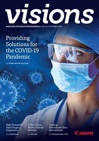
  • 1. MAGAZINEFORHEALTHPROFESSIONALS//NO.35//SEPTEMBER2020 Working together to understand your needs and challenges drives valuable outcomes that positively impact you and your patient’s future. Canon Medical’s visions and commitment to improving life for all, lies at the heart of everything we do. By partnering to focus on what matters, together we can deliver intelligent, high quality solutions. With Canon Medical, true innovation is made possible. Read more about our Mobile imaging solutions in the article on pages 10-15 or visit our website: https://eu.medical.canon Providing Solutions for the COVID-19 Pandemic 10 //MOBILE IMAGING SOLUTIONS A New Canon Medical Family Member 50 //EYE CARE MAGAZINE FOR HEALTH PROFESSIONALS //NO. 35 //SEPTEMBER 2020 Leading Solutions for Your Interventions 56 //INTERVENTIONAL X-RAY High-Resolution Hand Surgery Diagnostics 16 //ULTRASOUND
  • 2. Works are generally classified into two categories: Full length articles (e.g. clinical added value of new/special applications & technologies) and Short contributions (e.g. system testimonials, case reports, technical notes). General guidelines for authors Copyright©2015SocialMediaExaminer® All articles should be double-spaced. Full length articles Full length articles should generally include the following: - Author’s full name and highest academic degree, employer medical institution - Author’s biography (150 words) - Author’s passport-size photograph (suitable for publication); (image of 300 dpi) - 200-word abstract - Text including headline, sub-title, introduction and sections like: materials methods (which should include a full description of equipment used), results, discussion and references - Text approx 4 to 5 pages or 12.000 to 14.000 characters (not including figures, tables and photographs) - Correspondence address - Literature (no more than 10 references) - Separate, continuous numbered image- and table captions Short contributions Short contributions should generally include the following: - Author’s full name and highest academic degree - Author’s employer medical institution - Author’s biography (150 words) - Author’s passport-size photograph (suitable for publication); (image of 300 dpi) - Text including headline, sub-title, introduction and full description of methods materials/equipment used - Case Report or description of system improvements (Technical Notes) - Correspondence address - Literature (no more than 10 references) - Separate, continuous numbered image- and table captions Text The article should be saved in Microsoft Word (PC format) if possible, and, if not, in text only. Please indicate the software program and version used (Microsoft Word 2007, etc.) and whether it is a PC or Macintosh formatted document. If e-mailing, make sure to send it as an attachment, rather than embedded in the e-mail message. Symbols, formulas and abbreviations Symbols, Greek letters superscripts/Subscripts must to be identified clearly. Furthermore, the figure 1 (one) and the letter l (el) as well as the capital letter 0 and the figure 0 (zero) should be easy to differentiate. All abbreviations including units of measure, chemical names, technical or medical acronyms, names of organisations or institutions should be defined when they first appear in the text (e.g. congestive heart failure (CHF). Please refrain from using unfamiliar abbreviations, clinical slang or jargon. Images, art and tables Cite all figures and tables in text, preferably in consecutive order. Please include a caption for each figure. All captions for each figure, should be separate from the text, at the end of the manuscript on a separate page. Captions should avoid duplication of text material. Credit lines for artwork can appear at the end of the corresponding caption by stating: (Provided by first initial, last name). Black out (or give clear instructions which parts should be blackened out) of the images to not violate any data protection regulations (e.g. patient data) Do not embed figures, charts, or graphs into your document file. Please provide them as a separate file, as well as hard copy/correct .pdf file. Please use one the following formats: EPS , TIFF or JPEG. Arrows stuck onto images for purposes of delineation should be clearly visible and reproducible. Authors should indicate if they would like to have artwork returned. Each table should have a title, and all abbreviations should be spelled out or explained in a footnote. Style Title page should include full names, degrees and titles of authors, and affiliations (name of institution, city and state) for use in a by- line, as well as phone and fax numbers to facilitate sending edited copy back to author for approval. Define all symbols, abbreviations and acronyms on first reference. All manuscripts should be written in a third-person style, unless the article is specifically an editorial or first-hand review. References A maximum of 10 references is suggested. Complete references should be listed in order of citation in text, NOT alphabetically. Up to four authors will be listed; if there are five or more authors, only the first three will be listed, followed by et al. Within the text, reference numbers should appear as footnotes in parentheses or in superscript text at the end of each appropriate citation. Please do not use Microsoft Words endnote feature, as this causes major problems in the editing phase. In addition, if the reference is not in English, please indicate the language of publication. Journal example Oberhaensli RD, Galloway GJ, Hilton-Jones D, et al. The study of human organs by phosphorus-31 topical magnetic resonance spectroscopy. Br J Radiol 1987;60:367-373 Book example Welch KMA, Barkley GL. Biochemistry and pharmacology of cerebral ischemia. In: Barnett JHM, Stein BM, Mohr JP, Yatsu FM, eds. Stroke: pathophysiology, diagnosis and management. New York: Churchill Livingstone, 1986;75-90. Cover Image: A combination of two Stockphotos. VISIONS magazine is a publication of Canon Medical Europe and is offered free of charge to health professionals. The magazine is published twice a year. Registration to access full, previously published, digital editions can be done via the website: https://eu.medical.canon/visions-magazine. Canon Medical stores and uses personal data of the registration to send out the magazine and inform members about new developments. Members can customize preferences or opt-out, after registration. VISIONS magazine is covering Canon Medical’s European region and as such reflects products, technologies and services for this particular area. The mentioned products may not be available in other geographic regions. Please consult your Canon Medical representative sales office in case of any questions. No part of this publication may be reproduced in whole or in part, stored in an automated storage and retrieval system or transmitted in any manner whatsoever without written permission of the publisher. The opinions expressed in this publication are solely those of the authors and not necessarily those of Canon Medical. Canon Medical does not guarantee the accuracy or reliability of the information provided herein. News, articles and the full edition of VISIONS magazine are announced firstly, as pre-publication, via the dedicated VISIONS LinkedIn Group: https://www.linkedin.com/groups/3698045. In this group you can actively participate in discussions about the content and future direction of the magazine. Alphenix, Infinix-i, Aquilion, Aquilion ONE, Aquilion ONE PRISM, Aquilion ONE GENESIS, Aquilion PRIME, Aquilion Prime SP, Aquilion Lightning SP, Vantage Elan, Pianissimo, Vantage Orian, Vantage Galan, SURE VOI, Aplio, Viamo and Made for Life are trademarks of Canon Medical Systems Corporation. Xephilio is a trademark of Canon Inc. Olea Medical S.A.S. and ViTAL Images Inc. are Canon Group Companies. Publisher Canon Medical Systems Europe B.V. Zilverstraat 1, 2718 RP Zoetermeer The Netherlands +31 79 368 92 22 W: https://eu.medical.canon/ E: visions.eu@eu.medical.canon Editor-in-chief Jacqueline de Graaf jacqueline.degraaf@eu.medical.canon Design Layout Boerma Reclame boermareclame.com Printmanagement Printweb Media B.V. printweb.nl Photography Cojan van Toor www.cojanvantoor.nl Text contributions and editing Mélisande Rouger - Journalist and content manager Sara Sharp - The Creative Practice Folow us: © 2020 by Canon. All rights reserved. ISSN 1617-2876
  • 3. VISIONS 35 // 3 MR. NOBUYUKI HATAKEYAMA President and CEO Canon Medical Systems Europe Kind regards, //EDITORIAL © 2020 CANON MEDICAL SYSTEMS The coronavirus has presented us all with significant challenges. At Canon Medical, we remain committed to the safety and security of society and strive to minimize the impact of the COVID-19 outbreak, while maintaining customer service at the highest level for the benefit of the patients. We offer our deepest condolences to the impacted people, cities and countries . We also have the greatest admira- tion and respect for every healthcare profes- sional, who has been fighting the pandemic. After the outbreak, our team spirit in the company became even more robust. Our new European Management Team started during this challenging time but quickly developed a strong collective spirit. Our European offices aligned themselves instantly based on their local government policies trying to minimize the impact of the corona crisis. Minimizing social contact was vital in this. Thanks to the outstanding preparation of our IT colleagues, most of our employees were able to work from home and optimized the use of remote conferencing. Based on our Made for Life philosophy, Canon Medical has, of course, continue to support the healthcare society and contributed even more during the COVID-19 crisis. Through our Mobile Clinical Solutions with state-of-the-art equipment, we have been able to support hos- pitals and clinicians with their extremely high workloads. We also offer fixed rental solutions to meet local clinical needs. Inspired by a smart ‘container concept’, the Canon Medical CT Scan Unit is an impressive transportable imaging solution. It is equipped with an Aquilion Prime SP or Aquilion Lightning SP, and includes a separate operating room to lower the risk of person-to-person COVID-19 transmission. With a smart, compact design, it can be transported quickly and easily. This CT Scan Unit can be purchased or rented through a cost-efficient rental agreement. The heavy workflow is optimized thanks to the quality and innovation of our products, resulting in a more confident diagnosis. Remote connection to healthcare solutions is invaluable during the COVID-19 crisis. Especially when traveling becomes impossi- ble and access to hospitals is challenging or sometimes even impossible. Thanks to our InnerVision remote diagnostics, our highly trained engineers and application specialist are able to monitor your systems remotely and act immediately. By doing so, we can limit the interruption of your workflow of streamline patient care. You have our commitment, even more, during this challenging period. That is why we expanded our production capacity to meet the high and essential demand in the market. We have continued to expedite the delivery of parts, despite the challenges. Our mindset and philosophy are that nothing should stand between you and the critical work that you do.
  • 4. 4 // VISIONS 35 30New Diagnostic and Therapeutic Options in Eye Care EYE CARE //CONTENTS 16High-Resolution Sonography Redefines Hand Surgery Diagnostics ULTRASOUND 22Portable Performance, Perfect for the Peloton SPORTS MEDICINE, PARTNERSHIP 03 Editorial 06 News 09 President's Message 10 Providing Solutions for the Frontline of the COVID-19 Pandemic MOBILE IMAGING SOLUTIONS 16 High-Resolution Sonography Redefines Hand Surgery Diagnostics ULTRASOUND 22 Portable Performance, Perfect for the Peloton SPORTS MEDICINE, PARTNERSHIP 27 The Power of CTA and CTP for Characterization of Ischemia without Obstructive Stenosis COMPUTED TOMOGRAPHY 30 New Diagnostic and Therapeutic Options in Eye Care EYE CARE 35 Vantage Orian Opens New Perspectives in all Fields MAGNETIC RESONANCE 38 Shaping Biomedical Research of the Future MULTI-MODALITY 42 Alphenix 4D CT – Cutting-Edge Innovation in one Single Room INTERVENTIONAL X-RAY 47 The Alphenix 4D CT – More Treatments in a More Accurate Manner INTERVENTIONAL X-RAY 10Providing Solutions for the Frontline of the COVID-19 Pandemic MOBILE IMAGING SOLUTIONS
  • 5. VISIONS 35 // 5 62Canon Medical in Iceland: Landspitali Experiences MULTI-MODALITY 68Artificial Intelligence to Boost MR Imaging Quality and Productivity MAGNETIC RESONANCE 70Revealing the Secrets of Animal Mummies with Canon Technology COMPUTED TOMOGRAPHY 56Leading Solutions for Your Interventions INTERVENTIONAL X-RAY © 2020 CANON MEDICAL SYSTEMS 50 Eye Care: A New Canon Medical Family Member EYE CARE 56 Leading Solutions for Your Interventions INTERVENTIONAL X-RAY 62 Canon Medical in Iceland: Landspitali Experiences MULTI-MODALITY 68 Artificial Intelligence to Boost MR Imaging Quality and Productivity MAGNETIC RESONANCE 70 Revealing the Secrets of Animal Mummies with Canon Technology COMPUTED TOMOGRAPHY 76 First Vantage Orian 1.5T / Encore Upgrade Installed in Europe MAGNETIC RESONANCE 78 How a Flash of Feeling Becomes a Memory Forever CANON EUROPE LTD. 80 The Largest Radiology Center in Korea Increases the Success Rate of Prostate Artery Embolization (PAE) INTERVENTIONAL X-RAY 84 The Aplio i800 Takes Exotic Animals Imaging to the Next Level ULTRASOUND 90 The first Aquilion ONE / PRISM Edition in Europe COMPUTED TOMOGRAPHY
  • 6. 6 // VISIONS 35 The role for CT and Ultrasound in the acute stage of the COVID-19 disease is clear. For MR, the role is mainly in post-COVID care for patients with neurologic-, vascular- and/or cardiac pathologies. This role was discussed by leading radiology clinicians in Europe and the USA in our webinar. The panel comprised of Prof. Saba (University of Cagliari, Italy), Prof. Lima (John Hopkins University, USA), and Prof. Puig (Comparative Medicine and Bioimage Center of Catalonia, Spain) and was headed by Prof. Dousset (Bordeaux University Hospital, France). The panel discussed the situation in their respective countries and their observations of the disease with MR. Furthermore, the panel discussed the need for standardized protocols to follow up certain patients. If you are interested to learn more about the significant role the experts see for MR in the post- COVID19 world, please join the webinar here. // A reality where patients are optimally protected, risks are minimized and resources need to be optimized. Three European hospitals have joined forces in the fight against COVID-19, using CT scanners in particular, and now share their initial approach on trying to control the virus, how the hospitals managed and are still managing to cope during the pandemic and of course how CT scanners contributed to the battle against the virus. Interesting thoughts, opinions and learning curves are shared by Dr. Russell Bull (Royal Bournemouth Hospital,UK), Prof. Catherine Roy (Strasbourg University Hospital, France), Prof. Mickaël Ohana (Strasbourg University Hospital, France), Dr. Monique Brink (Radboud University Medical Center, The Netherlands) and Prof. Mathias Prokop (Radboud University Medical Center, The Netherlands) throughout the webinar ‘’COVID-19 Impact and Experiences’’. // Watch leading clinicians in Europe and the USA discuss the importance of MRI in a post- COVID-19 world. The COVID-19 crisis is perhaps the first global pandemic that has called upon the radiology com- munity to help with the delivery of crucial frontline patient care. It is abundantly clear that it is now an absolute necessity to prepare for working in the new (imaging) reality. Webinar: The Role of MR in the Post-COVID-19 Era CT Webinar: COVID-19 Impact and Experiences //NEWS Visit our website or scan the QR code: https://global.medical.canon/ service-support/ ➔ COVID-19 Resources Watch the video on our YouTube channel or by scanning the QR code: https://www.youtube.com/ watch?v=o2P4umCi6WE
  • 7. VISIONS 35 // 7 Creu Blanca strives to deliver the best point of care combined with optimal patient comfort without compromise. Dr. Alomar, Head of the Radiology Department and RD at the medical center, continuously adopts early-stage innovative technologies towards achieving this goal. The demand for simultaneously high resolution and fast imaging techniques continually increases, and AiCE could deliver both benefits without compromises. Compressed SPEEDER (CS) is another advanced technology that has been installed on both Canon Medical MR systems. This advanced software enables faster acquisition for various body parts, while adding AiCE to Compressed SPEEDER acquisition will combine the benefits to provide fast imaging with high spatial resolution. Clinica Creu Blanca private medical center in Barcelona, Spain, is the first to benefit from deep learning technologies in clinical practice with Canon Medical Systems’ groundbreaking Advanced Intelligent Clear-IQ Engine (AiCE) installed on two advanced MR systems - Canon Medical’s Vantage Orian and Vantage Galan. First European Clinical MR Site with Advanced Intelligent Clear-IQ Engine (AiCE) © 2020 CANON MEDICAL SYSTEMS An important concern for healthcare professionals is to consider the possible long backlog of patients requiring scans in a post- COVID period. Flexible technologies that can meet the dynamic needs of the present and future will be required. AiCE and Compressed SPEEDER can play an essential role in supporting smooth and fast operation and reducing waiting times. Canon Medical Systems is carrying out many installations of AiCE CS across Europe. Stay tuned to get clinical insights from early adopters! // System Vantage Orian 1.5T Protocol Knee SAG PD FatSAT Resolution 0.5 x 0.5 mm Slice thickness 2.5 mm Scan Time 1’22’’ Technologies CS x 3, AiCE, MSOFT Coil 16ch Flex SPEEDER System Vantage Orian 1.5T Protocol Ankle SAG PD FatSAT Resolution 0.5 x 0.5 mm Slice thickness 2 mm Scan Time 1’36’’ Technologies CS x 3, AiCE, MSOFT Coil 16ch Flex SPEEDER Visit our website: https://eu.medical.canon/ magnetic-resonance-aice/
  • 8. 8 // VISIONS 35 //NEWS TOKYO, February 19, 2020—Canon Inc. announced today that eight Canon Group product designs were recognized by iF International Forum Design GmbH with prestigious 2020 iF Design Awards. https://global.canon/en/news/2020/20200219.html Canon Designs Recognized with Internationally Renowned iF Design Awards for 26th Consecutive Year * Not available in Japan. This year marks Canon’s 26th consecutive year of winning iF Design Awards. Encouraged by the recognition of the Company’s design excellence, Canon will continue striv- ing to realize products that combine the highest levels of performance and design. Established in 1953, the iF Design Awards are recognized internation- ally as one of the most prestigious awards within the field of design, with outstanding industrial designs cho- sen from all over the world each year. This year over 7,200 entries from 56 countries and regions were judged by internationally active design experts across seven disciplines: product, packaging, communication, interior architecture, professional concept, service design and architecture. // Moto GP is the premier class of motorcycle and is the oldest established motorsport world championship. The race circuit in Spielberg and the team of the Emergency Medical Service sports Austria are well known for their high safety standards and ranked within the top 3 motorsport locations worldwide. For the Moto GP events, a CT Scan Unit was installed within the paddock, next to the medical center and besides the rescue helicopter and the medical car. In case of emergency on the circuit, drivers are able to get the best possible on-site care. Within 15 seconds, a full body scan can be performed and provides fast and precise data for the medical team. In case of fractures, cranio cerebral trauma or internal injuries, even if the driver is unconscious and unresponsive, the medical team At the RedBull Ring in Spielberg, Austria, the very first mobile CT scanner was used to increase the safety for drivers and take the diagnostics options to a new level. Fit to Race – Premiere For a Mobile CT in Moto GP can decide whether the patient is fit for transport. Another huge benefit is that the data can be sent to the nearby hospital in advance, allowing doctors to prepare for the patient arriving by helicopter in just a few minutes. Keeping in mind that we’re still in the middle of a global pandemic, Canon Medical makes sure that the unit is completely Corona proof thanks to separate air circuits for doctors and patients. All features and benefits combined takes medical care in the racing industry to a new level. Want to know more about the CT Scan Unit? You can read all about this solution in the article on pages 10-15 in this VISIONS edition! // iF Design Award 2020 (Product) winners: The EOS R System Imaging system Sumire Prime Fixed-focal-length cinema lenses for PL mount cameras OCT-A1 Optical coherence tomography (Eye Care, Canon Medical) IVY REC Compact digital camera (INSPiC REC in Japan) Arizona 1300 series* UV flatbed printer (Arizona 1380GT pictured) XF705 Professional handheld 4K UHD camcorder EF 400mm f/2.8L IS III USM EF 600mm f/4L IS III USM Super telephoto lenses (EF 400mm f/2.8L IS III USM pictured) PowerShot G5 X Mark II Compact digital camera
  • 9. VISIONS 35 // 9 “The fight against COVID-19” © 2020 CANON MEDICAL SYSTEMS I would like to start this message by acknowledging the frontline workers who continue to sacrifice so much in the fight against COVID-19. These have been unprecedented times for all of us, but I am particularly aware of the dedication and resilience you have consistently brought to your duties, and for that I offer my deepest thanks. I know that for many of us, the hope is to get back to normal as soon as possible, but I don’t believe that ‘normal’ will ever mean the same again. And nor should it. The health care industry, along with many other sectors, has had to change irrevocably since COVID-19 began making its way around the world. We have had to adapt - in many ways for the better - and I am inspired by the speed, agility, and conscientiousness shown by our colleagues and partners during this time. TOSHIO TAKIGUCHI President and Chief Executive Officer Canon Medical Systems Corporation In recent months, I have been lucky enough to hear from a number of customers regarding their approach to managing the pandemic - specifically with our solutions. I have been heartened by the insights they’ve shared, and I believe that the topics discussed are extremely pertinent to the future of our industry, and I hope you will get as much out of it as I have. As always, I hope that you and your loved ones are staying safe and well, and I look forward to updating you with our latest developments in due course. PRESIDENT’S MESSAGE
  • 10. 10 // VISIONS 35 PRODUCT //MOBILE IMAGING SOLUTIONS //CT Scan Unit, Mobile Trailer, Aquilion Prime SP, Aquilion Lightning SP, AiCE, COVID-19 Providing Solutions for the Frontline of the COVID-19 Pandemic CT can play a critical role in managing patients with infectious disease, but its use in this context requires specific safety and workflow protocols. One essential for health professionals during the COVID-19 pandemic is to be able to carry out CT scans, especially due to the potentially catastrophic effects of this virus on internal tissues and organs, particularly the lungs. As a consequence, there has been a sharp rise in demand for CT capacity in many hospitals, as well as extra emphasis on the importance of effective, thorough and rapid disinfection and recognition of the need to isolate scanning processes. Canon Medical Systems Europe have developed a quick and flexible response unit to meet this increased demand. VISIONS explores how this benefits healthcare institutions that are under pressure in dealing with an unprecedented public health crisis. Canon Medical’s CT Scan Unit.
  • 11. VISIONS 35 // 11 C anon Medical’s Refurbished and Mobile Imaging Solutions Group, is a well-established entity in the European market for mobile and rental solutions. Within weeks of the start of the COVID-19 crisis, the team had developed a CT Scan Unit for deployment in emergency responses. The first units were manufactured and delivered to customers in Germany, Italy and Norway. Key priorities for times of crisis The CT Scan Units are designed with some specific and key objectives in mind. Firstly, to provide hospitals with the capability to isolate CT space to keep patients and staff safe through the use of deployable imaging units. And secondly, to deliver confident diagnoses through Artificial Intelligence (AI)-based imaging for improved visualization and tech- nology and particularly fast imaging for patients with short breath-hold capacity. With the breathing difficulties associated with severe Coronavirus cases, this is extremely important. Mobile Trailer and CT Scan Unit Solutions “Our Mobile Trailers and CT Scan Units provide successful solutions for equipment procurement and capacity planning - relocatable units with flexible rental and leasing solutions.” says Johan Vochteloo, Canon Medical’s Director Refurbished © 2020 CANON MEDICAL SYSTEMS //GNEU20029 and Mobile Imaging Solutions. “Both our rental solutions are available for short- and long-term needs, and designed to be parked on site and up and running within one hour. All that is required to take advantage of the Mobile Trailer or CT Scan Unit solution, is sufficient parking and power facilities. With a European service network, we aim to provide 100% uptime. Our application staff provide full explanation, training and set-up of the equipment that is tailored to the customer’s needs.” CT Scan Unit “Our new CT Scan Unit is a fully equipped and shielded CT imaging facility. The CT Scan Unit contains separate, control-, scan- and preparation rooms, with fully separated airflows that support efficiency and comfort of patients and healthcare professionals, and meet all safety regulations at all times,” Johan continues. “The Unit is equipped with state-of- the-art imaging equipment to ensure premium image quality, fast scanning and superior workflow, with a choice between Canon Medical’s Aquilion Prime SP or Aquilion Lightning SP CT systems. Advanced technology, such as Advanced intelli- gent Clear IQ Engine (AiCE), Canon Medical’s innovative Deep Learning Reconstruction technology, can also be provided to guarantee that the best possible imaging results are available - even under emergency conditions.” “We invite you to discuss your clinical needs for mobile imaging solutions with us and we will bring together all the resources to provide scalable solutions to meet your evolving demands.” Johan Vochteloo, Director Refurbished and Mobile Imaging Solutions at Canon Medical Systems Europe.
  • 12. 12 // VISIONS 35 Based on a 12.19 m (42 feet reefer unit) shipping container, the CT Scan Unit is a 114.3 cm (45-inches) high Cube Container with a twist-lock system. It is engineered in such a way that it can be transported as one unit by road, rail, sea or air quickly and easily, to anywhere that it is needed. Unloading and set-up is achieved in a minimum amount of time. The Unit has hospital-grade walls and flooring, 360º lead-shielding, and a separated airflow for patient and operator rooms, making working easy, safe and affordable at any time. The Unit can easily be included in a tent environ- ment or combined with portacabins to create a temporary building facility. At the forefront of disease control The Leiden University Medical Center (LUMC) was one of the first hospitals in The Netherlands able to adapt to the crisis with extra CT capacity isolated from the rest of the Medical Center. “At the beginning of March 2020, we contacted Canon Medical Systems for help with a CT imaging facility for extra capacity in the growing COVID-19 crisis,” said Dr. Daan Katchaki, Manager Radiology at the LUMC. “We needed a lung CT scanner. It had to be good, quick, easy-to-install, easy-to-use, and easy-to-clean. At that time, we did not have a prepared room in our building and separated patient-rout- ing was not yet specified in regulations. However, the pandemic was evolving rapidly, and we needed fast service from Canon Medical Systems. They reacted almost immedi- ately to our needs with several possible solutions. One of the options proposed was an Aquilion Prime SP CT scanner in a trailer which was immediately available – It was exactly what we needed, and within a matter of days, the solution was transported and installed.” “As we have been Aquilion CT users since 2001, it was so easy to use the Aquilion Prime SP,” he adds. “Scan times are short and both spatial and temporal resolution are more than sufficient to diagnose lung infections.” The LUMC simultaneously built an internal department specifically for managing patients from the COVID-19 crisis. This part of the Medical Center is isolated from the rest. “With this in place, we installed and started using another Canon Medical Systems CT scanner within the building,“ says Dr. Katchaki. “We were able to switch from the Aquilion Prime SP in the Canon Medical mobile unit to an Aquilion Lightning SP (also an 80-detector-row-CT) in our building. We were delighted to have been able to use the Aquilion Prime SP CT Scan Unit in the first six weeks, when the COVID-19 outbreak was evolving so quickly. Excellent cooperation between our Hospital LUMC staff and the Canon Medical Systems staff has made our rapid response to the crisis possible.” “The Aquilion Prime SP was exactly what we needed and within a matter of days, the solution was transported and installed.” Dr. Daan Katchaki, Manager Radiology at the Leiden University Medical Center (LUMC). Control room of the CT Scan Units.
  • 13. VISIONS 35 // 13 A dynamic response Core to the success of any disease control strategy is the use of reliable, validated methods and technologies to successfully detect and manage the disease. Ever since the beginning of the COVID-19 pandemic, urgent, smooth and innovative collaboration between Canon Medical Systems and healthcare professionals throughout Europe has enabled both to work together fast to help contain the initial spread of the virus. “Canon Medical Systems’ Refurbished and Mobile Imaging Solutions has been able to offer a remarkably swift, dynamic and valuable response to the crisis, because it works so closely with customers and is tuned to their needs, even when they change drastically - within hours in this case!” says Johan. Refurbished and Mobile Imaging Solutions recently expanded team and new facility in the headquarters of Canon Medical Systems Europe in Zoetermeer, the Netherlands, have adapted their workflow to absorb the demands of a global health crisis within days. Refurbishing medical equipment is already sub- ject to many strict regulations and legal aspects. The Canon Medical Department has succeeded in increasing production capacity to meet the high demand, enabling delivery of multi- ple CT Scan Units per week. “We are continuously busy developing and implementing new technologies and extending our mobile imaging solu- tions to be able to improve our services for our customers – healthcare professionals, as well as the patients that they treat. At a time when you’re being called to do the impossi- ble, you deserve an imaging partner that innovates solutions that are ‘Made Possible’,” Johan concludes. © 2020 CANON MEDICAL SYSTEMS //GNEU20029 CT Scan Unit being transported from Canon Medical Systems Europe’s headquarters to the customer. Fast installation - Deployable imaging solution with uncompromising workflow and imaging performance. - Quick and easy transport anywhere needed by road, rail, sea or air. - Unloading and set-up in a minimum of time. - The CT Scan Unit can easily be included in a tent environ- ment or combined with portacabins to create a temporary building facility, or connected directly to the hospital. Safe and comfortable - Enables work to be efficient, comfortable and safe at all times. - Hospital-grade walls and floor installations. - Separate air flows for patient and operator rooms. High performance - State-of-the-art 80/160-row imaging equipment. - Reliable diagnoses with short scanning times and a superior workflow. - Advanced AI-based technology available to enable the best possible imaging results.
  • 14. 14 // VISIONS 35 Mobile Trailer Our Mobile Trailers are developed with the highest safety regulations and to create a comfortable environment for patient and staff. To ensure that we create the most comfortable environment, we developed a Mobile Trailer that has a good working-space, including a separate control room, specified to GDPR regulations, with air conditioning, patient monitors, outside surroundings monitors, telephone and PACS connections, contrast control panel, comfortable chairs for the operators, and privacy glass. The Mobile Trailer features a very spacious scan room, which is fully air-conditioned and can be monitored easily. It also has wall- and ceiling art, dimmable lighting that can be changed to different colors, and calming music to help the patient feel more comfortable and relaxed. The wide bore medical equipment provides a non-claus- trophobic experience for the patient. Patients restricted to beds can be maneuvered into the trailer via the patient lift. The trailer has a waiting and dressing room, washing facil- ity, panic button, and stair entrance for those not confined to a bed that can also be monitored. The Mobile Trailer is equipped with Canon Medical’s Aquilion Prime SP with Advanced intelligent Clear IQ Engine (AiCE), HII Vital data processing, Contrast Dual injector, Life Pulse, contrast oven and washing facility. The deployment model ensures that Mobile Trailer can be quickly transported to wherever needed. Before delivery, Canon Medical carries out a site survey to ensure seamless installation prior to delivery. Our Mobile Trailer solutions can provide excellent support for customers to meet peaks in demand, extend capacity for a longer period, cover a period of planned downtime, or as a rapidly deployed back-up. For governmental screening programs, these are a fantastic solution. Canon Medical’s Mobile Trailer in front of the hospital ‘Krankenhaus der Barmherzigen Schwestern Ried’ in Austria.
  • 15. VISIONS 35 // 15© 2020 CANON MEDICAL SYSTEMS //GNEU20029 “After delivery, we could immediately work in the trailer. The spacious area around the CT and the couch offers sufficient freedom for movements even for interventional procedures. Thanks to the patient couch lift, the system is also suitable for patients with limited mobility. Also the post-processing solution and the complete PACS connection is integrated in Mobile Trailer unit.” Dr. Claus Kölblinger, Head of Radiology Department at Krankenhaus der Barmherzigen Schwestern Ried, Austria. Control room in the Mobile Trailer. We aim for 100% uptime and 100% satisfaction through our outstanding European customer and service support. Renting our equipment enables clinical teams to access the latest technological developments and enhancements, while the responsibility for servicing the equipment lies with us, as per the rental agreement. Renting equipment can be a rewarding solution to fulfil immediate clinical needs while avoiding larger capital investments. //
  • 16. 16 // VISIONS 35 INTERVIEW, CLINICAL CASE //ULTRASOUND //Aplio i800 High-Resolution Sonography Redefines Hand Surgery Diagnostics The Handchirurgie Seefeld (hand surgery) clinic in Zürich, Switzerland, relies on high-tech medicine with the state-of-the-art Aplio i800 ultrasound diagnostic system of Canon Medical. T he major challenge in hand surgery is the precise handling of delicate structures. There is hardly any other specialty where elements of plastic surgery, orthopae- dic surgery and micro surgery are so closely related and deal with extremely complex and small anatomical structures. Therefore, diagnosis with sub-millimetre-level accuracy has a significant impact on optimal treatment success. Specialists like Dr. Sebastian Kluge, FMH professional association specialist doctor for surgery and hand surgery, ultrasound diagnostics, musculoskeletal system SGUM (Swiss Society of Ultrasound in Medicine), use the high-tech Aplio i800 sonography device to redefine the approach to a highly differentiated field. “The Aplio presents structures and situations in a spatial resolution which would not be possible for any CT or MRI, whilst simultaneously providing the possibility of dynamic assessment,” he explains. Dr. Sebastian Kluge, FMH professional association specialist doctor for surgery and hand surgery, ultrasound diagnostics, musculoskeletal system SGUM (Swiss Society of Ultrasound in Medicine), appreciates the huge advan- tages of sonography for hand surgery because it provides insights like no other diagnostic method and therefore redefines the causes of diagnosis. Dr. Sebastian Kluge started his medical career around 20 years ago at the Berufsgenossenschaftliche Unfallklinik (occupational accident clinic) in Ludwigshafen, where he completed civilian service after finishing school and discovered his interest in hand surgery. “Until that point I was all set on studying drums and piano. It was only my fascination with hand surgery that made me embark on a career in medicine.” Today he is one of the most well-known specialists in his field, and his expertise makes him highly respected by his patients and his medical colleagues alike. He has delivered lectures and ultrasound seminars around the world and worked with fellow specialists to write a standard textbook entitled “Ultraschalldiagnostik der Hand” (Diagnostic Ultrasound of the hand).
  • 17. VISIONS 35 // 17 Ultra-high frequency redefines causes of diagnoses The Aplio i800 sonography device from Canon Medical is an important “hand tool” in his daily routine. He decided to invest because “this system offers previously unforeseen new possibilities in hand surgery.” By this he means in particular the extremely high resolution that is possible in combination with the 24 MHz probe, therefore enabling the differentiation of structures down to a sub-millime- © 2020 CANON MEDICAL SYSTEMS //ULEU200076 “The presentation of soft tissue and bone surface and their dynamic interaction with a resolution of 24 MHz is stagger- ing and proves the huge potential of sonography.” Dr. Sebastian Kluge, FMH professional association specialist doctor for surgery and hand surgery, ultrasound diagnostics, musculoskeletal system SGUM (Swiss Society of Ultrasound in Medicine). tre level. In particular, where the small distance between the skin surface and bone means that a high penetration depth is not needed, the system displays a sonographic image in the highest resolution. Dr. Kluge therefore sees big advantages for high-frequency ultrasound in rheumatology, neurology and orthopaedics. Dr. Kluge’s specialties are post-traumatic changes in the forearm, wrist and fingers, the non-operative and operative treatment of nerve compression syndromes and inflamma- tory-degenerative and arthrotic changes in the hand and finger joints, including joint replacement. “The spectrum of causes problems and the range of treatment options available are immense”, the doctor explains. “In this respect, various suspected diagnoses need to be isolated from each other extremely precisely and treatments need to be planned and implemented with the utmost precision,” he added. The Aplio i800 is ideally suited for all of this work. This is because while sonography was previously carried out at sound frequencies of between 15 MHz and 18 MHz, Canon Medical offers matrix technology in the ultra-high frequencies of 24 MHz and 33 MHz. “The resulting increase in spatial resolution not only allows the person carrying out the examination to take the very important step of moving from a suspected diagnosis to absolute diagnostic certainty for many matters, it actually redefines the cause of certain diagnoses.” High-resolution imaging allows for high- precision interventions The extremely high resolution of the sonography system provides valuable support even for ultrasound-controlled interventions. The most common indication in hand surgery is stenosing tenosynovitis. This imbalance between the annular pulley width and flexor tendon thickness may result in clicking of the affected finger. When attempting non-oper- ative treatment, a cortisone injection can even be given with ultrasound support. This results in a lower rate of side effects due to more precise placement of the needle in the digital canal. However, the method only really comes into play in the event of differentiated hand surgery interventions such as occult wrist ganglions, which are “destroyed” under sono- graphic guidance, so in many cases the complaints can be per- manently eliminated even without surgery. “In these special cases, the immense spatial resolution even allows for targeted ultrasound-controlled anaesthesia of the nerve supplying the wrist joint capsule, which makes this a pain-free intervention. Even minimally-invasive carpal tunnel operations can now be done under sonographic guidance.”
  • 18. 18 // VISIONS 35 Another example for the usefulness of high-resolution ultrasound can be seen in the case of post-operative compli- cations. Implant protrusions, which may occur occasionally after surgery on radial fractures, for example, may cause irritation of adjacent flexor and extensor tendons. This often results in an extensor tendon rupture, usually of the long extensor tendon in the thumb. “The ideal method for localising any implant-related complications of this kind is ultrasound”, says Dr. Kluge. In these cases, CT shows the implant protrusion but not the soft tissue trauma caused, while in an MR scan the soft tissue injury is frequently covered by metal artefacts. “In this case, sonography allows for optimum differentiation of bone surface, soft tissue and implant – in high resolution”, Dr. Kluge summarised. “We not only have a responsibility to the patient, but also to our colleagues.” Patients are also repeatedly referred by fellow hand surgeons for ultrasound diagnosis – usually with very precise and differentiated problems. “In this case in par- ticular, the expectations of a precise diagnosis are extremely high, because our colleagues base their further treatment on this diagnosis”, said Dr. Kluge. can be precisely measured before the operation and nerve transplants can be planned or ordered in the correct size. In his experience, “the Aplio i800 is also an extremely user- friendly system”. An example of this is “when the program is selected, the system also changes to an optimised key assignment in the control panel, which means that there is no need to search for functions unnecessarily.” However, the perfect integration of the device into the practice’s work processes is also important for Dr. Kluge. The ultrasound device is integrated into the practice’s information system via a wireless network. After the hand specialist has carried out a clinical examination on a patient, he can access all of the patient information necessary for the examination on the Aplio i800 and then connect further diagnostics there immediately. The results are then automatically transferred to the practice’s software and are available there for further processing. “An accelerated workflow is just as important for us as com- prehensive clinical examination is for our patients. Suspected clinical diagnosis are often able to be confirmed or excluded during the initial consultation thanks to the sonography. In combination with the 24 MHz ultrasound probe, structures can be shown precisely and without artefacts. Optimal ease-of-use makes work processes easier The hand specialist has got to know the advantages of ultrasound tech- nology “from the bottom up”. In the wards where he completed his medical training in Frankfurt, Bern and Zurich, great importance was always placed on sonography. This is why he gives lectures and seminars on diagnostic and treatment options with the aid of modern sonography systems. Dr. Kluge gets to the heart of the matter when he says “the processing of the sound information and the resulting image quality in the new Canon Aplio systems are phenomenal.” They can be used to produce very exact findings. Even for the finest structures such as digital nerves it is possible to differ- entiate between intact and destroyed nerve fibres. This is particularly helpful when planning nerve reconstructions, because the length and thickness of a nerve transplant which is needed
  • 19. VISIONS 35 // 19© 2020 CANON MEDICAL SYSTEMS //ULEU200076 Canon Medical’s Aplio i800. Stenosing tenosynovitis. This is one of the most common conditions in the hand. In addition to primary causes such as the thickening of the A1 annular tendon at the level of the joint at the base of the finger (left), the condition can also be caused by synovitis of the flexor tendons. Subcutaneous Dupuytren's nodules can also radiate into the digital canal or the A1 annular pulley and result in a secondary constriction of the digital canal (right). A differentiation, which is relevant for the therapeutic approach, can be made reliably using sonography. In many cases, there is absolutely no need for additional imaging procedures such as CT and MR, which in the latter case also results in cost reductions and reduced radiation for the patient from CT”, the hand specialist explains. Thanks to the iDMS (intelligent Dynamic Micro-Slice) function of the matrix probes from Canon Medical, the crystal elements can be aligned so that a very narrow and focus-independent sound field is produced. “This “sharpening (of) imaging slice thick- ness” makes it possible to focus on even the smallest joint spaces between individual carpal bones”, stresses Dr. Kluge. A new look for micro-vascular situations “In addition to the usual Doppler and Power Doppler func- tions, with Canon there is an additional Doppler algorithm available, which can show the micro-vascular supply to a region very specifically”, adds the hand specialist. This “Superb Micro-vascular Imaging” (SMI) for contrast-free examination of micro-vessels provides insights that would otherwise be difficult or impossible to achieve. As in subtrac- tion angiography, the surrounding tissue can also be hidden and the focus can be placed solely on the micro-vascular supply. Inflammatory joint conditions such as synovitis can be presented in a highly differentiated manner using the sonog- raphy device from Canon Medical. “Thanks to the quantifica- tion of the synovial circulation, inflammatory joint changes can be diagnosed and the activity of the condition can be assessed. The response to medication, which may be pre- scribed for rheumatoid arthritis for example, can therefore be monitored and the dose can be adjusted if necessary. Specific changes in the cartilage surface allow additional conclusions to be drawn about causal crystal deposition diseases.” Dr. Sebastian Kluge is also fascinated with the efficiency of the Superb Micro-vascular Imaging in the detection of glo- mus tumours, which are frequently located under the finger nail and can be very painful for patients. The Aplio i800 captures the hyper- vascularisation that occurs: “The coloured display means that the increased vascular supply to these tumour areas is positively high- lighted from their surroundings.” Sonography will remain exciting in future Sonographic imaging has played a key role in the life of doctor and passionate music lover Sebastian Kluge for around 20 years, and to some extent he thinks of it as a work of art. He has been using it for a good 15 years as a hand surgeon and knows that “this diagnostic technology offers enormous opportunities that have by no means been exhausted yet”. He looks forward to the next developments, which he is monitoring keenly. He has already been able to scrutinise the Aplio i800 in advance as part of his own advanced training events. He thinks there is great potential in the further definition of software algorithms and image processing, and also in the further miniaturisation of the systems. He said “mak- ing high tech ultrasound systems like the Aplio i800 from Canon Medical portable would be a vision that would be worth achieving in many respects, not least because it would mean that high-frequency imaging could even be used in ultrasound-guided operations on the hand without having to be tied to a single operation site.” //
  • 20. 20 // VISIONS 35 De Quervain's tenosynovitis. This is an entrapment syndrome of the first extensor tendon compartment. The tendons of the abductor pollicis longus and extensor pollicis brevis muscles are compromised and can barely be differentiated from each other as individual structures anymore, while the extensor retinaculum displays a halo-like thickening (left). Some patients present with an additional partition of the first extensor tendon compart- ment (subsheath), which can also be easily distinguished using sonography (right). Knowledge of this is important for treatment, as patients with a subsheath do not respond as well to non-surgical treatments. Differentiation of tumours. Ultrasound also allows for reliable differentiation of many masses in the hand (left). This has become established not only for periph- eral nerve tumours. Giant cell tumours of the tendon sheath and pigmented villonodular synovitis (PVNS) emanat- ing from the joints are histologically identical and can only be differentiated based on their cause. This has relevant effects on the therapeutic approach, because in the case of PVNS (below) the joint in question also needs to be cleared intraoperatively in order to minimise the risk of the tumour relapsing.
  • 21. VISIONS 35 // 21 Extensor hood lesions. They are a frequent cause of persistent swelling and pain following trauma in the joint at the base of the finger. In many cases, subluxations of the respective extensor tendon(s) occur. This means that there can already be a clinical presumption of the diagnosis. However, sometimes these clinical indications are more subtle, so the correct diagnosis can often only be made using imaging procedures. In comparison with magnetic resonance imaging, the advantage of sonography lies in the higher resolution and the possibility of a dynamic examination. © 2020 CANON MEDICAL SYSTEMS //ULEU200076 Ganglia of the digital canal. Ganglia are benign tumours that may occur in both the wrist and the digital canal. They are filled with a clear, gel-like, viscous fluid. They can easily be differentiated due to their homogeneous reflex patterns, the sound amplification remote from the swelling and a shadow at the edge of the cyst (Dr.ip-off phenomenon). On the digital canal they can mainly be found at the level of the A1 and A2 annular pulley (above) and if non-surgical treatment is requested they can even be treated using ultrasound-guided needle aspiration (left). Implant-related complications. Postoperative complications caused by osteosynthesis materials which have been inserted are not uncommon. In the case of radial fractures treated using surgery in particular, prominent plate designs or excess screw length may result in irritation and a rupture of the flexor and extensor tendons. In this situation, the diagnostic advantage of sonography in comparison with CT relates to the simultaneous presentation of the cortex, relevant implant protrusion and soft issue, and the advantage over MRI relates to the absence of metal artefacts.
  • 22.
  • 23. VISIONS 35 // 23 INTERVIEW //SPORTS MEDICINE, PARTNERSHIP //Deceuninck - Quick-Step, The Wolfpack, Viamo sv7 © 2020 CANON MEDICAL SYSTEMS //GNEU20031 There’s no doubt that professional cycling is an endurance sport like no other. Races, such as the infamous Tour de France, cover unimaginable distances and torturous terrains that stretch these elite athletes to their mental and physical limits. For the 28 riders of Belgium’s celebrated Deceuninck – Quick-Step World Tour Cycling Team, this is a way of life. Since the team’s inception in 2003 they have racked up an incredible 700 UCI victories, includ- ing 19 Monuments, 4 World Road Championships, 6 World ITT Championships, 4 World TTT Championships, 2 World Cups and an Olympic Title. Interview with Patrick Lefevere (CEO), Koen Pelgrim (Head Coach) and Dr. Toon Cruyt (Head of Medical) from Cycling Team Deceuninck – Quick-Step. Portable Performance, Perfect for the Peloton T he Wolfpack, as they are affection- ately known, takes its nickname from a race report written by Sports Director Brian Holm who concluded “we are the wolfpack and we don’t take pris- oners.” But more than just an inside joke, Team CEO Patrick Lefevere believes that the name describes their ‘family’ well: “We race together, win together and live together – and nobody is ever left behind. The team is stronger than the single individuals.” This extends to the support network around The Wolfpack who attend to their performance, health and wellbeing all year round, as well as in the peloton.
  • 24. 24 // VISIONS 35 Head Coach, Koen Pelgrim and Head of Medical, Dr. Toon Cruyt, lead this team of professionals who play a critical role in the Deceuninck – Quick-Step’s success. “I’m responsible for designing training programs for our riders and follow up their performance, both in training and racing,” explains Koen. “It involves a variety of things like decision making and planning of team training camps and collaborating with my colleagues on the race programs of our riders.” Dr. Cruyt’s priority is the physi- cal health of the riders and he regularly deals with the inevitable injuries that occur in such a gruelling elite sport. Some are fairly straightforward (such as abrasions, saddle sores and muscle strain), but there are also more serious injuries, like fractures and concussions, that might put a rider out of action long- term if not treated swiftly. Together, they manage the needs of the riders through a demanding and strenuous race calendar. “Cycling on a professional level is always seeking the limits and every day the riders take big risks,” says Dr. Cruyt. “When a cyclist is seriously injured during a race, it has a big impact on the whole team. In such moments we are confronted with the ultrasound scanner, which is around the size of a tablet computer and can be used to give an immediate diagnosis on muscle injuries, detecting tears and the presence of fluid. “It has significantly evolved the way we look after the team,” says Dr. Cruyt. “For example, in case of localised muscle pain, with ultrasound you can make a differential diagnosis of hematoma or muscle strain or muscle tear. In case of hematoma you can let the rider start the next day, in case of a muscle tear we stop him. Being able to use the scanner in a quick and efficient manner, at the point of care, wherever we are, can make a huge difference to a rider’s recovery.” The timeframes within which the stages take place means that as well as being on the road and in remote locations, Deceuninck – Quick-Step are also against the clock. By the time the cyclists have completed a stage and have returned to their hotel, it’s late and the following morning starts early, so it’s an incredibly narrow window of opportunity in which to find and access the nearest ultrasound facilities. Dr. Cruyt gives the example of a rider who crashed and suffered a swollen elbow, which was x-rayed in hospital, fact that cycling is a dangerous sport.” However, he has been attending to the injuries of cycling teams for over twenty years, rising to the challenges of being a ‘medic on the move’ and now has the support of an extended team through a partnership with Canon Medical Systems Europe. This gives Dr. Cruyt and his fellow medical profes- sionals full access to the most up-to- date diagnostic imaging equipment. A handheld tool for medics on the move The whole team shares an appreciation of the enormous role that technology now plays in addressing the unique scenarios that team cycling regularly presents. When following the rid- ers and administering on-the-spot treatment, doctors can encounter the sort of unconventional and unusual locations that make hospital visits difficult. In the event of an accident, pro cyclists will often have a quick check-up with the race doctor (who follows the peloton) and then resume cycling to finish the stage, rather than lose their place in the race. Afterwards, Dr. Cruyt and his team make a more detailed examination, using tools like Canon Medical’s Viamo sv7 portable “The team is stronger than the single individuals.” Patrick Lefevere, CEO Deceuninck – Quick-Step.
  • 25. VISIONS 35 // 25 with the race. The portable ultrasound meant that they could examine him immediately and, to his relief, they discovered no muscle injury, “He could start Paris-Nice with the guarantee that his efforts were not going to aggravate his injury and that time would heal his discomfort.” Recovery times are understandably variable, but Dr. Cruyt describes professional cyclists as “very tough”. Common abrasions and contusions mean that they will usually continue as normal unless they have other symp- toms. A broken collarbone may require osteosynthesis (having the bones fixed with plates, screws or wires) and see the rider back on the rollers after three days, cycling after a week and racing in around three weeks. but showed nothing untoward. Yet, the next day the rider could not bend his elbow. “With Canon Medical’s Viamo sv7 we can check if there is a traumatic bursitis or if there is an intra-articular bleeding which can be an indication for further imaging of the elbow,” he explains. “Most of the injuries in cycling are multiple injuries and it’s often only one or two days later that some painful sites become manifest. © 2020 CANON MEDICAL SYSTEMS //GNEU20031 At this point the ultrasound is the number one way to reassure the cyclist that it won’t do any harm to race a few days with pain, and that it will only get better.” Diagnosis, recovery and long- term planning Knowledge is most definitely power in performance sport and having a tool which can provide the team medics with immediate insight allows them to effectively and accurately manage the capabilities of the riders as they work through the season. For example, Dr. Cruyt treated a rider who had arrived at the 2020 Paris-Nice race earlier this year having crashed at a race the previous weekend. He had a large and painful hematoma on his left hip but was still insistent on going ahead Koen Pelgrim, Head Coach Deceuninck – Quick-Step.
  • 26. 26 // VISIONS 35 More serious fractures (femur, vertebrae and the like) can have a recovery period of up to six months. However, preparations and objectives for the team are put in place well in advance and early diagnoses support riders to make a smooth tran- sition back into the training and racing schedule. Even this year, with the chal- lenges brought forth by Covid-19, Koen Pelgrim feels prepared and optimistic. “The main objectives remain pretty much the same as in a normal year, with the highlights being the grand tours and the classics,” he explains. We prepare with a training camp and then the further preparation depend- ing on the race. For the Tour de France for example, riders will do a big block of racing at the altitude camp to be 90-95% ready, followed by a few prepa- ration races early August to fine tune.” Beyond the normal scope of medical partnerships, Canon Medical have also been able to provide Deceuninck – Quick-Step with a Canon EOS 90D camera and Canon EF 70-200mm and EF 50mm f/1.4 USM lenses to support their content creation throughout the season. Fans will be treated to creative and beautifully captured images and videos from wherever The Wolfpack are. It’s an additional dimension to the relationship made possible by belong- ing to a global family with exceptional imaging technology at its very heart. The team are in superb hands, who have access to the very best tools available to elite athletes. For team CEO Patrick Lefevere this, along with the confidence and drive to succeed are a winning combination. “The biggest challenge of the team is to reinvent ourselves every year, to keep things fresh and keep everyone motivated to be the best that they can be.” // “The Viamo has significantly evolved the way we look after the team,” With Canon Medical’s ultrasound scanner we can make a huge difference to a rider’s recovery.” Dr. Toon Cruyt, Head of Medical, Deceuninck – Quick-Step. © Deceuninck - Quick-Step Cycling Team. Watch out for The Wolfpack on Twitter (@deceuninck_qst) and keep up with the latest team news on their website: www.deceuninck-quickstep.com
  • 27. VISIONS 35 // 27 SCIENTIFIC //COMPUTED TOMOGRAPHY //Aquilion ONE, CORE320, CTA, CTP, CAD, SPECT The Power of CTA and CTP for Characterization of Ischemia without Obstructive Stenosis Patients presenting with symptoms or other signs of myocardial ischemia in the absence of any significant epicardial coronary stenosis are at present underdiagnosed and undertreated as the cur- rent diagnostic pathway remains oriented towards finding obstructive coronary disease. Combined anatomic and functional imaging provided with coronary CT angiography (CTA) and myocardial CT perfusion (CTP) could address an important need to better identify these patients. A large percentage of patients undergoing invasive coronary angiography do not show significant obstructive coronary artery disease (CAD), despite their symptoms of chest pain1 . This finding, called ischemia without obstructive stenosis or INOCA, is reported in up to two thirds of patients and is more common in women1 . Given the absence of any significant stenosis, these patients are not candidates for revascularization, are often falsely reassured and are frequently not offered any specific treatment or follow-up. Yet, studies have consistently shown that these patients are still at elevated risk for cardiovascular events, indicating the need for adequate and timely recogni- tion of this condition2 . Coronary CTA has become an established imaging test for the clinical work-up of patients presenting with chest pain3 . During the same session also myocardial perfusion imaging can be performed to assess the presence of ischemia, a strat- egy that further enhances the diagnostic accuracy of cardiac CT and is increasingly applied4 . In the unique global multicenter CORE320 trial, 381 patients with suspected or known CAD were enrolled to undergo both coronary CTA and stress CTP using the Aquilion ONE from Canon Medical Systems in addition to invasive coronary angiography and SPECT perfusion imaging, regardless of stenosis severity5 . The trial confirmed high accuracy of combined coronary CTA and CTP obtained with wide area detector CT to identify patients with flow-limiting CAD as defined by a significant stenosis on invasive coronary angiography and a corre- sponding perfusion defect on SPECT imaging5 . The CATCH2 trial built on to this experience and, again using the Aquilion ONE, showed in 600 patients that a post-discharge diagnostic strategy of coronary CTA and CTP safely reduced the need for invasive examination and treatment in patients suspected of CAD6 . “The combination of CTA/ CTP can potentially be a more sensitive, more informative and fully non-invasive method to assess CAD as compared to invasive coronary angiography and SPECT imaging.” Joanne D. Schuijf, PhD © 2020 CANON MEDICAL SYSTEMS //CTEU200152 Combined anatomic and functional imaging may however also provide important clues for diagnosis and management in those patients who present with chest pain complaints in the absence of any significant epicardial lesions. Recently an ancillary investigation of the CORE320 trial, specifically addressing this question, was published in Radiology with its imaging findings featured on the cover7 . The primary objective of the study team was to explore the prevalence of INOCA in this high risk population using CTA and CTP. In addition, advantage was taken of another aspect of cardiac CT, namely that information on coronary atherosclerosis and plaque can simultaneously be derived in addition to the mere detection of coronary stenosis. Thus, patients with INOCA identified on CT were further characterized in terms of their clinical characteristics as well as extent and type of coronary plaques.
  • 28. 28 // VISIONS 35 ization, which has become almost standard on CTA, cannot be performed using invasive coronary angiography. Also, CTP imaging has a higher sensitivity to detect ischemia and can identify abnormalities in myocardial perfusion even in the presence of only moderate coronary narrowings. While at present SPECT imaging followed by diagnostic invasive coronary angiography is still more common in clinical practice to manage patients with suspected chest pain, the observations from this ancillary study re-affirm that the combination of CTA/CTP can potentially be a more sensitive, more informative and fully non-invasive method to assess CAD. Moreover, given the completely non-invasive and fast nature of CT, patients tend to strongly favor CT over other cardiac examinations8 . More recently, also dynamic CTP has been introduced and further developed allowing the quantification of absolute myocardial blood flow and coronary flow reserve which may further enhance the potential of CTA/CTP for clinical decision making9,10 . Figure 1. Patient with atypical angina. The presence of any significant stenosis was ruled out on both coronary CTA and invasive coronary angiog- raphy. On CTP, a defect corresponding to the LAD territory was identified during stress but not during rest, indicating the presence of ischemia and providing a potential explanation for the patient’s recurrent symptoms. In the entire study cohort, INOCA was identified on combined CTA and CTP in approximately one of every 10 patients. An image example is shown in Figure 1. While men showed a higher percentage of obstructive CAD, INOCA was seen in twice as much women. In addition, a link with increased body mass index was observed. Interestingly, patients with INOCA showed a higher plaque burden as compared to patients with normal perfusion on CTP and no stenosis on CTA. Also, high-risk features like positive remodeling and low attenuation plaque were more often seen. Ischemia on CTP in patients with completely normal coronary arteries (without any evidence of plaque) was very rare. These findings suggest that patients with INOCA should be treated medically not only to control their chest pain symptoms, but also to slow down the build-up of atherosclerotic plaques. Analyses were also performed using invasive coronary angiography and SPECT imaging. Overall, findings were comparable yet less informative. Detailed plaque character- “The combination of CTA and CTP, as can be easily performed with the Aquilion ONE, provides a promising opportunity for detailed and non-invasive CAD phenotyping.” “Non-invasive imagers could play a crucial role in recognizing phenotypes beyond obstructive CAD and guide referring physicians in management decisions.”
  • 29. VISIONS 35 // 29© 2020 CANON MEDICAL SYSTEMS //CTEU200152 Listen to the podcast by Radiology Editor David A. Bluemke, MD, PhD, providing a clear explanation of the CORE320 study, the value of wide area detector CT by Canon for cardiac imaging and the concept of INOCA in addition to the main study findings. Link to the full article in Radiology. Joanne D. Schuijf, PhD Clinical Research Manager, Global RDC, Canon Medical Systems Europe. References 1 Kunadian V, Chieffo A, Camici PG, et al. An EAPCI Expert Consensus Document on Ischaemia with Non-Obstructive Coronary Arteries in Collaboration with European Society of Cardiology Working Group on Coronary Pathophysiology Microcirculation Endorsed by Coronary Vasomotor Disorders International Study Group. EuroIntervention 2020. 2 Bairey Merz CN, Pepine CJ, Walsh MN, Fleg JL. Ischemia and No Obstructive Coronary Artery Disease (INOCA): Developing Evidence-Based Therapies and Research Agenda for the Next Decade. Circulation 2017;135:1075-92. 3 Knuuti J, Wijns W, Saraste A, et al. 2019 ESC Guidelines for the diagnosis and management of chronic coronary syndromes. Eur Heart J 2019;41:407-77. 4 Patel AR, Bamberg F, Branch K, et al. Society of cardiovascular computed tomography expert consensus document on myocardial computed tomography perfusion imaging. J Cardiovasc Comput Tomogr 2020;14:87-100. 5 Rochitte CE, George RT, Chen MY, et al. Computed tomography angiography and perfusion to assess coronary artery stenosis causing perfusion defects by single photon emission computed tomography: the CORE320 study. Eur Heart J 2014;35:1120-30. 6 Sorgaard MH, Linde JJ, Kuhl JT, et al. Value of Myocardial Perfusion Assessment With Coronary Computed Tomography Angiography in Patients With Recent Acute-Onset Chest Pain. JACC Cardiovascular imaging 2018;11:1611-21. 7 Schuijf JD, Matheson MB, Ostovaneh MR, et al. Ischemia and No Obstructive Stenosis (INOCA) at CT Angiography, CT Myocardial Perfusion, Invasive Coronary Angiography, and SPECT: The CORE320 Study. Radiology 2020;294:61-73. 8 Minhas A, Dewey M, Vavere AL, et al. Patient Preferences for Coronary CT Angiography with Stress Perfusion, SPECT, or Invasive Coronary Angiography. Radiology 2019;291:340-8. 9 Kikuchi Y, Oyama-Manabe N, Naya M, et al. Quantification of myocardial blood flow using dynamic 320-row multi-detector CT as compared with (1)(5)O-H(2)O PET. European radiology 2014;24:1547-56. 10 Tsuneta S, Oyama-Manabe N, Kameda H, et al. Improvement of image quality on low-dose dynamic myocardial perfusion computed tomography with a novel 4-dimensional similarity filter. Medicine (Baltimore) 2020;99:e20804. Summary The combination of coronary CTA and CTP as can be easily performed with the Aquilion ONE series provides a prom- ising opportunity for detailed and non-invasive coronary artery disease phenotyping, including the characterization of ischemia without obstructive stenosis. Correct identifica- tion of the latter is important as these patients do not benefit from revascularization but require optimal medical therapy to improve their prognosis. As senior author and principal investigator of the CORE320 trial Dr João Lima (Johns Hopkins School of Medicine, Baltimore, USA) points out, non-invasive imagers could play a crucial role in recognizing phenotypes beyond obstruc- tive CAD and guide referring physicians in management decisions. To this end, the combination of CTA and CTP with wide area detector CT will be an important tool. //
  • 30.
  • 31. VISIONS 35 // 31 INTERVIEW //EYE CARE //XEPHILIO OCT-S1, OCT-A1, WIDE FIELD SWEPT SOURCE © 2020 CANON MEDICAL SYSTEMS //ECEU20002 Canon Medical’s Xephilio OCT-S1 and OCT-A1 will help improve eye care by enabling to scan larger areas of the retina towards the periphery and the vitreous from the cortical to the mid-vitreous, according to Prof. Paulo E. Stanga, a consultant ophthalmologist and vitreoretinal surgeon at London Vision Clinic, UK. VISIONS spoke with Prof. Paulo E. Stanga, London Vision Clinic, UK. New Diagnostic and Therapeutic Options in Eye Care T hese systems have recently been installed at his London Vision Clinic and their numerous benefits - ease of use, fast acquisition time and high imaging resolution - could help foster screening and reduce reliance on fundus fluorescein angiography in many scenarios, he believes. The devices have renewed the interest of the team in retinal and vitreous imaging, as they bring new diagnostic opportunities and thera- peutic options, he told Visions in an exclusive interview.
  • 32. 32 // VISIONS 35 Could you describe your team at London Vision Clinic? My clinical team consists of a retina research assistant, a lead retinal nurse and eleven other nurses and ophthalmic tech- nicians shared between my retinal service and the corneal laser service here at LVC. We are supported by a team of patient care coordinators, clinic coordinators and the management team. The introduction of Canon Medical’s OCT-S1 and OCT-A1 has renewed the team’s enthusiasm in retinal and vitreous imaging, specially in high myopia, a condi- tion that affects a significant percentage of my patients through my partnership with world-renowned Prof. Reinstein and his corneal laser service and clinic. How many people in your staff cur- rently use the latest Canon Medical Xephilio OCT devices? My retina research assistant Abi Orr and I routinely use these systems, and they are increasingly used by our team of oph- thalmic technicians and nurses to scan patients under the care of the corneal laser service - which consists of three surgeons who are supported by optometrists. Was any specific training required to use the Xephilio OCT systems? The Canon Medical Eye Care Team has been collaborative by visiting London Vision Clinic to initially train me and a These devices may also perhaps allow us to reduce our dependency on fundus fluorescein angiography. Widefield OCT angiography is changing the way we assess and diagnose diabetes. What kind of procedures do you perform with the Canon Medical Xephilio OCT-S1 and OCT-A1? With the Xephilio OCT-S1, we mainly use the Widefield 3D vitreous, Radial OCT, Multi-cross (grid) and Cross OCT as well as Widefield OCT Angiography (OCT-A) scanning protocols. And with the Xephilio OCT-A1, we mainly use the High-resolution Topography Maps (3um)and OCT-A. What are the main advantages for you and your patients? Imaged patients will benefit from using the new Canon Wide Field Swept Source and high resolution imaging modalities, resulting in additional information that improve diagnosis and management. few other team members on the latest Xephilio OCT system. As it is very intui- tive and user-friendly, we have carried out ourselves further in-house training for the rest of the clinical team. What improvements do you expect for your daily practice with this new equipment? The Xephilio OCT-S1 and OCT-A1 will allow us to improve patient care by allowing us to scan larger areas of the retina and not just the posterior pole. The systems will help us image the retina using higher resolution and image not only the vitreoretinal interface but also deeper from the cortical towards the mid-vitreous. Post Treatment Single-scan Wide-Field OCT Angiogram. Perfused and non-perfused retinal areas are clearly defined without the need of intravenous dye. Note that the residual fibrosed superior mid-peripheral NVE remains perfused and the residual minimally fibrosed temporal mid-peripheral NVE is perfused.
  • 33. VISIONS 35 // 33 How would you describe your collaboration with Canon Medical Systems? The collaboration with Canon Medical Eye Care Team is going very well. We have regular video calls and email correspon- dence and, where possible, in-person meetings to discuss our experience with the Xephilio OCT systems currently at London Vision Clinic. Ori Zahavi, Clinical Specialist within Canon Medical Eye Care, is always quick to respond to any queries and happy to provide advice on how we can improve our imaging techniques and use the system to its full potential. The Eye Care Team is very receptive of any recommendations/feedback regard- ing how the system could be improved in the near future and we find this not only essential in the company we collaborate with but also, at the end of the day, to bring potentially significant benefit for our patients. We are very happy with Canon Medical’s systems and we are looking forward to developments to be made in the near future as part of our collaboration with the Eye Care Team. For example, these technological advances increase our understanding of which patients suffering from an acute Posterior Vitreous Detachment (PVD) are at risk of developing new or additional retinal tears and perhaps which patients with peripheral retinal lesions may benefit from prophylactic laser retinopexy. The imaging capabilities of these devices significantly facilitate the assessment of the cortical vitreous, the mid-peripheral retina and areas of retinal non-perfusion. Highmyopiaisnotoriouslydifficultto imageusingOCTtechnologyduetothe presenceofartefacts.However,these aresignificantlydiminishedwiththe XephilioOCT-S1,partlythankstothe23 mmscanlengthandthe5.3mmdeep imagingwindow.Ialsoexpectfewerdia- beticpatientswillneedtoundergofundus fluoresceinangiographyasaresult. Do you think the Xephilio OCT-S1 and OCT-A1 offer new unexplored possibilities? Absolutely. The capabilities of the Xephilio OCT-S1 to acquire single-scan wide field OCT angiograms and to improve visualisation of the mid- peripheral retina, the vitreoretinal interface and the deeper cortical vitreous should bring new diagnostic opportunities and therapeutic options. © 2020 CANON MEDICAL SYSTEMS //ECEU20002 I am excited to see many opportunities for further development thanks to our partnership with the Canon Medical Eye Care Team. Do you foresee any change in eye care patient management with these systems? Xephilio OCT-A1 is easy to use with a very fast acquisition time and high imaging resolution. This could be beneficial for screening purposes and clinics with a high number of patients on a daily basis. Xephilio OCT-S1 could be of significant benefit in the care of patients with retinal vasculopathies, such as diabetic retinopathy, vein occlusions or vascu- litis, and reduce their dependence on invasive, costly and time-consuming fundus fluorescein angiography. By definition a wider scan area facilitate earlier detection and treatment of a retinal pathology. Do you have any particular research projects involving the OCT-S1 and OCT-A1? Amongst others, we are currently look- ing into in-vivo cross sectional, en-face and angiographic optical coherence tomography imaging of the cortical and core vitreous, the vitreoretinal interface, the neuroretina and choroid in a popula- tion of patients attending our clinic for refractive surgery or vitreoretinal care. “The OCT-S1 and OCT-A1 has renewed the team’s enthusiasm in retinal and vitreous imaging.” Prof. Paulo E. Stanga, consultant ophthalmologist and vitreoretinal surgeon, London Vision Clinic, UK.
  • 34. 34 // VISIONS 35 Biography Prof. Paulo Eduardo Stanga, Consultant Ophthalmologist Vitreoretinal Surgeon, is Retina Lead Retina Service Partner at London Vision Clinic. He has over 30 years experience in Ophthalmology and 21 years in the NHS, having left both the NHS and the University of Manchester in 2019. Since 2019, he has been practicing oph- thalmology and continuing his research activities at the London Vision Clinic, with which he partnered with in 2017. Prof. Stanga specialises in vitreo-retinal disorders that require either medical, laser or surgical treatment, ocular trauma and cataract surgery. Since 1993, he has worked in the development and appli- cation of new outpatient and surgical therapies technologies. He is Principal Investigator (PI) on industry-sponsored trials and Scientific Advisor to commer- cial companies that develop pharmaco- logical/surgical therapies, diagnostic/ therapeutic equipment. He introduced OCT into clinical setting (UK, 1998), Pascal® Laser (EU, 2006), pio- neered Optos® Ultra Wide-Field Imaging (Intl., 2006) and helped introduce Swept- Source OCT (EU, 2012). He developed the new Hypersonic Vitrectomy technology for Bausch and Lomb Health from lab bench in 2012 to FDA 510(k) clearance in human in 2017. A year later, he intro- duced the technology in the EU. While still in the NHS, Prof. Stanga was PI for a pilot study of the Argus II in Age- related Macular Degeneration (AMD). He successfully implanted the first ever electronic retinal implants in AMD as part of a trial. He also had a pivotal role for NHS England to fund and evaluate Argus II in advanced Retinitis Pigmentosa, the first use of artificial vision in the NHS in 2016. He performed the first Argus II implantations in Retinitis Pigmentosa in several coun- tries and helped set up artificial vision programmes, for example in Russia. Prof. Stanga is a former Principal Investigator (PI) and Surgeon in a Gene Therapy Study in Retinitis Pigmentosa (RPGR), the results of which have recently been published in Nature Medicine. He continues his activities in the field of Gene Therapy in Age-related Macular Degeneration (AMD) at the London Vision Clinic. His current research interests are in vitreo-retinal imaging, laser-tissue interaction, laser photocoagulation techniques, development of hypersonic vitrectomy, electronic retinal implants/ artificial vision, gene and anti-comple- ment therapy and photo-biomodulation. Prof. Stanga actively publishes in peer-reviewed scientific journals with an H-Index of 30 and an RG score of 36.89, indicating a score higher than 95% of all ResearchGate member’s scores. He has received several distinctions, including the prestigious Achievement Award of the American Academy of Ophthalmology (AAO). //
  • 35. VISIONS 35 // 35 Vantage Orian Opens New Perspectives in all Fields At the Schön Klinik Neustadt – situated right on the Baltic Sea coast between Kiel and Lübeck – 1,150 employees care for around 20,000 inpatients from Germany and other countries. From a medical perspective, the academic teaching hospital of the University of Lübeck offers highly skilled specialised services in eight medical expert centres. One of these is the Clinic for Diagnostic and Interventional Radiology. Next to their focus on musculoskeletal radiology for orthopedics trauma surgery and spinal surgery, the clinic also provides quality radiological services in the fields of oncology, cardiology, gastroenterology, neurology vascular and abdominal surgery. “We provide our clinical partners a broad spectrum of radiological investigations,” said Dr. Thorsten Leibecke, Senior Radiologists in the Clinic for Diagnostic and Interventional Radiology, “including whole-body imaging and guideline-based multiparametric prostate imaging as part of tumour diagnostics.” H is team have been using the Vantage Orian from Canon Medical since the end of November 2018. “We opted for this machine following an intensive selection process in July 2018,” says Dr. Thorsten Leibecke. “In addition to the strong gradients required for the neuro- and cardio-imaging frequently performed in our clinic, it needed to be a 1.5 Tesla system due to e.g. the long implants in scoliosis surgery.” Spinal surgery is – next to arthro- plasty - a key focus in the medical work done in the Schön Klinik Neustadt. “We are very pleased with the Vantage Orian, equipped with cutting-edge technology.” The Senior Physician and his colleagues were also impressed with the reduced energy consumption thanks to the ECO Mode. “When purchasing new equipment, we consciously support any opportunity to save energy,” said Dr. Leibecke. Patients and staff are now benefiting from the Vantage Orian in their day-to-day clinical work. The dockable patient couch and the wider gantry opening allow personnel to move all patients into position without any problem. This applies particularly to patients with ankylosing spondylitis or pro- nounced kyphoscoliosis. “For the roughly 1,500 bedridden patients who are examined by MRI each year, it makes a big difference whether we need to move patients twice or four times, as was previously the case with an MR Compatible trolly,” said Dr. Leibecke. “The Vantage Orian’s Dockable Table means we can avoid 3,000 repositioning procedures per year, which also places considerably less strain on our personnel.” The patients themselves are also much more comfortable. Overweight patients and those with claustrophobia also find the wider opening to be significantly more pleasant. “I would not have thought that an 11 cm wider opening would make much of a difference,” said Dr. Leibecke. Performing MR studies in Vantage Orian also impresses in a number of technical aspects. For example, the use of flexible coils ensures that the image quality is at least as good as when dedicated coils are used. The coils can be used for any area of the extremities, and are therefore flexible in use. The Pianissimo and Pianissimo Zen technologies make the Vantage Orian one of the quietest MRIs available. “Some sequences, such as 4D angiography of the head, can be performed without any of the typical loud MRI noises,” said Dr. Leibecke. He also highlights the benefit of being able to pause the measurements without having to restart the entire sequence. “This spares the patients from having to lie down for so long,” he said. He adds that the homogeneous mag- netic field, even in the off-centre areas, allows the arm to be positioned beside the body for wrist Imaging. Dr. Leibecke’s assessment after the first period with the Vantage Orian is that the training, service and application were and remain outstanding. “So far, the whole operation has run smoothly and we haven’t had any unexpected downtimes whatsoever,” he enthuses. “And this has also represented a significant expansion to our portfolio.” PRODUCT //MAGNETIC RESONANCE //Vantage Orian, Pianissimo Zen, EasyTech CardioLine+ Dr. Thorsten Leibecke © 2020 CANON MEDICAL SYSTEMS //MREU20045
  • 36. 36 // VISIONS 35 For example, radiologists at the Schön Klinik Neustadt are increasingly performing angiograms without contrast agent. Amongst others, techniques such as FBI (Fresh Blood Imaging), Time-SLIP, 3D PCA, 3D TOF, mASTAR (time-re- solved), and FSBB (Flow Sensitive Black Blood) are routinely used here. The cerebral 3D TOF angiography often carried out in daily clinical practice impresses with its excellent depiction even of peripheral arterial vessels. Arterial Spin Labelling (ASL) also offers a native method for illustrating cerebral perfusion on the Vantage Orian. In musculoskeletal radiology, the range of features has also been expanded with “cartilage mapping” and Ultrashort Echo-time (UTE) imaging. The so-called UTE sequences help to identify even very discrete findings of pathologically altered tendon and ligament structures which would not vbe clearly shown by conventional sequence with longer echo-time (TE). This is possible with the Vantage Orian because UTE sequence allows for ultra-short TE’s with a spoiled gradient echo sequence. In addition, thanks to its EasyTech CardioLine+ technology, the system can inde- pendently detect a total of 14 different heart axes in line with the Society of Cardiology guidelines. This significantly simplifies and speeds up cardiac MRI investigations. For abdominal diagnostics, free-breathing protocols in particular make the investigations easier, both for the tech- nician and the patient. Regardless of which Vantage Orian technology the radiol- ogist has used until now, whether working in a specific team or between disciplines, the high expectations for MRI are met in all fields. “In Canon Medical we have a reliable and forward-thinking partner that we can trust,” said Dr. Leibecke. “We look forward to continuing our excellent working partnership.” // TE = 96 ms TE = 40 ms 0.095 ms – 4.4 ms Dr. Thorsten Leibecke Senior Physician for Diagnostic and Interventional Radiology, Schön Klinik Neustadt, Germany MSK Images - UTE - Depiction of the tendons.
  • 37. VISIONS 35 // 37© 2020 CANON MEDICAL SYSTEMS //MREU20045 Cartilage Imaging of knee: Color-coded T2 Map axial / color-coded T2 Map sagital / Pd-Fatsat axial / T2-map greyscale. Multistation imaging with automated stitching: T1w 3D fatsat / T1W 2D / T2w 2d fatsat. MSK, excellent off-center homogeneity: axial, para-coronal and para-sagital PD fatsat of the shoulder. MR-Angiography: 3D TOF axial and sagital projection.
  • 38. 38 // VISIONS 35 Shaping Biomedical Research of the Future The Comparative Medicine and Bioimage Centre of Catalonia (CMCiB) is a unique, new facility for research and training in biomedicine in Europe. Located in Barcelona, Spain, it is a facility of the Institute for Health Science Research Germans Trias i Pujol (IGTP) – a public health research center in Northern Spain. This Institute is associated with the Germans Trias i Pujol University Hospital (HUGTiP), a tertiary hospital which serves a catchment of 1.2 million people, and is part of a network of leading biomedicine research centers in southern Europe. RESEARCH //MULTI-MODALITY //Vantage Galan 3T / Saturn X Gradient, Alphenix Core +, Olea Medical, ViTAL Imaging, post-processing Dr. Manel Puig, Director of IGTP, Sara Capdevila, Technical Director at CMCiB and Dr. Josep Puig, Imaging Scientific Advisor at CMCiB. T he CMCiB employs around 20 professionals to give support to the Research Community, consisting of more than 150 research- ers from different international Institutions. CMCiB carries out a full range of biomedical studies that range from basic biomedical, pre-clinical and clinical research to new surgical techniques, all of which comply with European standards for responsible research. Its custom-built facilities are equipped with the most recent tech- nology, which now includes several state-of-the-art imaging systems from Canon: the Vantage Galan 3T / Saturn X Gradient MRI and Alphenix Core + Interventional System, as well as Olea Medical® and ViTAL Imaging post-processing software. Redefining research Bioimaging is key in achieving the CMCiB’s overall aim to contribute to the Medicine of the future. While performing responsible research by reducing, replacing and refining the use of animal models in medical- and health research. “Our goal is to help shape preclinical research of the future,” remarked Dr. Manel Puig, Director of IGTP. “We promote the use of new animal models and bioimaging in combination with computer models, and so we have invested in the best tools available.” “We are a support center for bio- medical research. Each project in bioimaging is a challenge that requires a diverse team of professionals, from veterinarians responsible for anesthesia and animal health; to engineers and physicists, who program the sequences; expert diagnostic imaging technicians and radiologists with research specialisms,” said Sara Capdevila, Technical Director at the CMCiB. “Together with Canon Medical’s professional group, we are developing “Our goal is to help shape preclinical research of the future and so we have invested in the best tools available.” Dr. Manel Puig, Director of Institute for health science research Germans Trias i Pujol (IGTP).
  • 39. VISIONS 35 // 39© 2020 CANON MEDICAL SYSTEMS //MMEU2001 and optimizing new imaging protocols that allow us to respond to different physio-pathological processes from a global perspective. The development of new protocols requires innovation that we simply do not have within the University Hospital teams.” Superior MRI capacity supports breakthroughs The installation of the MR system the Vantage Galan 3T / Saturn X Gradient, means that CMCiB will become one of the research centers leading tech- nological change in the healthcare system in Europe. “Imaging is a very powerful tool that, together with Artificial Intelligence (AI), will form the medicine of the future. We are currently carrying out projects in the fields of cardiovascular, neurovas- cular, musculoskeletal and abdominal research,” explained Dr. Josep Puig, Imaging Scientific Advisor. “We support national and international research projects in cardiac regen- eration after a heart attack, through personalized stem cell treatment, as well as cardiac mapping studies, and the treatment of arrhythmias. In the field of Neurosciences, we are also conducting studies towards creating a valid stroke model to train profes- sionals in thrombectomy, to reduce the sequelae derived from stroke and other brain disorders.” “The combination of high-end MRI using the Vantage Galan 3T / Saturn X Gradient and Interventional Radiology allows us to support the design and validation of new intravascular med- ical devices,” added Sara Capdevila. “In recent months, the CMCiB has also been key in supporting research on Covid-19 through the creation of a pig model of Acute Respiratory Distress Syndrome (ARDS). During the peak of the pandemic in Barcelona, our center worked to validate up to ten new emergency ventilation devices in vivo. With Interventional Radiology imaging, we validated that the lung image corresponded to the pathology caused by SARS-CoV-2.” Versatility The experts at the CMCiB have found their experience so far with the Vantage Galan 3T / Saturn X Gradient MR system very good. “We have been pleasantly surprised by the capacity of the system to adapt to any parametric change in improving the acquisition protocol, as well as the speed in the exploration time of the different anatomical areas. The resolution of the images that we obtain for cardiology, neurology and MSK are of excellent quality, and the equipment is adapted to each of the acquisitions we need to make, regard- less of how our approach may vary. It is also important to highlight the speed of the acquisition process. The flexible 16ch Flex SPEEDER coils give high ver- satility for the capture of the different images,” said Dr. Josep Puig. “In addition, we appreciate the technical service from Canon, which is excellent,” he continued. “An MRI dedicated to research can bring great professional challenges, as each project requires customization of acquisition sequences that are not standardized at the level of care services. We have had to adjust some aspects of protocols to cover all the needs derived from projects related to cardiac pathology, but have continually received pro- active and efficient support from Canon experts.” Comparative Medicine and Bioimage Centre of Catalonia (CMCiB), Barcelona, Spain.
  • 40. 40 // VISIONS 35 Broader horizons Dr. Josep Puig anticipates that it will be possible to develop procedures, such as T2 mapping sequence applied to cardiac studies or diffusion tensor for brain studies, with the Vantage Galan 3T / Saturn X Gradient. “However, this is just the beginning, as by applying AI to images, machine learning techniques will create predic- tive anatomical models for both early diagnosis of diseases and for planning of surgeries, through 3D-models. Mathematical modelling will enable us to better understand the influence of anatomical structure, and the force rela- tionships between structures, for better decision-making in medical therapies.” Leading in Vascular Interventional Radiology The CMCiB is one of the few centers in Europe that has Canon Alphenix Core + technology to support preclinical biomedical research. This positions it as a global center for the development and validation of new medical devices and new surgical techniques that require Vascular Interventional Radiology. For example, in cases of cardiovascular interventions and in organ transplants. Its strategy revolves around having equipment for translational research of excellence, in addition to being able to carry out multimodal studies through various equipment and facilities. “Having all these imaging technol- ogies in a single center, along with experimental operating rooms that are equipped for minimally invasive sur- gery, robotic surgery, and working with animal models, as well as the diversity of expertise that we have from medical, veterinary, engineering and physical professionals, make CMCiB a unique center for developing healthcare inno- vation for the future,” explained Sara Capdevila .“With this equipment in our workplace, we can tackle highly com- petitive projects and advance medicine in our own hospital and other hospitals in Europe, as well as biotechnology companies and companies in the field of innovation, such as the development of medical devices.” The Alphenix Core + at the CMCiB is already used for peripheral angio- graphic and interventional cardiology procedures combined with cardiac mapping. One of the most important challenges for the institute in the next year is the development of a porcine model of vascular thrombectomy to create a training plan for neurosur- geons to provide the care system with more professionals prepared for rapid action in case of stroke. “The sharpness and definition of the images derived from the Alphenix Core + are remarkable. This level of quality is possible thanks to the noise reduction technology,” said Dr. Josep Puig. “The professionals working with “The Alphenix Core + is quite unique in the field of preclinical research in Europe, and its features will bring a clear quantitative improvement in the research that we carry out.” Sara Capdevila, Technical Director at the Comparative Medicine and Bioimage Centre of Catalonia (CMCiB).
  • 41. VISIONS 35 // 41 our new system have already reported a very good preliminary experience. Its C-arm facilitates the development of complex procedures with optimal access range and full coverage. The full multi-tasking capability of the system console makes work more efficient. The Alphenix Core + is designed to minimize unnecessary dose exposure, while improving the overall workflow. Dose reduction is a critical issue at our center. The integrated dose manage- ment program offered by the Alphenix Core + considerably reduces the dose for the operators and staff. In addition, the ability to superimpose a three-di- mensional volume on fluoroscopic images contributes to performing interventional procedures with more confidence and safety.” “We anticipate that our workflow will improve with the Alphenix Core + in our equipment suite. It is quite unique in the field of preclinical research in Europe, and its features will bring a clear quantitative improvement in the research that we carry out,” added Dr. Josep Puig. Advanced post-processing software Both the ViTAL Imaging and Olea Medical software that is installed at the CMCiB contribute to significantly streamline post-processing. Olea Medical technology developed an interface that allows a large number of parameters to be obtained from advanced and multimodal MR proto- cols. “The ease and speed of use of all Olea Medical’s functions, regardless of the complexity of the acquisition parameters, is remarkable,” added Dr. Josep Puig. Ambitious research As the CMCiB explore the full poten- tial of the new equipment, they are able to share the challenges of ambitious research projects, such as stroke studies, with Canon Medical’s professional specialists. “Canon Medical’s experts have been alongside our professionals at all times, working as a team to tackle the projects that have emerged and the challenges that have been raised. We count on them as support in the new challenges we will be facing, and we hope to be part of the improve- ment processes of the bioimaging techniques of the future.” // “Canon Medical’s experts have been alongside our professionals at all times, working as a team to tackle the projects that have emerged and the challenges that have been raised.” Dr. Josep Puig, Imaging Scientific Advisor at CMCiB © 2020 CANON MEDICAL SYSTEMS //MMEU2001 “The progressive and continuous development of Canon software offer provides CMCiB with a wide coverage in terms of advanced image post- processing. ViTAL software offers the possibility of concentrating a large part of the image post-process- ing analysis. “ViTAL’s set of applications are per- fectly adapted to the post-processing of acquisition protocols of different ana- tomical areas, even beyond the field of MR,” said Dr. Josep Puig. “The three- dimensional reconstructions that it provides, as well as the multiparamet- ric and semi-automatic quantification, are characteristics that we have found particularly impressive.”