SlideShare a Scribd company logo
Spot Fluoro - Reducing Dose
in Interventional Procedures
Superiority of Lung
Subtraction Above
Dual Energy
Creating New Standards
in Ultrasound Cardiology
Vantage ELAN Perfectly Combines
Performance and Affordability 23
X-RAY
COMPUTED TOMOGRAPHY
ULTRASOUND
MAGNETIC RESONANCE
ISSN 1617-2876
VISIONSFEBRUARY 2014
Caspar Berger Caspar Berger - Bone at Beelden aan Zee
Vita Subsidiaria Calva/Self-portrait 31 (excerpt)
Caspar Berger, 2014
Material: Print, argmented reality dimensions: 102 x 72
This artwork is about the question
of whether we can rethink life,
faced with the possibility of science
cracking the code of life, of being
able to make and fix life. If we were
offered the choice, would we dare
to be immortal?
Download the ‘Caspar Berger
Skeleton’ app, point your mobile
device at the artwork and give the
skull a check-up!
About the Bone Exhibition
For his project ‘Skeleton’, award-winning Dutch artist, Caspar Berger, made an exact copy of
his own skeleton using a Toshiba Aquilion CT scanner and 3D-print technology, which he then
incorporated into a wide variety of creative interpretations. The result is a lifelike ‘interior’
portrait of the artist, although none of the individual works features his fully intact skeleton.
For ‘Bone’, Berger explored, for example, the concept of humans in fossil form. The hard
parts of his body were effectively ‘preserved’ in stone objects. The piece entitled “Do Not be
Afraid of Becoming a Bench/Self-Portrait 28” contains casts of all the bones of his skeleton.
While seated on the stone benches, which can be interpreted as symbolizing the possible
role of Man in the history of the World , the viewer can watch the video projection, entitled
‘Attraction/Self-Portrait 30’ which shows a reversed process of creation. In the video, silver-
grey balloons with cast parts of Berger’s skeleton attached descend from a cloudy sky.
Then the sun emerges, encouraging viewers to question if Man’s ‘place’ is actually between
heaven and Earth.
About Caspar Berger
Sculptor Caspar Berger (1965) studied at the AKI, Enschede and at the Jan van Eyck Academy
in Maastricht, the Netherlands. In his work Berger investigates the relationship between
the interior and exterior, between reality and image. Berger gives shape to his ideas using
silicone casts of models and of his own skin or even what’s underneath, leading to its end
result in bronze, silver and sometimes gold; more recently, video installations, concrete and
print. In 2013 he was awarded Singer prijs 2013 and recently he received the Sacha Tanja
Penning 2014.
www.casparberger.nl
Admission
Free
Museum Beelden aan Zee
Harteveltstraat 1
2586 EL Den Haag
T: +31 70 358 58 57
www.beeldenaanzee.nl
This voucher entitles free
admission for one person
to Museum Beelden aan Zee.
Valid until June 1st 2014
Hidden in the dunes of Scheveningen, like
a pearl in the sand, you will find museum
Beelden aan Zee, a truly unique museum in the
Netherlands that focuses on modern and con-
temporary international sculpture. Man - the
human image - is the leitmotiv of the collection
of Beelden aan Zee. The collection holds work
from sculptors as: Karel Appel, Atelier van
Lieshout, Fernando Botero, Cesar, Tony Cragg,
Igor Mitoraj, Jan Meefout and Ossip Zadkine.
As from January 31st 2014 the following expo-
sition can be seen at museum Beelden aan Zee:
• Caspar Berger - Bone.
• Yubi Kirindongo - Rebel in Art  Soul.
• George Minne - Voorbode van de Moderne
Kunst  Nick Ervinck - GNI-RI.
www.beeldenaanzee.nl
This artwork is made possible in part by: Toshiba Medical Systems Europe and Beyond Reality
Imprint
Publisher:
TOSHIBA Medical Systems Europe B.V.,
Zilverstraat 1
NL-2718 RP Zoetermeer
Tel.: +31 79 368 92 22
Fax: +31 79 368 94 44
Web: www.toshiba-medical.eu
Email: info@tmse.nl
Editor-in-chief:
Jack Hoogendoorn (jhoogendoorn@tmse.nl)
Modality coordinators:
CT: Roy Irwan
UL: Joerg Schlegel
XR: Jaco Terlouw
“Creating New Standards in Ultrasound Cardiology”“Customer Focus: Rigshospitalet, Copenhagen”
by The Creative Practice (www.thecreativepractice.com)
Design  Layout:
Boerma Reclame (www.boermareclame.com)
Printing:
Schefferdrukkerij (www.schefferdrukkerij.nl)
Subscription Service:
www.toshiba-medical.eu/visions
© 2014 by TOSHIBA Medical Systems Europe
All rights reserved
Museum Beelden aan Zee
Harteveltstraat 1
2586 EL Den Haag
T: +31 70 358 58 57
www.beeldenaanzee.nl
Photography:
Erik en Petra Hesmerg
VISIONS23 | 3
I grew up following the science fiction TV series,‘Star Trek’. The original series was broadcast in the Netherlands
between 1966 and 1969 and at that time, I was already intrigued by the foresight of the series’ Writer and
Producer, Gene Roddenberry, and his team, particularly on technology and medical science.
Devices in the series, such asThe Communicator, Hypospray and MedicalTricorder, were presented as everyday
utilitarian objects. Central characters, such as Captain James T. Kirk, used communicators to talk across galaxies
with the starship, ‘The USS Enterprise’. And the ship’s Chief Medical Officer, Dr. McCoy, collected physiological
information about patients and diagnosed diseases in just a couple of seconds. Further extensive clinical diag-
noses were possible on board the ship using even more futuristic and astonishing systems and technologies.
‘Completely logical’, I imagine Science Officer, Spock, would say.
Now, almost 50 years later, we consider mobile phones as an essential part of our lives, ourselves, and some-
times, even our personality. While they are true commodities, promising new concepts, technologies and
products are emerging on the healthcare horizon. For example, Google Glass1 that enables live transmission
of surgeries to medical colleagues and students. Or the Google Smart Contact Lens2 that can help people
with diabetes control blood sugar level by measuring glucose levels in their tears second-by-second. Last but
not least, the Scanadu Scout3. A scanner packed with sensors that enable anyone to conduct sophisticated
physical examinations in an instance. A first step in the realization of Dr. McCoy’s Medical Tricorder? These are
all fascinating developments that might have immediate impact on medicine, or might become important in
the very near future.
We, at Toshiba, follow all kinds of medical developments with great interest, but nevertheless, prefer to focus
on continuously creating leading innovations in our field of expertise: medical imaging and treatment. Every
year, we file thousands of patents, making innovation a key part ofToshiba’s fabric. And with each new product
and technology that we develop, we keep in mind that the best patient treatment starts with a fast, safe and
accurate examination to enable reliable diagnosis.
Our aim and dedication is to provide you with amazing, unprecedented, state-of-the-art imaging and treat-
ment products, systems and technologies that you never thought to be true. Or, as Dr. McCoy would say:
“It is medical imaging, Jim, but not as we know it”.
Kind regards,
Dear reader,
EDITORIAL
Jack Hoogendoorn
Sr. Manager Marketing Communications
Toshiba Medical Systems Europe BV
1 http://tinyurl.com/nfdfuwc
2 http://tinyurl.com/mfd8aua
3 http://www.scanadu.com
©2014 TOSHIBA MEDICAL SYSTEMS4 | VISIONS23
10	ECR 2014 abstracts
15	Time to ditch the chest X-ray?
A comparison with CT
17	 Usefulness of 320-row CT in the oil industry
20	 Medical Imaging Enters the Next Generation
24	 Planning a Hybrid Lab
29	Dual Energy CT in the PRIME Time
32	Dual Energy Raw Data Based Decomposition
Analysis on Aquilion ONE
38	Spot Fluoro – A novel innovative approach
to reduce the dose during interventional
procedures.
40	 Toshiba’s new Vantage ELAN Combines Strong
Performance and Affordability
20
24
40
Further enhancement and technical
innovation have taken Toshiba Medical
Systems’Aquilion ONE™ onto a new level.
There are many challenges that have to
be met to achieve an environment that
works well for all.
The impressive qualities of high performance,
affordability and enhanced patient comfort
define Toshiba’s new MR system.
CONTENT
Chest, Radiation Dose Reduction, AIDR3D
Research, environment, oil recovery
Product Introduction, Aquilion ONE Next Generation
Indications, planning, equipment
Dual Energy
Dual Energy
Dose Reduction, Intervention, Spot Fluoroscopy
Product Introduction, Vantage ELAN
VISIONS23 | 5
43	Single energy metal artifact reduction algorithm
for CT evaluation of periprosthetic soft tissues:
Clinical applications
46	Creating New Standards in Ultrasound Cardiology
49	Superiority of Lung Subtracton Above Dual Energy
52	Violation of longitudinal strain of the left
ventricular myocardium as a predictor of
positive stress echocardiography results
55	 CUSTOMER FOCUS: Rigshospitalet
67	Subtraction CTA of the brain detects
cerebral aneurysms	
68	Diagnosis and treatment of primary
sarcoma of the liver
74	320-row CT as a single and effective platform for
anatomical and functional evaluation of coronary
artery disease: the CORE320 trial	
76	 Secondlife Mobile CT
46
55
68
New Guidelines Incorporating 2D Wall Motion
Tracking to Assess Non-ST Elevation Acute
Coronary Syndrome Could Save Lives
The Radiology Department
at Rigshospitalet Copenhagen
in Denmark
This case report confirms the requirement for
accurate morphological verification of tumor
process before the start of the antitumor therapy.
03	 Editorial
06	 News
09	 Message from the President
VISIONS
SPECIAL
Orthopedics, SEMAR, metallic artifacts
2D WMT, NSTE-ACS, Guidelines
CTPA, Lungs, substraction
2D Speckle Tracking, Artida
Cerebral Aneurysms, Subtraction CTA, AIDR 3D
Histiocytic sarcoma, malignant histiocytosis, liver, differential diagnosis
Myocardial Perfusion, Coronary Angiography
Mobile CT, temporary imaging, Secondlife
©2014 TOSHIBA MEDICAL SYSTEMS6 | VISIONS23
The page on the right is part of the VISIONS Photo Page Series reflecting an eye for the beauty of
our planet, the environment and the direct surroundings where Toshiba’s systems are installed
by Toshiba and its customers. Not the actual imaging products but photos of sceneries, cities,
countries or other cultural aspects are highlighted on this photo page. The Photo Page is based
upon an idea of Prof. Edwin van Beek.
Every reader of VISIONS can participate and get their picture published. The submitted content should
include: high resolution (300dpi) image, photo of the hospital and a brief text, name of photographer
and Toshiba system(s) installed. The complete result is shown on the opposite page.
Send your pictures and texts to: jhoogendoorn@tmse.nl, Subject: Photo Page
►
NEWS
Edinburgh: Transforming Global Healthcare
Edinburgh is home to world-leading medical researchers
who work in partnership with the NHS and the private
sector. This close cooperation helps to accelerate
medical discoveries and transform them into treatments
for patients.
Toshiba is featured in a promotional clip from the
inspiring captial of Scotland. Have a look and pay spe-
cial attention to the frames from 1:23m-1:44m where
Dr. Ken Sutherland explains about Toshiba’s activities
in Edinburgh. http://tinyurl.com/punkwhp
Self-portrait forms a red thread throughout the work of
Caspar Berger. He became famous for self-portraits made
from direct casts of his body, previously taken from the
skin. This time the award-winning Dutch artist made an
exact copy of his own skeleton using a Toshiba Aquilion
CT scanner and 3D-print technology, which he then
incorporated into a wide variety of creative interpreta-
tions. The result is a lifelike ‘interior’ portrait of the artist,
although none of the individual works features his fully
intact skeleton. Curious how this is done? Have a look at:
http://tinyurl.com/pk9rbhv
In ‘Bone’, Berger explored the concept of humans in
fossil form. The hard parts of his body were effectively
‘preserved’ in stone objects. The piece entitled ‘Do Not
be Afraid of Becoming a Bench/Self-Portrait 28’contains
casts of all the bones of his skeleton. The exhibition
‘Bone’ can be seen at museum Beelden aan Zee, the
Netherlands from February till June 2014.
The Bone Exhibition
Vanitas/Self-portrait 35, 2014 (exerpt)
Material: sealing wax, wood
Dimensions: 140x85x80
Photography: Erik en Petra Hesmerg
www.casparberger.nl
www.beeldenaanzee.nl/en
Follow us
Admission
Free with the voucher on the
back of this magazine!
VISIONS23 | 7
Kruger National Park is one of the largest game reserves in Africa.
It covers an area of 19,633 km2 in the provinces of Limpopo and
Mpumalanga in northeastern South Africa, and extends 360 km from
north to south and 65 km from east to west. Areas of the park were first
protected by the government of the South African Republic in 1898,
and it became South Africa’s first national park in 1926.
Text Source: Wikipedia – Photography: Jaco Terlouw
Nelspruit Medi-Clinic offers a magnificent panoramic view of
the Drakensberg Mountain range in South Africa, and is conven-
iently situated within easy reach of major arterial traffic routes.
The hospital services a community that spreads as far as Belfast
to Hoedspruit to Swaziland to Maputo. The imaging facilities of
Drs Van Rensburg and Partners provide a full radiological service
to the patients and doctors of Nelspruit Medi-Clinic, offering all
diagnostic modalities including a Toshiba Aquilion64 CT scan-
ner and Toshiba Zexira/FPD medical diagnostic imaging system.
Text sources: www.mediclinic.co.za  http://vrprad.co.za/nelspruit-medi-clinic.html
Photography: Jaco Terlouw
www.toshiba-medical.eu ULTRASOUND CT MRI X-RAY SERVICES
Our lives and social environment are subject to constant change and create
ever-increasing needs and high demand for better medical solutions. We at
Toshiba aim to maximize the quality, safety, and efficiency of medical care,
supporting clinical practice with reliable quality products and innovative,
cutting-edge technologies.
The high image resolution and superior operability of our medical systems
creates new clinical value. While our advanced applications, supported by
highly reliable technologies, open the door to the next stage of medical care.
We will continue to provide a wide variety of leading-edge solutions for the
benefit of all people around the world, and seek to further development in the
field of healthcare following our basic commitments: “Improving the quality of
life”, “Lifelong commitment to innovation”, and “Achieving lifetime partnerships”.
Toshiba: Made for Life!
MADE FOR LIFE.
VISIONS23 | 9
PRESIDENT’S
MESSAGE
Satoshi Tsunakawa
President and Chief Executive Officer
Toshiba Medical Systems Corporation
Toshiba’s primary focus for FY2013 was to better
understand our customer and the needs of their
business in medical imaging – we always want to keep
the customers first.
In our CT business, we reached a very significant
milestone with the manufacture of our 30,000th CT
system in November 2013.We have continuously offered
leading, innovative products based on the clinical needs
of our customers since our first CT introduction. We
believe that lowering CT dose and improving workflow
are not choices that clinicians should have to make, so
Toshiba CT is putting customers first by providing the
industry’s best solutions to solve these challenges. We
introduced Toshiba’s most advanced dose reduction
technology, AIDR 3D, which enables our customers
to achieve the best clinical results at the lowest dose.
More recently we have introduced the suite of Adaptive
Diagnostic CT technology, which makes complex
examinations easier while simultaneously improving
diagnostic accuracy and reproducibility to solve clinical
challenges faced daily in routine practice.
In our X-Ray business, we had the worldwide
introduction of the DTS (Dose Tracking System) at RSNA
2013. This technology provides a real-time display of the
cumulative skin dose distribution during interventional
procedures. The push for developing this technology
came from the strong desire from the doctors to be
able to monitor and minimize patient radiation which
had risen sharply in recent years during interventions.
Our team listened to this VOC (Voice of Customers)
from around the world and successfully developed this
system – understanding our customers’ business needs
to protect their patients.
In the MRI business unit, we launched Vantage
Elan™, which is providing excellent image quality, while
achieving ease-of-use, in a quiet environment, and with
a very compact footprint. The operation of Vantage
Elan is simple for any level of user which means better
images for every customer while increasing operational
efficiency. Vantage Elan requires only 23m2, and
5 working days for the complete installation after system
delivery which means real savings for the customer.
Vantage Elan is the realization of the MRI team’s
design goal of “No Compromises in Image Quality”,
in spite of its compactness. I am confident that this
system will become the new standard of the next
generation of MRI systems.
Our Ultrasound team launched a new technology
called SMI (Superb Micro vascular Imaging). This new
algorithm isolates and removes clutter while preserving
the underlying hemodynamic flow information. This
technology allows you to see the flow information that
is behind the clutter and visualize extremely low velocity
flows that are typically obscured with conventional
Color Doppler. See the unseen – A giant step towards
achieving our goal of “Picture Perfect Ultrasound” starts
from here.
Finally, last but not least, as of October 1st, healthcare
has become one of the three business pillars of the
Toshiba Corporation, andTMSC will be the core member
of the new Healthcare Systems  Services Group.
We will be working closely with the new established
Healthcare Business Development Division in Toshiba
Corporation. The mission of the Healthcare Group is to
extend the scope of business from medical diagnostics
to new healthcare related business, including disease
prevention and patient care. With this initiative, and
carefully expanding our businesses, we are continuing
in our corporate philosophy to keep the customers and
more importantly their patients first.
“We always keep our
customers first.”
By focusing on low dose, high-quality imaging technologies
for accurate diagnosis and treatment, Toshiba continues to
improve the quality of life for all people.
©2014 TOSHIBA MEDICAL SYSTEMS10 | VISIONS23
1) Prof. F. Barkhof
A 3T MRI system brings major advantages in neuroradiology compared to the 1.5T systems. Our initial experience with
a latest generation 3T system, the Vantage Titan, indicates that the system provides increased patient comfort while
offering significant quality and diagnostic benefits for several clinical applications. In this presentation we will discuss
our initial clinical experience with the Vantage Titan 3T system, including clinical cases of time-of-flight MRA for evalu-
ation of the vasculature, isotropic 3D FLAIR in the assessment of glioma and multiple sclerosis. Moreover, flow-spoiled
black blood (FSBB), a proprietary Toshiba technique, is explored for the evaluation of microvasculature (lenticulostriate
arteries) in aging and dementia.
2) Prof. A. Blum
Total Hip Arthroplasty (THA) is frequently performed for the treatment of advanced degenerative joint disease of the
hip. Complication rates are low but the great number of hip prosthesis implanted renders radiologists prone to be
confronted to post surgical peri prosthetic complications.
Radiography remains the first step to assess a THA but CT-scans, MRI and sonography are now complementary tools
that might be useful for the etiological diagnosis of a painful THA. Common indications for CT of THA are a painful hip
prosthesis with normal radiographic findings, the assessment of osteolysis before surgical revision, the evaluation of
soft tissues complications and the measurement of acetabular cup placement.
Classic CT-scan is hampered by metal artifacts and is responsible for high radiation dose to the patient. Soft tissues
anomalies are hardly seen with classic CT-scan. When optimized to reduce metal artifacts using suitable acquisition
techniques and new algorithms, CT-scan is the most polyvalent technique to assess the components of the prosthesis,
the cement and the bone interface, the bone stock and the soft tissues. These new algorithms: iterative reconstruction
(AIDR 3D) and metal artifact reduction (SEMAR) software improve the image quality while reducing the dose. The
objective of this presentation is to show how to improve prosthesis evaluation with CT scan.
3) Prof. M. Prokop
Pulmonary embolism with CT is a great challenge because small thrombi can easily be missed (many small arteries),
differentiation partial volume making real embolus difficult. Furthermore, reduced lung perfusion almost invisible on
standard CT.
In this lecture, we will look into Dual Energy and Subtraction imaging techniques to detect perfusion defects, to help
localize small emboli, and to increase a prognostic value. Both advantages and down sides of each method will be
analysed and presented.
Satellite Symposia MR/CT
Friday, March 7, 12:30 - 13:30 hrs, Room B
Moderator: Prof. Dr. D. Hahn, Berlin, Germany
ECR 2014
Abstracts
Adaptive Diagnostics - Solving Clinical Challenges
Speaker 	 Time Slot	 Title
Prof. Dr. F. Barkhof, Amsterdam, The Netherlands	 12.30 - 12.50	Latest generation 3T MRI first experience
in Neuroradiology 1)
Prof. A. Blum, Nancy, France 	 12.50 - 13.10	A new reconstruction algorithm: Single Energy
Metal Artefacts Reduction (SEMAR), applied for
evaluation of hip protheses 2)
Prof. M. Prokop, Nijmegen, The Netherlands	 13.10 - 13.30	 Lung subtraction versus Dual Energy 3)
Prof. A. Blum
Prof. F. Barkhof
Prof M. Prokop
VISIONS23 | 11
Satellite Symposia ULTRASOUND
Saturday, March 8, 2014, 12.30 - 13.30 hrs, Room E1
Moderator:	 Prof. V. Mitkov, Russia
1) Dr. T. Fischer
Prostate: Men with an elevated serum level of prostate-specific antigen (PSA) or suspicious findings on digital rectal
examination (DRE) are examined by transrectal ultrasound (TRUS). For histological confirmation and therapeutic plan-
ning, ultrasound guided systematic biopsy of the prostate is performed. However, in a subgroup of patients with
elevated PSA levels, no malignancy is detected by biopsy or up to four biopsies are performed before prostate cancer is
detected. A negative biopsy therefore does not exclude prostate cancer. In consequence, unnecessary biopsies with an
increase of complications are performed in healthy men. Real-time MR/US image fusion may enhance cancer detection
rates of TRUS-guided biopsies and contributes to lesion characterization by state-of-the-art US techniques. The study
presented here for the first time compares state-of-the art US techniques after contrast medium administration with
MRI and histology as the standard of reference.
Scrotum: Ultrasound is the imaging modality of choice for the examination of the scrotum. The propblem of tumour
/ haematoma misinterpretation can result in unnecessary orchiectomy. CEUS for the testis can be currently recom-
mended for the differentiation between hypovascular and avascular lesions (benign).
Learning objects:
- To investigate whether multiparametric magnetic resonance imaging (MRI) allows lesion localization in prostate can-
cer in patients scheduled for MR/US fusion biopsy and whether the findings correlate with new ultrasound techniques
- Prostate lesions were classified on the basis of MRI and US (B-mode scan, power Doppler, elastography/TDI, CEUS)
- Targeted biopsies were performed in the MR/US fusionmode
- Indications for the use of CEUS in focal testicular lesions, secmental infartion, after trauma and abscess formation in
severe epididymoorchitis.
2) Prof. A. Lim
This talk will encompass the latest technological developments of the Aplio 500 with particular focus on a few advanced
applications for abdominal and small parts ultrasound.These newest developments include the improved visualisation
of the microvasculature without the need for contrast and its potential clinical applications will be discussed. There
have also been significant improvement in specialist applications such as elastography and also improved sensitivity in
high frequency contrast technology. The latter now also allows reliable detection of the sentinel lymph node and their
clinical impact will be outlined and illustrated.
Expanding Clinical Boundries in Ultrasound
Speaker 	 Time Slot	 Title
Dr. T. Fischer, Berlin, Germany 	 12.30 - 12.50	Genitourinary Ultrasound: Advanced Diagnostics
in Prostate Cancer and Scrotal Lesions 1)
Prof. A. Lim, London, United Kingdom	 12.50 - 13.10	Abdominal and high-frequency Ultrasound
imaging: Latest technologies for improved
lesion detection and characterisation 2)
Prof. J. Hata, Kawasaki, Japan 	 13.10 - 13.30	Seeing the Unseen - New Clinical Findings
by Novel Imaging Techniques 3)
Dr. T. Fischer
Prof. A. Lim
BRINGING INNOVATION TO LIFE
©2014 TOSHIBA MEDICAL SYSTEMS12 | VISIONS23
3) Prof. J. Hata
In this session, the clinical experience of Superb microflow imaging (SMI), which is the novel application of Aplio 500
from Toshiba, is discussed. SMI is a unique ultrasound Doppler imaging employing multi-dimensional filter that makes
it possible to depict minute vessels with slow velocity, without using contrast agents. In other words, the advantages
of SMI are; 1.high frame rate, 2. high sensitivity, 3. high resolution, and 4. less motion artifact.
Therefore, SMI is useful in various situations encountered in daily practice. The evaluation of the density and the shape
of tumor vessels with SMI help us diagnose and differentiate tumorous lesions and also could be the non-invasive
modality for the assessment of therapeutic effect of chemotherapy. Since SMI clearly visualizes minute vessels, it can
also be used to evaluate the disease activity in inflammatory disorders, even in bowel inflammation such as ulcerative
colitis, Crohn’s disease, and so forth. Furthermore, the diagnosis of organ ischemia can be done accurately with SMI,
which is expressed as the focal defect of minute vessels in a real-time fashion, with high spatial resolution. In addition,
SMI provides more sensitive view of minute vessels after the injection of contrast agent, which helps us to obtain the
sustained dynamic image of microflow even after the first pass of the contrast agent.
In conclusion, SMI, which provides the dynamic image of microvessels, is an essential tool for the diagnosis and the
evaluation of various clinical conditions.
Toshiba Workshop
Sessions will be offered Friday, March 7 thru Sunday, March 9. Times as listed below in room Lounge 6 - Level 01
(max 20 seats).
Workshops are open to any ECR 2014 attendees. On-line registration in the “myuserArea”of the ECR web page.
Toshiba is offering a series of educational sessions for radiologists throughout ECR 2014. Each mentored workshop is
preceded by a short lecture providing an overview on data acquisition, clinical use and image interpretation. Clinical
cases will be analyzed and interpreted by participants on the workstations provided. The maximum number of
participants per session is 20 (2 participants per workstation). CME credits will be provided, and the workshop language
is English.
Date		 Time Slot	 Title
March 7, 2013	 1	 10:00 - 11:00	 Ultrasound - Elastography Quantification, guest speaker: Dr. V. Cantisani, Rome, Italy
March 7, 2013	 2	 11:15 - 12:15	 Ultrasound - Fly Thru Acquisition and Reconstructions, guest speaker: Dr. B. Smith, London, UK
March 7, 2013	 3	 12:30 - 13:30	 Ultrasound - DCEUS in a multicenter study, guest speaker: Dr. N. Lassau, Paris, France
March 7, 2013	 4	 13:45 - 14:45	 Ultrasound - Elastography Quantification, guest speaker: Dr. V. Cantisani, Rome, Italy
March 7, 2013	 5	 15:00 - 16:00	 Ultrasound - Fly Thru Acquisition and Reconstructions, guest speaker: Dr. B. Smith, London, UK
March 7, 2013	 6	 16:15 - 17:15	 Ultrasound - DCEUS in a multicenter study, guest speaker: Dr. N. Lassau, Paris, France
March 8, 2013	 7	 10:00 - 11:00	 CT - Ultra low dose brain perfusion, guest speaker: Dr. E. Smit, Nijmegen, The Netherlands
March 8, 2013	 8	 11:15 - 12:15	 ViTAL - Lung Density Analysis, guest speaker: Dr. S. Niehues, Berlin, Germany
March 8, 2013	 9	 13:45 - 14:45	 CT - Myocardial perfusion, guest speaker: Dr. K. Kofoed, Copenhagen, Denmark
March 8, 2013 10 15:00 - 16:00		 CT - Liver perfusion, guest speaker: Dr. H. Schöllnast, Graz, Austria
March 8, 2013 11 16:15 - 17:15		 CT - Ultra Low Cardiac CTA, guest speaker: Dr. R. Bull, Bournemouth, UK
March 9, 2013 12 10:00 - 11:00		 ViTAL - TAVR planning, guest speaker: Dr. S. Niehues, Berlin, Germany
March 9, 2013 13 11:15 - 12:15		 CT - Ultra Low Cardiac CTA, guest speaker: Dr. R. Bull, Bournemouth, UK
March 9, 2013 14 12:30 - 13:30		 ViTAL - Lung Density Analysis, guest speakers: Dr. S. Niehues, Berlin, Germany
March 9, 2013 15 13:45 - 14:45		 CT - Liver perfusion, guest speaker: Dr. H. Schöllnast, Graz, Austria
March 9, 2013 16 15:00 - 16:00		 CT - Myocardial perfusion, guest speaker: Dr. K. Kofoed, Copenhagen, Denmark
March 9, 2013 17 16:15 - 17:15		 CT - Ultra low dose brain perfusion, guest speaker: Dr. E. Smit, Nijmegen, The Netherlands
Prof. J. Hata
VISIONS23 | 13
BRINGING INNOVATION TO LIFE
DCEUS Quantification Workshop Bracco/Toshiba
Saturday, March 8, 2014, 12.30 - 13.20 hrs, Room Lounge 1 - Level 01 (max 30 seats)
Moderator:	 Prof. Jean-Michel Correas, Paris, France
Quantification of Renal Perfusion
Bracco and Toshiba offer a 50 minutes educational session for radiologists on quantification of Dynamic Contrast
Enhanced Ultrasound (DCEUS) based on the use of the Bracco quantification softwareVueBox™ and contrast-enhanced
ultrasound sequences acquired with the Toshiba Aplio 500. Brief introductionary lectures will provide an overview of
concepts in DCEUS quantification and in renal perfusion. In the hands on session, the participants will perform their
own analysis ofToshiba Aplio 500 clips under the supervision of Prof. Jean-Michel Correas using laptops equipped with
the VueBox software.
Floor plan is an indication.
Please follow the signage
TOSHIBA IWS.
Want to be up-to-date on
Diagnostic Imaging Innovations?
www.toshiba-medical.eu ULTRASOUND CT MRI X-RAY SERVICES
Adaptive Diagnostics - Solving Clinical Challenges
Date:	 Friday, March 7, 2014
Time:	 12.30 - 13.30 hrs
Room:	 Room B
Quantification of Renal Perfusion (Toshiba  Bracco)
Date:	 Saturday, March 8, 2014
Time:	 10.30 - 11.30 hrs
Room:	 Room Lounge 1 - Level 01
Expanding Clinical Boundries in Ultrasound
Date:	 Saturday, March 8, 2014
Time:	 12.30 - 13.30 hrs
Room:	 Room E1
Toshiba Workshops for the duration of ECR
Room:	 Software Demonstration Room, Lounge 6 - Level 01
Sessions open to any ECR 2014 attendees.
On-line registration in the “myuserArea” of the ECR web page.
Join the Toshiba Sessions
CME
credited!
Earn
1 CME
credit!
Earn
1 CME
credit!
VISIONS23 | 15
MATERIALS AND METHODS
We examined 13 patients with suspected pulmonary
lesions, following chest radiography with ultralow-dose
CT acquired on an Aquilion ONE scanner. Images were
acquired at 135 kV and 10 mA, with 0.5-mm slice thick-
ness and rotation time of 350 msec.
Three experienced chest radiologists rated both sets
of images for pathological findings on a three-point
scale, and image quality was rated according to nine
criteria of the European guidelines for chest CT and chest
radiographs.
RESULTS
Both image quality and the ability to detect and exclude
pathology was strikingly better for ultralow-dose CT than
for radiography. The radiation dose for CT and conven-
tional radiographs was comparable and the laboratory
time only marginally longer for CT.
In summary, we found the following:
• 33 relevant findings including 19 masses, nodules, and
micronodules.
• Detection sensitivity for all observers and all findings
was 18% ± 3% for radiography and 89% ± 2% for
ultralow-dose CT.
• The positive predictive value for radiography was 37%,
including 31 false-positive findings, while the positive
predictive value for CT was 98% with two false-positive
findings.
• Image quality for all readers was significantly higher for
the CT images (p  0.05).
• The average effective dose was 0.05 mSv for chest radi-
ography and 0.105 mSv for CT.
• The average laboratory time was five minutes for chest
radiography versus seven minutes for CT.
Time to ditch the chest X-ray?
A comparison with CT
It is widely accepted that Computer Tomography (CT) is both more time consuming
and expensive than chest radiography. Furthermore, CT incurs a considerably higher
radiation dose than the conventional chest X-ray. Ideally we wish to combine the low-
dose of the chest X-ray with the high image quality of CT, but this ambitious aim has,
untilnow,notbeenpossible.Inthisstudywecomparedconventionalchestradiography
to 320-detector-row CT Aquilion ONE™ using advanced iterative reconstruction (AIDR
3D) in terms of image quality, radiation dose and laboratory time.
Dr. T.M. Aaløkken
J.F. Kristiansen MSc
Dr. A. Günther
A.C.T. Martinsen PhD
1-4) Oslo University
Hospital, Norway
CTEU130074
CLINICAL CASE COMPUTED TOMOGRAPHY
Chest, Radiation Dose Reduction, AIDR3D
Figure 1: Above is a false-negative chest X-ray.
Figure 2: an ultralow-dose 3-mm axial chest CT of the same
patient acquired on a 320-detector-row scanner (Aquilion
ONE) and reconstructed with AIDR 3D shows a 7-mm nodule.
Dr. T.M.Aaløkken 1), J.F. Kristiansen MSc 2), Dr. A. Günther 3), A.C.T. Martinsen PhD 4)
©2014 TOSHIBA MEDICAL SYSTEMS16 | VISIONS23
CONCLUSION
The study demonstrated that the diagnostic yield from
ultralowdoseCTisfarsuperiortothatofchestradiographs.
Chest CT has traditionally incurred an effective radia-
tion dose of approximately 4 mSv. Iterative reconstruction
techniques now enable CT imaging at much lower doses
and CT has become a fast and accessible modality. In this
light the only remaining advantage of chest radiography
is the low expense.
This work was presented at RSNA 2012. Moderator
Dr. Narinder Paul from the University of Toronto rose the
issue of an increase in findings with CT potentially leading
to additional tests, costs, and risks.
Figure 3: Axial (previous page) and Coronal (above)
maximum-intensity projection (MIP) show a 7-mm nodule
in the left lung and some atelectasis in the right lung.
AIDR 3D is an advanced iterative reconstruction algorithm that
reduces noise in both the three dimensional reconstruction data and
raw data domains.
With the integration of AIDR 3D into SUREExposure controls, radiation
exposure is automatically reduced before the scan, ensuring that the
lowest possible dose is employed for the specific diagnostic objective
irrespective of the size or shape of the patient. AIDR 3D integrated in
SUREExposure 3D Adaptive can be applied to all acquisition modes for
routine clinical use and is able to remove up to 50% of image noise
resulting, which corresponds to a dose reduction of up to 75% by the
same Standard Deviation (SD) for noise.
Clinical results show that AIDR 3D substantially reduces noise, while
preserving sharp details, providing “natural looking” images. As well
this new technology is especially good in artifact suppression (streaks,
beam-hardening, etc.). Reconstruction performed with AIDR 3D per-
mits substantial dose reduction when compared to scans performed
with traditional Filtered Back Projection (FBP) techniques. The AIDR 3D
algorithmisdesignedtoworkinboththerawdataandreconstruction
domainsandoptimizesimagequalityforeachparticularbodyregion.
AIDR 3D - ADAPTIVE ITERATIVE DOSE REDUCTION 3D
Key features of AIDR 3D
- Full integration in scan protocols
for improved workflow
- Dose reduction in clinical setting
by up to 75%
- Minimal penalty in reconstruction
times
- Noise reduction and improved
Spatial Resolution
- Superb artifact suppression
VISIONS23 | 17
As a world leader in energy technology and innovation,
HOOS works with companies to enhance the recovery
of unconventional oil. The goal is to help oil producers
engage in sustainable development of Alberta’s
resources through technologies that reduce costs,
improve recovery, and decrease environmental impact.
Research in this area is facilitated by the Aquilion ONE,
which was purchased primarily as support for applied
RD in petroleum recovery and advanced materials.
This article describes the use of CT with Aquilion ONE
for research at Tech Futures.
COMPUTED TOMOGRAPHY AT TECH FUTURES
Through computed tomography, non-destructive exami-
nation of internal structure is accomplished and high-res-
olution 3D images of items like geological core samples
and sand packs are created. A wide variety of reconstruc-
tion algorithms and post-processing options are used
at Tech Futures to suit the material being scanned and
the imaging needs of the customer. Accurate extrac-
tion of quantitative information from advanced image
processing, along with 2D and 3D reconstructions, is
often paramount. Quantitative information is delivered
to clients by way of customized calibrations for each
object or experiment scanned. Targeted noise-reduction
techniques provide methods to acquire the best reso-
lution possible. Specifically, this involves repeat scans
and averaging data whenever possible to reduce noise.
Further discrimination of materials can be done through
dual energy scanning, which exploits subtle differences
in attenuation spectra.
Tech Futures’ current strategy is to design CT experi-
ments conducive to the energy level and geometry of
the Aquilion ONE. Low-attenuation vessels and materials
suitable for X-ray use are considered in the experimen-
tal design stage. The speed of the scanner, for both the
acquisition and reconstruction of data, makes it possible
to track processes that change on a scale of minutes.
The usefulness of a 320-row CT
scanner in the oil industry
Alberta Innovates -Technology Futures (Tech Futures) is part of an integrated provincial
system that works to further research and innovation in Alberta, Canada.The priority of
the corporation is to accelerate the development and growth of new and existing
industries in agriculture, forestry, energy, environment, and health. To help meet
research needs in Alberta, the Heavy Oil  Oil Sands (HOOS) group at Tech Futures
acquired an Aquilion ONE™ CT scanner.
CTEU130078
Shauna Cameron, P.Ag,
Imaging Specialist
1) Alberta Innovates –
Technology Futures.
Alberta Innovates - Technology Futures, Edmonton, Canada
Shauna Cameron, P.Ag 1)
This is invaluable for fluid flow experiments in cores and
sand packs.
Efforts are also made to carefully register experiments
when repeat scans are taken in order to successfully
subtract images and uncover small changes in density.
Orbital sync has been a useful tool in securing registra-
tion. This function ensures that the source and detector
are located in precisely the same starting position for each
repeat scan allowing the images to be either subtracted
for discrimination of tiny density changes or averaged for
noise reduction.
Radiation dose reduction is not critical to scans
carried out at Tech Futures since subjects are not living.
In fact, objects or experiments are scanned repeatedly
at the highest available settings in order to extract the
best quantitative information. Often, protocols are
designed so objects are scanned in short lengths, with
multiple volumes necessary to cover the desired length.
This technique permits the highest allowable dose to
penetrate objects much denser than the human body.
OIL RECOVERY FROM ROCK: DIGITAL ROCKS
One application for advanced imaging with Tech Futures’
scanner is digital rocks, which is an emerging concept in
characterizing reservoirs by obtaining physical properties
directly from images of rocks. The technology combines
OIL INDUSTRY COMPUTED TOMOGRAPHY
Research, environment, oil recovery
data can be very useful in studying pore architecture and
physics of the processes happening during enhanced oil
recovery (EOR). It is especially important for unconven-
tional resources like shale, carbonate, tight gas, and coal
seam gas. One of the most interesting aspects at this time
is characterizing bitumen-bearing carbonates, especially
fracture characterization.
Digital rocks enables Tech Futures to engage in a
unique up-scaling effort of merging micro and macro
imaging to observe a wide range of fluids, conditions,
and detailed behaviour in EOR processes. The advantage
of the technique is the considerable cost savings over
securing individual laboratory analysis for each desired
property. The procedure involves first obtaining full core
mm-scale resolution scans with the Aquilion ONE scan-
ner to identify sites representative of a range of image
properties. This is followed by micro CT scans (approxi-
mately 10 micron resolution), available from various out-
sourced labs, which are suitable for sand packs or other
very small samples. Imaging with sufficient resolution to
resolve individual pores in a sand pack or a core is inevita-
bly restricted to a very small volume. Simulation software
is used to upscale the results of pore scale multi-phase
flow simulations to obtain flow properties at each site
and look for correlations between flow properties and
CT at different scales (mm to micron resolution) with
X-ray and scanning-electron microscopy (SEM) to create
images of the internal structure of the pore spaces as
well as minerals and organic matter within them. Such
Figure 2: Axial image of Alberta Carbonate Core
Dinosaurs first appeared 225 million years ago and died out
65 million years ago. Today most dinosaur fossils are found
by doing fieldwork, but also during excavations for tunnels,
buildings and drilling for oil wells. The Aquilion ONE at
Alberta Innovates - Technology Futures scans these fossils
to provide detailed information to paleontologists on
the structures of the animals. Below are two examples of
dinosaur fossils scanned with Aquilion ONE.
3D Image of a
Juvenile Ceratopsian
3D image of the Snout (Antorbital
Region) of a Tyrannosaur
225 TO 65 MILLION YEARS IN THE MAKING
Specimens are from the collections of the University of
Alberta, Laboratory of Vertebrate Paleontology
AQUILION ONE ASSISTS IN MEASURING PERMAFROST
Water is trapped as ice in the permafrost of the world’s artic regions. If the Earth
is warming, the amount of water in the permafrost should decrease as the ice
melts. How can we measure the ice in permafrost? The Aquilion ONE at Alberta
Innovates - Technology Futures, is doing just such measurements. Permafrost
cores are scanned and 3D volume rendered images are produced depicting ice
and sediment. Quantitative measurements can also be made to determine the
percentage of ice in each core. By imaging cores from the same region over many
years, the change in the amount of ice can be measured.
Three images from CT data. (A) Axial slice; (B) 3D reconstruction; (C) Longitudinal
slice. Soil is white, ice is gray and gas is black. Calmels, F., Froese D.G., and Clavano,
W. Cryostratigraphic record of permafrost degradation and recovery following
historic (1898-1992) surface disturbances in the Klondike region, central Yukon
Territory. Can. J. Earth Sci. 49: 938–952
18 | VISIONS23 ©2014 TOSHIBA MEDICAL SYSTEMS
VISIONS23 | 19
image properties. Properties are then mapped back onto
the macro CT scan using the correlations and core scale
flow simulations are performed with the results to make
predictions for recovery from the oil reservoir.
OIL RECOVERY FROM SAND: IMAGING SAND PACKS
Other HOOS applications of the Tech Futures scanner
include calculating porosities and saturations in sand
packs, tracking solute in sand packs, testing solute uptake
in oil, studying turbulent flow in water as well as laminar
flow in oil, monitoring sand failure or production, and
investigating the waterflood process used in oil recovery
at the scale of sand grain pores.
CONCLUSION
The use of CT to image and quantify materials greatly
assists scientists in determining potential sites for com-
mercial oil extraction in unconventional materials such
as carbonates and oil sands. This is a very interesting and
different application of CT from its usual medical uses.
Figure 3: Merging macro and micro scanning to observe
a range of conditions to determine the behaviour of
enhanced oil recovery.
Figure 4: A sagittal section through a waterflood experiment
in a cylindrical sand pack. Flow is from left to right. Blue indi-
cates “unswept” regions with oil saturations of about 90%.
Red indicates where water has displaced about 25% of the
oil. This “synthetic radiograph” was obtained by averaging
a number of sagittal slices near the axis of a cylindrical pack,
to reduce noise and improve the spatial resolution.
Research is facilitated by the Aquilion ONE, which was purchased in 2013,
primarily as support for applied RD in petroleum recovery and advanced
materials. This CT system was an upgrade to an Aquilion 64, which had been
installed in the Edmonton facility in 2007.
The decision to purchase the original Aquilion 64 in 2007 was based partly on its
ability to monitor dynamic processes. Another deciding factor was the presence
of a local service base, which enables Tech Futures to obtain same-day service
calls to address instrument problems. While the energy range of the medical
scanner is lower than that of the industrial CT scanner HOOS used previously,
the trade-off of higher energies and longer scan times for greater speed and local
service was deemed favourable.
The speed of the scanner, for both the acquisition and reconstruction of data,
makes it possible to track processes that change on a scale of minutes. This is
invaluable to fluid flow experiments in cores and sand packs. The wide area
coverage allows cores to be scanned in fewer sections and enhances the
measurements of dynamic processes.
LOCAL SERVICE SEALS THE DEAL
Core experiments on the Aquilion ONE
Flow Experiments on the Aquilion 64
CTEU130078
©2014 TOSHIBA MEDICAL SYSTEMS20 | VISIONS23
AQUILION ONE NEXT GENERATION
The latest evolution of the leading dynamic volume CT
system sees increased ease of use for radiologists and
radiographers, better patient safety and comfort as
well as outstanding image clarity. Like its predecessor,
the next generation of the Aquilion ONE system has
the ability to scan entire organs in a single gantry rota-
tion. The 16 cm detector makes it possible to capture
morphology at a single moment in time - be it a heart,
foot, or an infant’s chest - and eliminates movement
artefacts. Patient comfort and safety are optimized by a
larger gantry aperture and a newly developed Quantum
VI detector, providing higher light output for optimized
dose reduction.
The new CT is entering the market at a time when
Toshiba Medical Systems is set to become an ever more
important‘player’within theToshiba Corporation and the
corporation’s healthcare sector already has plans for fur-
ther expansion. Satoshi Tsunakawa, President and CEO
of Toshiba Medical Systems Corp., Japan, emphasizes
that the main aim of the business is to further extend
the scope from medical diagnostics to new business to
include disease prevention and patient care. Toshiba is
already a key player in diagnostic imaging and has already
sold 30,000 CT machines worldwide but believes there
are further opportunities within the sector. “In order to
realise this vision, our target for revenue in this year is 10%
growth over last year,” Tsunakawa adds.
Medical Imaging Enters
the Next Generation
The world’s best scanner just got even better. The performance of Toshiba Medical
Systems’Aquilion ONE™ has impressed radiologists in recent years but further
enhancement and technical innovation have taken it onto a new level and added yet
another dimension to CT imaging.
NEW COMPUTED TOMOGRAPHY
Product Introduction, Aquilion ONE Next Generation
The next generation of
Aquilion ONE offers enhanced
workflow and patient comfort
along with innovative
new imaging tools.
Ground-breaking Aquilion ONE Technology
VISIONS23 | 21CTEU140086
At the heart of that growth – and a critical component
in the evolution of the new Aquilion ONE – is the strong
ethos of innovation within Toshiba. Henk Zomer, Senior
Manager of the CT Business Unit at Toshiba Medical
Systems Europe, says that innovation is a strong theme
running through the development of CT within Toshiba
which has seen the company become the CT market
leader in Japan. At present, it is in third place on the
global stage but has clearly-defined aims to become the
number one CT manufacturer in the world. “Innovation
never stops,” Zomer points out, and adds “it is a never-
ending challenge between highly professional creative
users and our engineers.”
ADAPTIVE DIAGNOSTICS
What sets the next generation of Aquilion ONE apart
is its flexibility and performance, making use of new
innovations like Adaptive Diagnostics, including Dual
Energy raw data analysis, Variable Helical Pitch, SEMAR
(Single-Energy Metal Artifact Reduction) and new
SURESubtraction applications. Adaptive Diagnostics is
Toshiba’s patient-centric suite of unique imaging solu-
tions to simplify complex protocols and ensure consist-
ent quality of results and simplifying workflows.
Meanwhile Dual Energy scanning is helping to bring
greater consistency to clinical results. While anatomical
structures attenuate X-rays differently, Dual Energy raw
data analysis increases the amount of information avail-
able from CT imaging.
In addition the SURESubtraction applications provide
clinical solutions to the challenges faced in everyday
clinical practice: the brain subtraction algorithm enables
accurate subtraction of the skull and medical implants;
the neck subtraction deformable registration algorithm
creates high-resolution images freed of bone structures;
the lung subtraction provides iodine maps of the lung
parenchyma with exceptional high contrast-to-noise
ratio; and ortho subtraction ensures accurate subtraction
of skeletal structures and calcified plaques.
With a constant focus on radiation dose, patient and
staff safety, Toshiba developed fully integrated AIDR 3D
(Adaptive Iterative Dose Reduction). AIDR 3D assists the
radiologist in automatically saving dose on every exami-
nation while maintaining excellent diagnostic image
quality at a radiation dose suitable for each patient.
A major challenge in CT remains the interpretation of
scans from patients with metallic implants. This is where
SEMAR technology plays a role by employing a sophis-
ticated reconstruction algorithm to eliminate artefacts
caused by metal while still improving visualisation of the
implant. SEMAR can be used in routine low dose standard
volume acquisitions and the combination with AIDR 3D
provides excellent image quality.
INITIAL CLINICAL EXPERIENCE WITH
THE NEW AQUILION ONE
Radiologists who have experienced the Aquilion ONE in
recent years have been impressed with the next genera-
tion of this dynamic volume CT system.
ImageQualityandWorkflow
Consultant radiologist Dr. Russell Bull from the Royal
Bournemouth Hospital in the south of England has been
using an Aquilion ONE since 2009. At that time, he recalls,
the facility to cover a whole organ for the first time was
a major leap forward in scanning for his department. But
as he points out: “None of these advances matter unless
they change the diagnosis or experience of the patient,
the throughput or cost of procedure. If it is just clever
that does not matter, but the Aquilion ONE completely
Satoshi Tsunakawa, President and CEO, Toshiba Medical
System Corporation, Japan
Dr. Russell Bull is particularly impressed by the ease of use,
image quality and integrated dose reduction of the new
Aquilion ONE.
©2014 TOSHIBA MEDICAL SYSTEMS22 | VISIONS23
revolutionised our practice.” He saw improved efficiency
and throughput. “It increased scan efficiency and the
volumetric scanning allowed use of much lower doses
of contrast agent.”
“We wondered whether it could ever get any better,”
he says, “but it did, we have now the Aquilion ONE Next
Generation.” He is particularly pleased with the new
Quantum Vi Detector which he says has improved detec-
tor efficiency.“The improvements we have seen are large;
much better patient experience from the large bore,
very large patients receive low radiation dose and image
quality is again better. There are fewer artefacts and less
IV contrast is needed. Furthermore we have the ability
to scan a wider range of patients as well seeing much
faster reporting. As a result, the workflow has improved
enormously.”
“We always thought that our Aquilion ONE was the
‘world’s best scanner’but it is clear that the Aquilion ONE
Next Generation is now the ‘world’s best scanner’. It is
unbeatable for ease of use, image quality, low radiation
and is completely revolutionising what we do.”
Matthew Benbow, who is a SuperintendentTechnologist
at Royal Bournemouth Hospital, stated that the new
Aquilion ONE has made important differences to his
team.
The wider bore has made it more comfortable for
patients, especially for those whose movements are
restricted and those who are unable to place their arms
above their heads. Aquilion ONE has been ergonomi-
cally designed for fast patient throughput and increased
patient comfort, with the technology aligned in such a
way to ensure that examinations are conducted with
minimal steps.
Benbow said: “Our Aquilion ONE with motorized
lateral table slide function allows the patient to be
positioned without physical strain. This is an invaluable
feature safeguarding the health, safety and wellbeing of
radiographers from a lifting perspective”.
SEMARMetalArtefactReduction
The Aquilion ONE has clear benefits across a whole range
of innovative applications ranging from muscular-skeletal
to the lungs. Prof. Alain Blum, Head of the Radiology
Department at Service d’Imagerie Guilloz – C.H.U. Hôpital
Central Nancy, France, believes that the new system offers
“a huge step forward for the evaluation of soft tissues.”
Especially in the postoperative evaluation of patients with
prosthesis, CT will play an increasing role. Since the classic
CT scan is hampered by metal artefacts, new algorithms
such as iterative reconstruction and SEMAR will definitely
help to minimize those artefacts while presenting a better
image quality – at a much lower dose than ever before.
A preliminary study of 68 patients with hip prosthe-
sis recently conducted by Prof. Blum demonstrates that
newly released Adaptive Diagnostics software for the
Aquilion ONE significantly improves image quality, espe-
cially in those patients with bilateral hip prosthesis. In
addition, he states a 30 percent increase in the detection
rate of peri-articular pseudo-tumours.
Summarizing the advantages of the new developments
Prof. Blum says: “CT is the only technique providing
detailed information on the implants, bone and the
soft tissue. By using iterative reconstruction and SEMAR,
Superintendent Technologist Matthew Benbow values the
enhanced comfort Aquilion ONE offers for both his patients
and his staff.
Prof. Alain Blum sees a significant increase in image quality
with the new metal artefact reduction technology SEMAR,
especially in patients with bilateral hip prosthesis.
VISIONS23 | 23
artefacts are significantly reduced and diagnostic image
quality is dramatically improved.This will surely lead to an
increase of CT-examinations for patients with hip pros-
thesis, so far difficult to examine.”
SURESUBTRACTION LUNG IODINE MAPPING
Prof. Mathias Prokop, Chairman of the Department
of Radiology at Radboud University Medical Center
in Nijmegen in The Netherlands, has evaluated
SURESubtraction Lung.
This newly developed Adaptive Diagnostics software
creates iodine maps by subtracting a low-dose pre-
contrast CT from a CT angiogram of the lung.“The iodine
maps created by SURESubtraction on the Aquilion ONE,”
Prof. Prokop points out, “have greatly improved signal
and higher spatial resolution compared to iodine maps
derived from dual energy acquisitions. SURESubtraction
Lung shows perfusion defects and thus provides direct
insight in the effect of embolism on lung perfusion.
By providing a perfusion map of the lung it can help
SURESubtraction Lung provides iodine maps of the lung
parenchyma with a high contrast/noise ratio. The color
overlay allows easy identification of hypo-perfused areas.
SEMAR removes artefacts caused by metal and improves visualization of the implant, supporting bone
and adjacent soft tissues for clearer and more confident diagnosis.
Prof. Matthias Prokop points out that SURESubtraction
Lung creates excellent iodine maps without increase
of radiation dose.
clinicians find small emboli they could have missed oth-
erwise. It also has enormous unexplored potential, for
example with regard to diagnosing the activity of inter-
stitial lung disease, or evaluating the interaction between
aeration and perfusion in diseases like emphysema or
small airway disease.”
In acute and chronic pulmonary embolism stud-
ies SURESubtraction Lung creates excellent iodine maps
without increase of radiation dose.“SURESubtraction Lung,”
Prof. Prokop underlines,“is an easy to learn technique with
simple scanning procedures and fast processing times
that do not burden the workflow. In short, a very powerful
technique with outstanding image quality.”
SUMMARY
The next generation of Aquilion ONE marks a major step
forward in scanning technology enhancing the whole CT
experience for the patient and the health professional.
With reduced radiation dose, less contrast agent, quicker
delivery of results, improved imaging and increased
versatility it offers a major advantage in technology and
helps lead to better patient diagnosis and outcomes.
CTEU140086
©2014 TOSHIBA MEDICAL SYSTEMS24 | VISIONS23
Whilst it may seem a simple task to combine an imaging suite with a surgical one,
there are many challenges that have to be met to achieve an environment that works
well for all.
Angela Nightingale 1)
Angela Nightingale
1) Toshiba Medical
Systems Ltd, Crawley,
United Kingdom
PRACTICAL X-RAY
Indications, planning, equipment
Advanced technology is changing the way surgery is
being performed, with minimally invasive techniques
being applied to many areas. To undertake these com-
plex procedures a new ‘Hybrid’ operating and imaging
environment has been developed which combines a full
operating theatre with high level surgical facilities and
advanced imaging equipment.
There needs to be a great deal of thought given
to whether it is even appropriate, so there should be
detailed analysis of what procedures will be undertaken,
in what numbers and by whom, to ensure that the maxi-
mum use will be made of such a major financial invest-
ment. In some cases it may be more appropriate and
financially prudent to install separate facilities.
If the clinical and financial indications are positive,
then all the stakeholders must be identified at a very early
stage, their input sought and a room usage plan devel-
oped. These high technology rooms require too large an
investment to be used on a part time basis, so care should
be taken to maximise use.
Planning a Hybrid Lab
VISIONS23 | 25
CLINICAL INDICATIONS FOR A HYBRID LAB
Cardiac
Initially developed for Paediatric Cardiology, use of hybrid
labs has expanded into adult surgery with coronary revas-
cularisation, trans catheter valve replacement (TAVI) and
repair,leftventricularassistdevices(LVAD’s)andaorticstent
placement ideally performed in such an environment.
Tavi
Trans catheter replacement of aortic valves is still limited
to patients at high risk during conventional surgical tech-
niques. The European Societies of Cardiac Surgery and
Cardiology have recommended the hybrid environment
as the ideal for these new less invasive techniques.
Figure 1: Tavi procedure picture
Congenital heart disease
In certain groups of patients and conditions the combi-
nation of imaging and a percutaneous approach reduces
the challenge of navigating complex anatomy, bypass
time, overall risk and therefore improves outcomes.
Coronary artery disease
Primary diagnosis will always be via CT or a conventional
Cath, but in cases of graft failures research suggests
that 13-20% could be diagnosed and then immediately
repaired, but currently the two procedures are generally
regarded as separate options. A hybrid approach can
decrease morbidity and mortality when compared with
conventional surgery.
Endovascular aortic repair
Endovascular repair of the descending aorta (EVAR) is a
well-established technique with a higher survival rate than
open surgery, but only recently has the same technique
been applied to the rest of the aorta. This is often com-
bined with open surgery, a situation for which a hybrid lab
is ideally suited and minimises risk for the patient.
XREU130018
Figure 2: Evar procedure image
Pacemaker and icd implantation
The hybrid lab offers better imaging and superior angula-
tion than a mobile unit and higher infection control than
a conventional lab, minimising risk.
EP
Theoretically the combined use of surgical epicardial
and interventional endocardial approaches for atrial
fibrillation can offer advantages over conventional EP
treatment.
Non Cardiac
The last two decades have seen a paradigm shift in the
treatment of vascular diseases, from traditional open
surgical repairs (OSR) to percutaneous interventions.
Neither the classic operating room nor the conventional
angiography suite is optimal for both.
Thoracic aortic aneurysm
Diseases of the thoracic aorta are currently commonly
being treated by transfemoral endovascular procedures,
avoiding the inherent morbidity of other more invasive
procedures and involving a considerably shorter recovery
period.
Abdominal aortic aneurysm
When aortic repairs are combined with revascularisation
or embolisation of other vessels the need for a combined
imaging and surgical facility becomes essential.
Limb ischaemia
Endovascular treatment has also gaining in acceptance
for the treatment of chronic limb ischemia. Many of these
patients have multilevel disease so that iliac and femoral
revascularisation is often needed, sometimes in conjunc-
tion with the popliteal segment.
©2014 TOSHIBA MEDICAL SYSTEMS26 | VISIONS23
Neuro-interventional
For Neuro-interventional procedures, a hybrid theatre
can provide clinical benefits in stroke, aneurysm, trauma,
paediatric and more complex cases. New procedures,
such as endovascular neurosurgery, are evolving as
the latest imaging equipment can provide advanced
image guidance to allow more accurate positioning of
Catheters, stents, coils and guide wires.
PLANNING A HYBRID THEATRE
Addressing all clinical needs
For the successful development of a hybrid lab the roles
of each of the stake holding groups need to be under-
stood and taken into account. The key internal people
who should be involved on hybrid suite planning com-
mittees include the surgeons and cardiologists who will
use the facility, the theatre and imaging lab staff, surgical
nurses, IT staff, facilities/estates manager, infection con-
trol personnel, hospital engineers and directorate man-
agement. External stakeholders are major equipment
manufacturers, the architect, M and E consultants, struc-
tural engineers, electricians and IT.
Location, location, location
It is generally simpler to put a hybrid room close to or in
an Operating Theatre environment as the infrastructure
to support its use is close at hand, rather than try and
reproduce a full surgical environment in an open depart-
ment. In a theatre, positive pressure ventilation, sterilisa-
tion equipment, scrubs, etc., are all available so do not
need to be duplicated.
Size does matter
A hybrid lab needs to be substantially larger than either a
conventional surgical theatre or an interventional imag-
ing room as, unlike an imaging only room, there needs to
be space to park the imaging equipment out of the way
of the surgical area.
Physical room considerations
Interventional imaging equipment may suspended from
the ceiling, located on the floor, or both, so the floor and
ceiling strength needs to be determined, before a choice
of equipment is made. Ceiling mounted systems require
a minimum and maximum height for installation which
may be difficult to achieve in an existing space.
Ancillary rooms
These should include patient preparation and recovery
areas, clean and dirty utility, staff areas and offices. If the
Hybrid room is located in the same area as other operat-
ing theatres, this will allow sharing of some of these facili-
ties, reducing cost and increasing usage rates.
Construction access
Typically the imaging equipment in a hybrid room is
delivered in one or two very large pieces which require
a delivery route with minimum door and ceiling heights
and widths, which should be taken into account when
looking at potential locations.
IN-ROOM PLANNING
Radiation protection
A radiation protection advisor should be consulted when
planning the layout of the area so that they can advise
on the level of protection required, which may include
lead shielding of wall/doors, radiation warning lights and
local rules.
When selecting the imaging equipment for the room,
input should be sought from radiology or cardiology staff
to look at clinical examination dose levels v image quality
and potential dose saving features of each system.
Figure 3: Aortic aneurysm repair
Figure 4: Avm embolisation image
VISIONS23 | 27XREU130018
Lighting
There are many different types of lighting that need to be
planned in this type of suite – general/ambient, general
surgical and procedure specific. The various users of the
room should suggest the lighting they need and the pro-
ject manager and building works contractor will plan a
scheme in conjunction with specialists if needed.
Anaesthetic considerations
In an ideal situation the positioning and type of these
facilities would suit every surgeon’s/anaesthetist’s
preference and procedure type, but this is rarely possi-
ble. Ceiling mounted units will provide the maximum
flexibility whilst keeping the floor clear of obstructions,
facilitating high levels of infection control and the best
access to the patient.
Air conditioning
Full compliance with current codes of practice and
guidelines for a surgical theatre will be required. Imaging
equipment has an temperature range for optimum reli-
ability and image quality and its heat output and that of
other equipment in the room will need to be included in
all air conditioning calculations. Specific surgical proce-
dures may also require specialist environmental condi-
tions and these should be taken into account.
It considerations
Hybrid surgical suites are often one of the most techno-
logically advanced rooms in a hospital, so the suite will
require multiple network points to access patient records,
previous studies and other data, which may also require
additional display monitors.
External AV conferencing is also frequently required
which will need to be planned for – microphone and
camera positions, network points, bandwidth etc. all have
to be part of the suite specification.
Infection control
It is important that the site’s IC department is consulted at
an early stage to ensure that their recommendations are
incorporated into the design of the suite and possibly the
choice of equipment within the lab.
Storage/catheters
Opinion on storage for Catheters, devices, consumables
and other theatre items varies widely and will largely
depend on local opinion and the predominant use of
the room. For imaging there may be a need to store
Catheters within the room and appropriate cabinets
will need to be installed to comply with local infection
controls regulations. If space permits it may be better to
have a storage area adjacent to and accessible from the
theatre itself.
ADDITIONAL EQUIPMENT
Specialized Equipment
The list of additional equipment that may be used in this
type of environment is almost infinite and will change
over time as new procedures are developed.
Imaging equipment choice
C-Arm
For the successful implementation of a hybrid suite the
choice of imaging equipment is of vital importance.
Increasingly complex procedures require high quality
imaging with a choice of acquisition and display modes
such as rotational and 3D angiography, CT like images
and stent enhancement as well as integration with other
diagnostic and treatment technologies. The first choice
to be made is between a mobile C-arm and a fixed system.
Mobile systems
This type of C-arm is in common use in Theatres and is
used for many surgical applications. However the power
output and image quality are insufficient to show the
fine vessels and guide wires that are used in hybrid
procedures.
Fixed systems
A fixed system has the ability to move quickly and eas-
ily to a park position and offers flexibility in positioning,
without compromising clinical working. Lateral move-
ment is desirable to allow easy access to both sides of
the patient with minimal table movement. The size of
the flat panel detector (FPD) and its housing should also
to be considered, as there is wide variance which will
impact positioning and dose levels.
Mono or biplane system
Any hybrid suite is a very crowded area and a biplane
system makes use of the theatre more complex,
with reduced patient access. Single plane systems
©2014 TOSHIBA MEDICAL SYSTEMS28 | VISIONS23
are generally the system of choice for most vascular,
cardiac, GI and orthopaedic suites. A biplane system may
be considered for dedicated neuroradiology, paediatric
cardiology or electrophysiology hybrid theatres, but
the actual use should be clarified with all stakeholders
to ensure that the need for Biplane imaging outweighs
the drawbacks of the room complexity.
Floor or ceiling mount
Ceiling mounted systems generally have a greater range
of movement, take up no floor space and are easier to
park away from the operating table. Floor-mounted sys-
tems have no overhead cabling or ceiling tracks above
the operative field, reducing the infection control burden.
Reconciling these two aspects is an individual choice, but
the majority of hospitals decide on ceiling mount as they
provide best access to the patient without the need to
move the table and the most flexible use of the theatre.
Table
The challenge in selecting a table for a hybrid theatre is
the compromise between imaging and surgical needs.
Surgery needs a breakable table that can be customised
with different tops and accessories to cater for the varying
types of procedures that will be undertaken, but imaging
requires a radiolucent floating table top which is not
available with surgical tables. For 3D and CT like imaging
the table needs to be integrated into the imaging system
and so the choice of tables is normally limited to the one
that is standard on the system and one or two theatre
type tables.
SUMMARY
Implementation of a hybrid surgical/imaging suite is a
complex process involving many different clinical and
non-clinical disciplines with different requirements and
priorities. Keys to a successful and economically viable
outcome are consultation, accurate understanding of the
purpose of the suite and detailed planning.
Procedure Mix Equipment Recommendations
Procedure
Majority
Endovascular
Primary
Pediatric
Endovascular
Primary
Neuro
Endovascular
Primary Adult
Endovascular
Greater than 70%
Wide Mix of Endovascular,
Open Vascular,
General Surgeries
Majority
Open Surgical
Hybrid Cath Table/Conventional
OR Table with Vascular and
Universal Tabletops
Hybrid Cath Table/
Tilting Angio Table/
Conventional Angio Table
Conventional OR Table
with Vascular and
Universal Tabletops
Table Type
Cardiovascular Systems
Single Plane Ceiling/
Single Plane Floor/
Biplane
Biplane
Biplane
Single Plane Ceiling/
Single Plane Floor/
Single Plane Ceiling/
Single Plane Floor/
Single Plane Ceiling/
Single Plane Floor/
Figure 5: Operation view (tumour arrowed)
VISIONS23 | 29
By virtue of the interaction of emitted xrays from the CT
tube with the k-edge of matter, CT has not had difficulty
differentiating anatomic structures with significantly dif-
ferent composition or atomic number. The attenuation
coefficient of photons varies significantly with differing
k-edge binding energies. Fat, bone or air for example will
not be mistaken on CT. The challenge arises for matter
with similar k-edge binding energies. Two materials will
behave differently at differing photon energies, depend-
ing on the elements they are composed. Matter with a
high atomic number such as calcium has a higher change
in attenuation at differing kVp energies than uric acid for
example, which has a lower atomic number.
By emitting two different photon energies, 135 and
80 kVp, matter with close k-edge binding energies can
be differentiated. Aquilion ONE and Aquilion PRIME
(Toshiba), are amongst the scanners that are leading the
way in this new era of CT technology.
PRACTICAL CONSIDERATIONS FOR DUAL ENERGY CT
1. Noise and image quality:
How to prevent noise on the low dose scan?
Although two separate volumes with separate energies
are acquired, the Toshiba’s DE technology automatically
adjusts the mA settings for each kVp to provide image
volumes with similar signal to noise ratios.
2. Coverage:
Complete coverage of a region is acquired at both
energies by using a very low pitch. Scan start times
are synchronized. With the z axis resolution of 16cm in
the Aquilion ONE, volumetric scanning of a solid organ
can be acquired with two temporally uniform data sets
provided. Any potential for motion artifact is eliminated
Dual Energy CT in
the PRIME Time
The storms of the ‘slice war’, ‘gantry rotation war’ and ‘dose war’ have calmed, for now.
Significant technologic advances are now available such as 320 detector row panels
with 16cm volume coverage and remarkable gantry rotation times (275ms) on the
Aquilion ONE™ (Toshiba). Toshiba AIDR 3D technology has shown the effective dose
exposures can be reduced by up to 75%. What is next for CT, what battle to choose
now? Huge advances in spatial and temporal resolution aren’t enough? CT now leaps
further into the realm of tissue characterisation like its rival MRI?
Dr. Orla Buckley 1)
Dr. Orla Buckley
1) Tallaght Hospital
Ireland
CTEU140081
TECHNOLOGY COMPUTED TOMOGRAPHY
Dual Energy
with this capability. Scanners with less detector rows take
advantage of a very low pitch to ensure coverage of the
required volume.
3. Radiation exposure:
Low pitch, dual energy exposures… Is this recipe for high
radiation exposure? Depending on the body mass of the
patient and the volume of the target organ, the mA can
be manually set to a higher or lower on the console prior
to scanning. Secondly, exposure can be manually turned
OFF in the upper part of gantry rotation that would
expose the ventral side of the patient and potentially
radiation sensitive areas such as breast tissue in females
(Fig. 1).
Figure 1: Tube exposure can be manually turned OFF in the
upper 180° of gantry rotation that would expose the ventral
side of the patient and potentially more radiation sensitive
areas such as breast tissue in females.
©2014 TOSHIBA MEDICAL SYSTEMS30 | VISIONS23
Figure 2a: Coronal CT images of the kidneys at low kV,
high mA (left), high kV, low mA (right)
Figure 2b
Although noise is reduced by automatic adjustment of
the mA settings on the low dose scan, the DE images will
often appear‘noisier’than the diagnostic standard expo-
sure. Radiologists need to recognize that the dataset will
provide images with low noise for general diagnosis in
addition to lower kVp data, which is used for the tissue
decomposition algorithm.
4. Post processing
Post processing of the image datasets produces not only
color-coded cross-sectional images but three-dimen-
sional, 360° rotational volume-rendered models. This is a
relatively automated process. As an example, in the set-
ting of analysis of renal stone composition, a two mate-
rial composition algorithm is utilized to separate stones
composed of uric acid from other compositions such as
calcium or struvite, Colour coding of the post processed
data set readily identifies the composition of the stone.
5. What can Dual Energy CT do?
By virtue of its ability to differentiate tissue composition
and behavior of matter at different energies, DECT can
characterise matter, can be used to generate virtual non
contrast data sets and can enhance depiction of iodine
containing structures at low energy exposures.
CLINICAL APPLICATIONS
Case 1
A 62 year old male patient presented to the emer-
gency department with renal colic. Background history
includes obesity, hypercholesterolaemia and a personal
history of renal stones. Dipstick urinalysis demonstrates
microscopic haematuria. Review of prior imaging dem-
onstrates a longstanding left lower pole renal stone
measuring 1cm. Discussion with the urology service
indicates prior failed lithotripsy.
CT KUB was ordered (Fig. 2). A dual energy proto-
col was performed to identify the composition of the
known stone in light of prior failed lithotripsy.
Fig. 2a and 2b are select coronal images from the
DECT acquisition. Fig. 2a illustrates the coronal CT
images of the left kidney at the high and low kVp with
adjusted mA. The 1 cm high attenuating renal calculus
is seen at the lower pole of the left kidney. Application
of the colour coded decomposition algorithm indicates
this stone is made of mixed composition.
10 % of renal stones are composed of uric acid.
Lithotripsy is of reduced therapeutic benefit in these
patients compared to other stone compositions and
urine alkalinisation can be a better therapeutic option.
At our institution, a National Urology centre, renal stone
characterization has had a big impact on stone man-
agement. Alternate pathways of treatment are suited
to different stones and DECT permits this characteriza-
tion. Some are suitable for lithotripsy, uric acid stones
response to urine alkalinisation and very high density
stones such as brushite stones may be refractory to all
non-invasive treatment and percutaneous nephrosto-
lithotomy is the best therapeutic option. Recognition
of the stone composition and selection of appropriate
therapy results in better clinical outcome, better patient
satisfaction as a result and also economic benefit.
Case 2
A 64 year old gentleman presented to the Rheumatology
clinic in August 2012 with a 20-year history of joint pain
and swelling. His symptoms had started in the right
second metatarsophalangeal joint (MTP). Subsequently
over years this progressed to involve his elbows, ankles,
knees and fingers and toes. At presentation he was tak-
ing NSAIDs, allopurinol and colchicine as prescribed by
his general practitioner.
Joint aspirate was non diagnostic. Ultrasound of
the small joints of the hands was suggestive of gout.
VISIONS23 | 31
Although the clinical picture was highly suggestive of
gout, prior to initiation of uric oxidase infusions, DECT
was performed to confirm the presence of uric acid
and quantitatively assess tophus burden so treatment
efficacy could be monitored on serial scanning.
DECT of the hands and feet was acquired as shown in
Fig. 3 and Fig. 4.
DECT allows earlier detection of gout before erosive
changes have taken place. Clinically challenging cases
where the cause of the inflamed joint is unclear and
serum biochemistry and ultrasound are equivocal often
occur and DECT can provide a definitive answer as the
presence of uric acid crystals or other causes. With the
advent of new costly uric acid pharmacologic agents
such as the new recombinant uric oxidase agents, there
is greater pressure to determine tophus burden before,
during and after treatment to try and prove efficacy of
treatment. The ability to perform volumetric analysis of
the tophus burden is of particular utility in quantifying
response to treatment.
References
1.	Nicolaou S, Yong-Hing C, Glea-Soler S, Hou DJ, Louis L, Munk P. Dual
Energy CT as a Potential New Diagnostic Tool in the Management of
Gout in the Acute Setting. AJR 2010 194: 1072-1078.
2.	Desai MA, Peterson JJ, Warren Garner H, Kansdorf MJ. Clinical Utility
of Dual Energy CT for Evaluation of Tophaceous Gout. Radiographics
2001; 31: 1365-1375.
3.	Coursey CA, Nelson RC, Boll DT, Paulson EK, Neville AM, Marin D, Gupta
RT, Schindera ST. Radiographics. 2010 Jul-Aug;30 (4):1037-55
Figure 3: Extensive uric acid deposits are demonstrated in these volume rendered images and
Multi Planar Reformat images. In these images acquired using dual energy with application
of a gout algorithm, uric acid crystals are coded in red. Gout is present at the second MCP in
particular where a volume of 0.22cm3 of crystal deposit is present.
Figure 4: Uric acid deposits colour coded in red at the
first MTP joint associated with underlying bone erosions
(osseus erosions better depicted on bone algorithm).
Volumetric quantitation of the uric acid deposits provided,
0.37 cm³ in this example.
CTEU140081
©2014 TOSHIBA MEDICAL SYSTEMS32 | VISIONS23
Dual energy CT, in
which the subject is
scanned at two dif-
ferent energies (tube
voltages), can be used
to perform material
decomposition based
on the differences in
material absorption
coefficients at dif-
ferent energies and
also to create refer-
ence material images
for various types of
analysis. Specifically,
it supports material
decomposition, the
creation of iodine
maps and virtual non-contrast images, effective atomic
number analysis and electron density analysis.
The basic requirements for dual energy analysis
are that the datasets must be temporally and spatially
matched. Specifically, there must be no temporal differ-
ence between the two scans and the tube orbits must
match. It is also important that there should be a large dif-
ference between the two energies and the exposure dose
should be adjusted so that the noise levels are the same.
Aquilion ONE supports dual energy imaging and
analysis using both volume scanning and helical scan-
ning. In volume scanning, it is possible to switch the tube
voltage between 135 kV and 80 kV in as little as 0.2 s. In
helical scanning, a mode in which exposure is performed
only from the patient’s back is provided to minimize X-ray
exposure to the breasts.
Dual Energy analysis methods can be broadly classi-
fied into two types: image-based analysis and raw data-
based analysis. In Aquilion ONE, it is possible to perform
Dual Energy Raw Data Based
Decomposition Analysis
on Aquilion ONE
Dual energy raw data based decomposition analysis has recently become available on
the Aquilion ONE™ CT scanner. Monochromatic images are generated using this tech-
nique. Research performed at other sites has indicated that monochromatic images
may have clinical value. In this article we outline our investigations into the use of this
technology in both phantom experiments and in two clinical cases.
Dr. Fuminari Tatsugami 1), Dr. Toru Higaki 2), Dr. Kazuo Awai 3)
Dr. Fuminari Tatsugami
Dr. Toru Higaki
Dr. Kazuo Awai
1) Department of
Diagnostic Radiology,
Hiroshima University
Hospital
2-3) Department of
Diagnostic Radiology,
Institute of Biomedical
Sciences, Hiroshima
University
TECHNOLOGY COMPUTED TOMOGRAPHY
Dual Energy
raw data-based analysis using volume scan data. Analysis
using raw data has been shown to produce more accu-
rate dual energy results than image based analysis.
RAW DATA-BASED ANALYSIS
In raw data-based analysis, the count values (raw data-
sets) acquired at two different energies are used for
calculation. Specifically, the human body is assumed to
be a mixture of two different materials such as water and
bone or water and iodine, and the content of each mate-
rial is calculated from the two count values (raw datasets)
obtained by dual energy scanning (Fig. 1). Since the Liner
attenuation coefficient of each material is known, the CT
Figure 1: Raw data-based dual energy analysis uses projec-
tion data from two different energies. Two raw datasets
based on water and bone densities are created and recon-
structed into volumes. These volumes are used to generate
monochromatic images.
VISIONS23 | 33CTEU130080
Figure 2: Beam hardening artifacts seen in a single energy 120kV image, image based dual energy image
and monochromatic raw data based dual energy image.
number at a given energy can be determined by combi-
nation with the calculated content, making it possible to
create monochromatic images (keV images).
Novel analysis software which utilizes raw data
based dual energy data available on the Aquilion
ONE includes reduction of beam hardening artifacts,
automatic generation of the best CNR images, virtual
non-contrast image, iodine map, effective atomic num-
ber and electron density imaging. Among these new
technologies, electron density images has gained atten-
tion of radiologists and radiation oncologists because
it is a promising method for generating more accurate
electron density images than conventional methods in
radiotherapy planning.
We performed phantom experiments to verify the
accuracy of this method.
PHANTOM EXPERIMENTS
Improvement of beam hardening artifacts
When two syringes containing a high-absorption mate-
rial (iodine with a CT number of approximately 1000 HU)
are placed in a phantom filled with water and scanned
at a single energy, severe beam hardening artifacts are
observed between the two syringes. The beam hard-
ening artifacts are still seen in the Virtual 120kV image
generated using image based dual energy analysis. In a
monochromatic image created by raw data-based dual
energy analysis, the beam hardening artifacts are signifi-
cantly reduced (Fig. 2).
Best CNR image
The use of monochromatic images makes it possible to
obtain an image with the highest possible contrast-to-
noise ratio in the region of interest (called the Best CNR
image). The results of comparison between image-based
analysis and raw data-based analysis showed that the
CNR was approximately 5% higher for raw data-based
analysis. It is considered that beam hardening artifacts in
high-absorption materials are reduced, resulting in more
accurate CT number measurement in the subject.
Virtual non-contrast image
Investigations using a water phantom containing
syringes filled with fat and with iodinated contrast
medium at seven different concentrations showed that
both the iodine map and virtual non-contrast image
obtained using the raw data-based analysis tool were
superior. In the raw data-based analysis, the influence
of beam hardening artifacts was reduced and the CT
number accuracy in the virtual non-contrast image was
improved.
Effective atomic number and electron density
It is possible to determine the effective atomic number
and the electron density of an object using the raw
Aquilion ONE
©2014 TOSHIBA MEDICAL SYSTEMS34 | VISIONS23
data-based analysis application. The fat and water in the
phantom were measured to determine their effective
atomic numbers.The measured values were 5.45 and 7.31,
respectively, which were quite close to the typical values of
5.92 for fat and 7.42 for water (Fig. 3).The higher the iodine
concentration, the higher the effective atomic number.
The measured electron density values for fat and water
were 3.11 and 3.35, respectively, which were also quite
close to the typical values of 3.09 for fat and 3.34 for water
(Fig. 4).
CLINICAL CASES
Raw Data based dual energy analysis methods were then
applied to two clinical cases to understand the clinical
value of this application.
Improvement of beam hardening artifacts
This 52-year-old man visited a local physician complain-
ing of narrowing of the visual fields. Physical examination
showed papilledema. A CT was requested to investigate
the cause of the visual disturbances. In Figure 5, a tumor
Figure 3: The measured values for water and fat were close to the typical values for these materials. For iodine,
the higher the concentration, the higher the effective Z value, the lower the concentration the closer the
effective Z value is to water.
Figure 4: The measured electron density values for water and fat were close to the typical values for
these materials. The iodine measurements were equivalent to water, regardless of concentration.
VISIONS23 | 35
Figure 5: Comparison of beam hardening artifacts in a dual energy scan of the brain.
The artifacts are reduced in the 70keV monochromatic image.
CTEU130080
Figure 6: The best CNR image was selected from a series of monochromatic
images to demonstrate the tumor and parenchyma.
©2014 TOSHIBA MEDICAL SYSTEMS36 | VISIONS23
Figure 7: Comparison of virtual non-contrast images indicates that the SD and CNR are
improved with raw data based analysis.
is seen at the left cerebellopontine angle, but band-like
beam hardening artifacts are superimposed on the
tumor at the skull base. These artifacts could be reduced
by creating a monochromatic image at 70 keV while
maintaining good image quality.
A tumor at the left cerebellopontine angle and hydro-
cephalus associated with the tumor were confirmed by
MRI.The patient was referred to the neurosurgery depart-
ment of our hospital.
Best CNR image
This 75-year-old man visited a local physician complain-
ing of speech difficulties, headache, and vomiting. CT
showed a metastatic tumor in the left parietal region
surrounded by hemorrhage in the left parietal lobe.
Monochromatic images in the range from 35 to 135 keV
were created, and the image with the optimal CNR for
evaluating the tumor and cerebral parenchyma was gen-
erated using the Best CNR tool. The CNR values for each
keV are displayed as a graph. The Best CNR image for this
patient was 63 keV (Fig 6).The patient was referred to the
neurosurgery department of our hospital.
Virtual non-contrast image
The data from the same patient was then used to
investigate virtual non contrast images. Figure 7 shows
the comparison of image-based and raw data-based
virtual non-contrast images. The noise level in the raw
data-based virtual non-contrast image was lower than
that in the image-based virtual non-contrast image. The
SD of the raw data-based virtual non-contrast image was
8.4, which was superior to the SD of 11.6 for the image-
based virtual non-contrast image. The CNR was also
improved to 3.5 from 2.0.
Effective atomic number and electron density
The data from the same patient was then used to investi-
gate effective atomic number and electron density. ROIs
were placed in the bone, hemorrhage, and tumor to cre-
ate effective atomic number images (Fig. 8).
The measured value for bone was 12.67, which was
close to the typical value of 13.8. The measured value
for hemorrhage was 6.75, which was lower than that of
water (7.42). Although the measured value for the tumor
was 8.69, such measurements are affected by the iodi-
nated contrast medium and may therefore vary depend-
ing on the primary lesion or the malignancy grade of
the tumor.
The measured electron density value for bone was
5.12, which was higher than that of hemorrhage (3.7) or
the tumor (3.58) (Fig. 9). The electron density values for
hemorrhage and the tumor were close to that of water
(3.34). Electron density images can be used to determine
electron density values directly from clinical images, and
it may therefore be possible to create more accurate
dose distribution maps for radiotherapy planning.
VISIONS23 | 37
Figure 8: Effective atomic number indicates that the hemorrhage has a value lower
than water (typical value:7.42). The value for tumor could indicate grade of malignancy.
Figure 9: Electron density image could potentially be used for radiotherapy planning
as the electron density values can be calculated from the clinical images.
CONCLUSION
The main advantages of dual energy technology using
Aquilion ONE are that volume data can be acquired over
a range of 16 cm in a single rotation, the tube current can
be adjusted for each tube voltage, and AIDR 3D can be
used in combination.
We have shown that monochromatic images can be
used to reduce beam hardening artifacts. In addition, it
is possible to use monochromatic images to measure
effective atomic number and electron density which
are expected to be employed in clinical practice in
the near future. Effective atomic number images have
the potential to be used to differentiate tumors, while
electron density measurements could be used for more
accurate radiotherapy planning.
We look forward to further investigations into the
clinical value of raw data based dual energy analysis.
©2014 TOSHIBA MEDICAL SYSTEMS38 | VISIONS23
The machine was designed to be very robust, but highly
ergonomical and user-friendly. This system is character-
ized by superior mobility of the C-arms and unique,
flexible lateral isocenter, which enables an optimal com-
bination of biplanar working projections, regardless of
the size and position of the region of interest.
The ‘jewel in the crown’ of the Infinix is, however,
the Spot Fluoroscopy function, which is, according to
Toshiba’s Development Team, an innovative system,
designed to save dose in interventional procedures.
Interventional procedures have become increasingly
complex, which results in a significant prolongation of
the fluoroscopic time during endovascular procedures.
That is why, dose-saving techniques have gained
more significance than ever before for patients and
medical staff.
Sport Fluoroscopy is a Toshiba-patented function that is
based on:
1. Asymmetric Virtual Collimation that allows a free
definition of any desired collimation based on Last
Image Hold.
2. Superimposition of Last Image Hold information to
keep anatomical or device relevant reference informa-
tion visible during Fluoroscopy.
3. Novel Automatic Brightness Control (ABC) technique
that avoids a dose increase, regardless of collimation.
The core of the new function is the flexible sensing
area of the ABC that adapts instantly to the predefined
collimated field of view. The activation of the Spot
Fluoroscopy by a simple, double mouse-click for each
projection is enough to define the area of interest,
required for exposure during Fluoroscopy. The locali-
zation, shape and size of this area can be redefined as
The Infinix™ VF-i/BP was installed in November 2011 at the Department of Radiology,
University Hospital in Uppsala, Sweden. Since then, more than 480 interventions in
the cerebrospinal blood vessels and more than 1000 cerebral angiographies have
been performed using the system. Additionally, more than 120 peripheral AVM were
treated under the same period. The system has also been used for neurosurgical
interventions,nerve root blockades and spinal myelographies.
1) Unit of
Neurointervention,
Center for Imaging,
University Hospital,
Uppsala, Sweden
2) Toshiba Medical Systems
Europe BV,
Zoetermeer,
The Netherlands
3) Toshiba Medical Systems
Corporation, Otawara,
Japan
TECHNOLOGY X-RAY
Dose Reduction, Intervention, Spot Fluoroscopy
Dr. Ljubisa Borota MD PhD 1), Andreas Patz 2), Takuya Sakaguchi 3)
many times as necessary using LIH (Last Image Hold).
Since the last image hold is superimposed over the
collimator blades, important anatomic landmarks are
not lost during Fluoroscopy. This easy-to-use function
reduces both the dose directly delivered to the patient
and the scattered radiation that the staff is exposed to.
Spot Fluoroscopy, coupled with the live zoom function,
enable superior visualization of the target vascular struc-
tures, without requiring an increased dose.
During the summer of 2013, the Neurointerventional
Team of the Uppsala University Hospital, together with
Toshiba’s engineers, performed large-scale measure-
ments of various dose parameters, analyzed these and
explored their impact on daily work.
Spot Fluoro - A Novel, Promising
Approach to Reduce the Dose in
Interventional Procedures
Dr. Ljubisa Borota
No radiation. Reference
image based on LIH.
No loss of information
Only radiation in
selected region of
interest. No increased
Entrance Dose
SpotFluoroadvantages
VISIONS23 | 39XREU140019
The results of the analysis showed statistically highly
significant reduction of the Dose Area Product (DAP) and
Dose Area Product Rate (DAP Rate) 1. The analysis also
showed that activation of the Spot Fluoro function did
not lead to any prolongation of the total Fluoroscopy
time or, in other words, that activation of the Spot Fluoro
did not have any negative impact on daily work 1. The
image quality was assessed by two experienced neuro-
radiologists. It was concluded that the steep angles of
working projections and massive bony structures of the
skull base surrounding the target might cause marginal
degradation of image quality.
Our results have clearly shown that reasonable
combination of conventional Fluoroscopy and Spot
Fluoroscopy can provide satisfactory image quality
during neurointerventional procedure with the lowest
possible dose to the patient and staff.
When I became Head of the Centers in 2010, I put a focus on developing our
Neuro-section. We changed our way of working and I have been lucky enough
to be able to recruit expert competence. Our vision is to contribute to good
health and good quality of life, and involves making the patients an integral
part of the process.
Since 2011, we have seen extensive development of Neurointerventional
Radiology at the Centre for Medical Imaging. The number of interventions
performed by the Neurointerventional Team has increased tremendously.
This increase corresponds with the installation of Toshiba’s Infinix i-BP sys-
tem. During the first phase of development in this specialist department, the
Neurointerventional Team focused on acquiring the knowledge and skills nec-
essary for treating a wide range of cerebrospinal vascular diseases. Despite the
factthattheTeamwasunderstaffed,theyachievedamazingresults.Thenum-
ber of interventions increased by 300% compared to before 2010. Toshiba’s
teamofengineerscontributedsignificantlytofastdevelopmentofTheCenter’sneurointerventionalservice.Itcould
be said that the Neurointervention Team and Toshiba’s engineers ‘grew up together’ during this time.
Since the beginning of the 2013, the Neurointervention Team and Toshiba’s Development Team worked hard
on the clinical implementation of Spot Fluoroscopy - a new innovative approach for reducing dosage in inter-
ventional procedures. This represented a new phase in the development of the neurointervention at The Center.
During the phase, Toshiba’s Development Team played a very important role. I am proud that the first results of
this common project have already been successfully demonstrated at the ABC/WIN (Anatomy Biology Clinical
Correlations Working Group in Interventional Neuroradiology) Congress 2014, in Val d’Isere, France, and now, in
extended form, at the ECR (European Congress of Radiology) in Vienna, Austria. I really hope that the scientific
collaboration between my department and Toshiba Medical Systems will continue in the same vein and with even
better scientific results.
Throughout 2013, the Neurointerventional Team has performed numerous demonstrations of the equipment for
other potential customers by performing neurointerventions and complex examinations of the cerebral blood
vessels, along with presentation of detailed descriptions of the system. I have received very positive feedback from
this. I also hope that the future relations will continue to be based on mutual benefit and be as pleasurable as they
have so far proved to be.
Dr. Shalabi - Head of department
Akademiska, Uppsala University,
Sweden.
DR. SHALABI LEADS THE IMAGING AND FUNCTIONAL MEDICAL CENTERS (BFC)
AT AKADEMISKA UNIVERSITY HOSPITAL IN UPPSALA, SWEDEN.
1. Borota L., (Uppsala, SWEDEN); Patz A. (Zoetermeer, NETHERLANDS); Saka-
guchiT. (Otawara, JAPAN): Spot Fluoro – A novel innovative approach to
reduce the dose in interventional Procedures, Advantages and Draw-
backs, ECR, Vienna, 7 – 10 March, 2014
Toshiba's VISIONS Magazine - issue 23

More Related Content

What's hot

Robohealth
RobohealthRobohealth
Robohealth
Paolo Donati
 
Dr. Dominick Maino Quoted in AOAFocus Article: Wearable Wonder
Dr. Dominick Maino Quoted in AOAFocus Article: Wearable WonderDr. Dominick Maino Quoted in AOAFocus Article: Wearable Wonder
Dr. Dominick Maino Quoted in AOAFocus Article: Wearable Wonder
Dominick Maino
 
Medical Design Excellence Awards 2013
Medical Design Excellence Awards 2013Medical Design Excellence Awards 2013
Medical Design Excellence Awards 2013
UBMCanon
 
Arab Health Congress 2016 - 3D Medical Printing Conference Brochure
Arab Health Congress 2016 - 3D Medical Printing Conference BrochureArab Health Congress 2016 - 3D Medical Printing Conference Brochure
Arab Health Congress 2016 - 3D Medical Printing Conference Brochure
Cheryl Prior
 
Connecting with hit
Connecting with hitConnecting with hit
Connecting with hitDavid Battle
 
Morpheus3d company introduction
Morpheus3d company introductionMorpheus3d company introduction
Morpheus3d company introduction
ㅁㅁㅁ
 

What's hot (7)

Robohealth
RobohealthRobohealth
Robohealth
 
Somatom sessions 30
Somatom sessions 30Somatom sessions 30
Somatom sessions 30
 
Dr. Dominick Maino Quoted in AOAFocus Article: Wearable Wonder
Dr. Dominick Maino Quoted in AOAFocus Article: Wearable WonderDr. Dominick Maino Quoted in AOAFocus Article: Wearable Wonder
Dr. Dominick Maino Quoted in AOAFocus Article: Wearable Wonder
 
Medical Design Excellence Awards 2013
Medical Design Excellence Awards 2013Medical Design Excellence Awards 2013
Medical Design Excellence Awards 2013
 
Arab Health Congress 2016 - 3D Medical Printing Conference Brochure
Arab Health Congress 2016 - 3D Medical Printing Conference BrochureArab Health Congress 2016 - 3D Medical Printing Conference Brochure
Arab Health Congress 2016 - 3D Medical Printing Conference Brochure
 
Connecting with hit
Connecting with hitConnecting with hit
Connecting with hit
 
Morpheus3d company introduction
Morpheus3d company introductionMorpheus3d company introduction
Morpheus3d company introduction
 

Similar to Toshiba's VISIONS Magazine - issue 23

Toshiba's VISIONS Magazine - issue 28
Toshiba's VISIONS Magazine - issue 28Toshiba's VISIONS Magazine - issue 28
Toshiba's VISIONS Magazine - issue 28
Canon Medical Systems Europe
 
FS30_4522_962_18711_LR_psd
FS30_4522_962_18711_LR_psdFS30_4522_962_18711_LR_psd
FS30_4522_962_18711_LR_psdJerry Duncan
 
Versatile ultrasound system could transform how doctors use medical imaging
Versatile ultrasound system could transform how doctors use medical imagingVersatile ultrasound system could transform how doctors use medical imaging
Versatile ultrasound system could transform how doctors use medical imaging
Apparao Mukkamala
 
Wavelength April 2015 Volume 19 No.1
Wavelength April 2015 Volume 19 No.1Wavelength April 2015 Volume 19 No.1
Wavelength April 2015 Volume 19 No.1Jerry Duncan
 
Nikolakopoulos 25+ 27 g evolution in bimanual primary vitrectomy for rd
Nikolakopoulos 25+ 27 g evolution in bimanual primary vitrectomy for rdNikolakopoulos 25+ 27 g evolution in bimanual primary vitrectomy for rd
Nikolakopoulos 25+ 27 g evolution in bimanual primary vitrectomy for rd
Thessaloniki International Vitreo-Retinal Summer School
 
Computer Tomography (CT Scan)
Computer Tomography (CT Scan)Computer Tomography (CT Scan)
Computer Tomography (CT Scan)
Likan Patra
 
What’s New & Happening at The RSNA Show?
What’s New & Happening at The RSNA Show?What’s New & Happening at The RSNA Show?
What’s New & Happening at The RSNA Show?
Atlantis Worldwide LLC
 
Ct head protocols
Ct head protocolsCt head protocols
Ct head protocols
Maajid Mohi ud din
 
Case study electronic equipments in hospital ece-131
Case study electronic equipments in hospital  ece-131Case study electronic equipments in hospital  ece-131
Case study electronic equipments in hospital ece-131
Pushkar N/A
 
SLOPE 1st workshop - presentation 1
SLOPE 1st workshop - presentation 1SLOPE 1st workshop - presentation 1
SLOPE 1st workshop - presentation 1
SLOPE Project
 
Neuroradiologia
NeuroradiologiaNeuroradiologia
Application of instrumentation in medical world
Application of instrumentation in medical worldApplication of instrumentation in medical world
Application of instrumentation in medical world
kanhaiya jha
 
Radiology & Medical Imaging
Radiology & Medical ImagingRadiology & Medical Imaging
Radiology & Medical Imaging
Madhuka Perera
 

Similar to Toshiba's VISIONS Magazine - issue 23 (20)

Toshiba Visions 23
Toshiba Visions 23Toshiba Visions 23
Toshiba Visions 23
 
Toshiba's VISIONS Magazine - issue 28
Toshiba's VISIONS Magazine - issue 28Toshiba's VISIONS Magazine - issue 28
Toshiba's VISIONS Magazine - issue 28
 
FS30_4522_962_18711_LR_psd
FS30_4522_962_18711_LR_psdFS30_4522_962_18711_LR_psd
FS30_4522_962_18711_LR_psd
 
Versatile ultrasound system could transform how doctors use medical imaging
Versatile ultrasound system could transform how doctors use medical imagingVersatile ultrasound system could transform how doctors use medical imaging
Versatile ultrasound system could transform how doctors use medical imaging
 
Wavelength April 2015 Volume 19 No.1
Wavelength April 2015 Volume 19 No.1Wavelength April 2015 Volume 19 No.1
Wavelength April 2015 Volume 19 No.1
 
Nikolakopoulos 25+ 27 g evolution in bimanual primary vitrectomy for rd
Nikolakopoulos 25+ 27 g evolution in bimanual primary vitrectomy for rdNikolakopoulos 25+ 27 g evolution in bimanual primary vitrectomy for rd
Nikolakopoulos 25+ 27 g evolution in bimanual primary vitrectomy for rd
 
CT SCAN
CT SCANCT SCAN
CT SCAN
 
Computer Tomography (CT Scan)
Computer Tomography (CT Scan)Computer Tomography (CT Scan)
Computer Tomography (CT Scan)
 
What’s New & Happening at The RSNA Show?
What’s New & Happening at The RSNA Show?What’s New & Happening at The RSNA Show?
What’s New & Happening at The RSNA Show?
 
Somatom sessions 32
Somatom sessions 32Somatom sessions 32
Somatom sessions 32
 
Segment Profile
Segment ProfileSegment Profile
Segment Profile
 
Ct head protocols
Ct head protocolsCt head protocols
Ct head protocols
 
Case study electronic equipments in hospital ece-131
Case study electronic equipments in hospital  ece-131Case study electronic equipments in hospital  ece-131
Case study electronic equipments in hospital ece-131
 
Somatom sessions 24
Somatom sessions 24Somatom sessions 24
Somatom sessions 24
 
SLOPE 1st workshop - presentation 1
SLOPE 1st workshop - presentation 1SLOPE 1st workshop - presentation 1
SLOPE 1st workshop - presentation 1
 
P3 Medical Physics
P3 Medical PhysicsP3 Medical Physics
P3 Medical Physics
 
Neuroradiologia
NeuroradiologiaNeuroradiologia
Neuroradiologia
 
PHILIPS - Field strength 2013
PHILIPS - Field strength 2013PHILIPS - Field strength 2013
PHILIPS - Field strength 2013
 
Application of instrumentation in medical world
Application of instrumentation in medical worldApplication of instrumentation in medical world
Application of instrumentation in medical world
 
Radiology & Medical Imaging
Radiology & Medical ImagingRadiology & Medical Imaging
Radiology & Medical Imaging
 

More from Canon Medical Systems Europe

Canon Medical Systems VISIONS Magazine - issue 37
Canon Medical Systems VISIONS Magazine - issue 37Canon Medical Systems VISIONS Magazine - issue 37
Canon Medical Systems VISIONS Magazine - issue 37
Canon Medical Systems Europe
 
VISIONS Veterinary Special
VISIONS Veterinary SpecialVISIONS Veterinary Special
VISIONS Veterinary Special
Canon Medical Systems Europe
 
Canon Medical Systems VISIONS Magazine - issue 35
Canon Medical Systems VISIONS Magazine - issue 35Canon Medical Systems VISIONS Magazine - issue 35
Canon Medical Systems VISIONS Magazine - issue 35
Canon Medical Systems Europe
 
VISIONS MR Special
VISIONS MR SpecialVISIONS MR Special
VISIONS MR Special
Canon Medical Systems Europe
 
Canon Medical Systems VISIONS Magazine - issue 34
Canon Medical Systems VISIONS Magazine - issue 34Canon Medical Systems VISIONS Magazine - issue 34
Canon Medical Systems VISIONS Magazine - issue 34
Canon Medical Systems Europe
 
Canon Medical Systems VISIONS Magazine - issue 33
Canon Medical Systems VISIONS Magazine - issue 33Canon Medical Systems VISIONS Magazine - issue 33
Canon Medical Systems VISIONS Magazine - issue 33
Canon Medical Systems Europe
 
Canon Medical Systems VISIONS Magazine - issue 32
Canon Medical Systems VISIONS Magazine - issue 32Canon Medical Systems VISIONS Magazine - issue 32
Canon Medical Systems VISIONS Magazine - issue 32
Canon Medical Systems Europe
 
Canon Medical Systems VISIONS Magazine - issue 31
Canon Medical Systems VISIONS Magazine - issue 31Canon Medical Systems VISIONS Magazine - issue 31
Canon Medical Systems VISIONS Magazine - issue 31
Canon Medical Systems Europe
 
Canon Medical Systems VISIONS Magazine - issue 30
Canon Medical Systems VISIONS Magazine - issue 30Canon Medical Systems VISIONS Magazine - issue 30
Canon Medical Systems VISIONS Magazine - issue 30
Canon Medical Systems Europe
 
Toshiba Medical's VISIONS Magazine - issue 29
Toshiba Medical's VISIONS Magazine - issue 29Toshiba Medical's VISIONS Magazine - issue 29
Toshiba Medical's VISIONS Magazine - issue 29
Canon Medical Systems Europe
 
Toshiba's VISIONS Magazine - issue 27
Toshiba's VISIONS Magazine - issue 27Toshiba's VISIONS Magazine - issue 27
Toshiba's VISIONS Magazine - issue 27
Canon Medical Systems Europe
 
Exercise Myocardial Performance - Dr. Guido E. Pieles
Exercise Myocardial Performance - Dr. Guido E. PielesExercise Myocardial Performance - Dr. Guido E. Pieles
Exercise Myocardial Performance - Dr. Guido E. Pieles
Canon Medical Systems Europe
 
Lung SURESubtraction in Everyday Practice - Dr R. Bull
Lung SURESubtraction in Everyday Practice - Dr R. BullLung SURESubtraction in Everyday Practice - Dr R. Bull
Lung SURESubtraction in Everyday Practice - Dr R. Bull
Canon Medical Systems Europe
 
Dual energy case study
Dual energy case studyDual energy case study
Dual energy case study
Canon Medical Systems Europe
 
First Clinical Results of Coronary CT Subtraction
First Clinical Results of Coronary CT SubtractionFirst Clinical Results of Coronary CT Subtraction
First Clinical Results of Coronary CT Subtraction
Canon Medical Systems Europe
 
White paper "Advances in ultrasound - smart fusion technology"
White paper "Advances in ultrasound - smart fusion technology"White paper "Advances in ultrasound - smart fusion technology"
White paper "Advances in ultrasound - smart fusion technology"
Canon Medical Systems Europe
 
White paper "coronary ct angiography with reduced radiation dose using the aq...
White paper "coronary ct angiography with reduced radiation dose using the aq...White paper "coronary ct angiography with reduced radiation dose using the aq...
White paper "coronary ct angiography with reduced radiation dose using the aq...
Canon Medical Systems Europe
 
White paper "the clinical utility of smi"
White paper "the clinical utility of smi"White paper "the clinical utility of smi"
White paper "the clinical utility of smi"
Canon Medical Systems Europe
 
White paper "SMI – a new technique for the analysis of the microvascular tree...
White paper "SMI – a new technique for the analysis of the microvascular tree...White paper "SMI – a new technique for the analysis of the microvascular tree...
White paper "SMI – a new technique for the analysis of the microvascular tree...
Canon Medical Systems Europe
 
Quantitative Elastography - A useful ultrasound tool for differentiating thyr...
Quantitative Elastography - A useful ultrasound tool for differentiating thyr...Quantitative Elastography - A useful ultrasound tool for differentiating thyr...
Quantitative Elastography - A useful ultrasound tool for differentiating thyr...
Canon Medical Systems Europe
 

More from Canon Medical Systems Europe (20)

Canon Medical Systems VISIONS Magazine - issue 37
Canon Medical Systems VISIONS Magazine - issue 37Canon Medical Systems VISIONS Magazine - issue 37
Canon Medical Systems VISIONS Magazine - issue 37
 
VISIONS Veterinary Special
VISIONS Veterinary SpecialVISIONS Veterinary Special
VISIONS Veterinary Special
 
Canon Medical Systems VISIONS Magazine - issue 35
Canon Medical Systems VISIONS Magazine - issue 35Canon Medical Systems VISIONS Magazine - issue 35
Canon Medical Systems VISIONS Magazine - issue 35
 
VISIONS MR Special
VISIONS MR SpecialVISIONS MR Special
VISIONS MR Special
 
Canon Medical Systems VISIONS Magazine - issue 34
Canon Medical Systems VISIONS Magazine - issue 34Canon Medical Systems VISIONS Magazine - issue 34
Canon Medical Systems VISIONS Magazine - issue 34
 
Canon Medical Systems VISIONS Magazine - issue 33
Canon Medical Systems VISIONS Magazine - issue 33Canon Medical Systems VISIONS Magazine - issue 33
Canon Medical Systems VISIONS Magazine - issue 33
 
Canon Medical Systems VISIONS Magazine - issue 32
Canon Medical Systems VISIONS Magazine - issue 32Canon Medical Systems VISIONS Magazine - issue 32
Canon Medical Systems VISIONS Magazine - issue 32
 
Canon Medical Systems VISIONS Magazine - issue 31
Canon Medical Systems VISIONS Magazine - issue 31Canon Medical Systems VISIONS Magazine - issue 31
Canon Medical Systems VISIONS Magazine - issue 31
 
Canon Medical Systems VISIONS Magazine - issue 30
Canon Medical Systems VISIONS Magazine - issue 30Canon Medical Systems VISIONS Magazine - issue 30
Canon Medical Systems VISIONS Magazine - issue 30
 
Toshiba Medical's VISIONS Magazine - issue 29
Toshiba Medical's VISIONS Magazine - issue 29Toshiba Medical's VISIONS Magazine - issue 29
Toshiba Medical's VISIONS Magazine - issue 29
 
Toshiba's VISIONS Magazine - issue 27
Toshiba's VISIONS Magazine - issue 27Toshiba's VISIONS Magazine - issue 27
Toshiba's VISIONS Magazine - issue 27
 
Exercise Myocardial Performance - Dr. Guido E. Pieles
Exercise Myocardial Performance - Dr. Guido E. PielesExercise Myocardial Performance - Dr. Guido E. Pieles
Exercise Myocardial Performance - Dr. Guido E. Pieles
 
Lung SURESubtraction in Everyday Practice - Dr R. Bull
Lung SURESubtraction in Everyday Practice - Dr R. BullLung SURESubtraction in Everyday Practice - Dr R. Bull
Lung SURESubtraction in Everyday Practice - Dr R. Bull
 
Dual energy case study
Dual energy case studyDual energy case study
Dual energy case study
 
First Clinical Results of Coronary CT Subtraction
First Clinical Results of Coronary CT SubtractionFirst Clinical Results of Coronary CT Subtraction
First Clinical Results of Coronary CT Subtraction
 
White paper "Advances in ultrasound - smart fusion technology"
White paper "Advances in ultrasound - smart fusion technology"White paper "Advances in ultrasound - smart fusion technology"
White paper "Advances in ultrasound - smart fusion technology"
 
White paper "coronary ct angiography with reduced radiation dose using the aq...
White paper "coronary ct angiography with reduced radiation dose using the aq...White paper "coronary ct angiography with reduced radiation dose using the aq...
White paper "coronary ct angiography with reduced radiation dose using the aq...
 
White paper "the clinical utility of smi"
White paper "the clinical utility of smi"White paper "the clinical utility of smi"
White paper "the clinical utility of smi"
 
White paper "SMI – a new technique for the analysis of the microvascular tree...
White paper "SMI – a new technique for the analysis of the microvascular tree...White paper "SMI – a new technique for the analysis of the microvascular tree...
White paper "SMI – a new technique for the analysis of the microvascular tree...
 
Quantitative Elastography - A useful ultrasound tool for differentiating thyr...
Quantitative Elastography - A useful ultrasound tool for differentiating thyr...Quantitative Elastography - A useful ultrasound tool for differentiating thyr...
Quantitative Elastography - A useful ultrasound tool for differentiating thyr...
 

Recently uploaded

Jaipur ❤cALL gIRLS 89O1183002 ❤ℂall Girls IN JaiPuR ESCORT SERVICE
Jaipur ❤cALL gIRLS 89O1183002 ❤ℂall Girls IN JaiPuR ESCORT SERVICEJaipur ❤cALL gIRLS 89O1183002 ❤ℂall Girls IN JaiPuR ESCORT SERVICE
Jaipur ❤cALL gIRLS 89O1183002 ❤ℂall Girls IN JaiPuR ESCORT SERVICE
ranishasharma67
 
ABDOMINAL COMPARTMENT SYSNDROME
ABDOMINAL COMPARTMENT SYSNDROMEABDOMINAL COMPARTMENT SYSNDROME
ABDOMINAL COMPARTMENT SYSNDROME
Rommel Luis III Israel
 
Essential Metrics for Palliative Care Management
Essential Metrics for Palliative Care ManagementEssential Metrics for Palliative Care Management
Essential Metrics for Palliative Care Management
Care Coordinations
 
Health Education on prevention of hypertension
Health Education on prevention of hypertensionHealth Education on prevention of hypertension
Health Education on prevention of hypertension
Radhika kulvi
 
ventilator, child on ventilator, newborn
ventilator, child on ventilator, newbornventilator, child on ventilator, newborn
ventilator, child on ventilator, newborn
Pooja Rani
 
Haridwar ❤CALL Girls 🔝 89011★83002 🔝 ❤ℂall Girls IN Haridwar ESCORT SERVICE❤
Haridwar ❤CALL Girls 🔝 89011★83002 🔝 ❤ℂall Girls IN Haridwar ESCORT SERVICE❤Haridwar ❤CALL Girls 🔝 89011★83002 🔝 ❤ℂall Girls IN Haridwar ESCORT SERVICE❤
Haridwar ❤CALL Girls 🔝 89011★83002 🔝 ❤ℂall Girls IN Haridwar ESCORT SERVICE❤
ranishasharma67
 
Yemen National Tuberculosis Program .ppt
Yemen National Tuberculosis Program .pptYemen National Tuberculosis Program .ppt
Yemen National Tuberculosis Program .ppt
Esam43
 
BOWEL ELIMINATION BY ANUSHRI SRIVASTAVA.pptx
BOWEL ELIMINATION BY ANUSHRI SRIVASTAVA.pptxBOWEL ELIMINATION BY ANUSHRI SRIVASTAVA.pptx
BOWEL ELIMINATION BY ANUSHRI SRIVASTAVA.pptx
AnushriSrivastav
 
10 Ideas for Enhancing Your Meeting Experience
10 Ideas for Enhancing Your Meeting Experience10 Ideas for Enhancing Your Meeting Experience
10 Ideas for Enhancing Your Meeting Experience
ranishasharma67
 
CHAPTER 1 SEMESTER V PREVENTIVE-PEDIATRICS.pdf
CHAPTER 1 SEMESTER V PREVENTIVE-PEDIATRICS.pdfCHAPTER 1 SEMESTER V PREVENTIVE-PEDIATRICS.pdf
CHAPTER 1 SEMESTER V PREVENTIVE-PEDIATRICS.pdf
Sachin Sharma
 
POLYCYSTIC OVARIAN SYNDROME (PCOS)......
POLYCYSTIC OVARIAN SYNDROME (PCOS)......POLYCYSTIC OVARIAN SYNDROME (PCOS)......
POLYCYSTIC OVARIAN SYNDROME (PCOS)......
Ameena Kadar
 
India Clinical Trials Market: Industry Size and Growth Trends [2030] Analyzed...
India Clinical Trials Market: Industry Size and Growth Trends [2030] Analyzed...India Clinical Trials Market: Industry Size and Growth Trends [2030] Analyzed...
India Clinical Trials Market: Industry Size and Growth Trends [2030] Analyzed...
Kumar Satyam
 
The Impact of Meeting: How It Can Change Your Life
The Impact of Meeting: How It Can Change Your LifeThe Impact of Meeting: How It Can Change Your Life
The Impact of Meeting: How It Can Change Your Life
ranishasharma67
 
CANCER CANCER CANCER CANCER CANCER CANCER
CANCER  CANCER  CANCER  CANCER  CANCER CANCERCANCER  CANCER  CANCER  CANCER  CANCER CANCER
CANCER CANCER CANCER CANCER CANCER CANCER
KRISTELLEGAMBOA2
 
Medical Technology Tackles New Health Care Demand - Research Report - March 2...
Medical Technology Tackles New Health Care Demand - Research Report - March 2...Medical Technology Tackles New Health Care Demand - Research Report - March 2...
Medical Technology Tackles New Health Care Demand - Research Report - March 2...
pchutichetpong
 
Antibiotic Stewardship by Anushri Srivastava.pptx
Antibiotic Stewardship by Anushri Srivastava.pptxAntibiotic Stewardship by Anushri Srivastava.pptx
Antibiotic Stewardship by Anushri Srivastava.pptx
AnushriSrivastav
 
VVIP Dehradun Girls 9719300533 Heat-bake { Dehradun } Genteel ℂall Serviℂe By...
VVIP Dehradun Girls 9719300533 Heat-bake { Dehradun } Genteel ℂall Serviℂe By...VVIP Dehradun Girls 9719300533 Heat-bake { Dehradun } Genteel ℂall Serviℂe By...
VVIP Dehradun Girls 9719300533 Heat-bake { Dehradun } Genteel ℂall Serviℂe By...
rajkumar669520
 
一比一原版纽约大学毕业证(NYU毕业证)成绩单留信认证
一比一原版纽约大学毕业证(NYU毕业证)成绩单留信认证一比一原版纽约大学毕业证(NYU毕业证)成绩单留信认证
一比一原版纽约大学毕业证(NYU毕业证)成绩单留信认证
o6ov5dqmf
 
Global launch of the Healthy Ageing and Prevention Index 2nd wave – alongside...
Global launch of the Healthy Ageing and Prevention Index 2nd wave – alongside...Global launch of the Healthy Ageing and Prevention Index 2nd wave – alongside...
Global launch of the Healthy Ageing and Prevention Index 2nd wave – alongside...
ILC- UK
 
Deep Leg Vein Thrombosis (DVT): Meaning, Causes, Symptoms, Treatment, and Mor...
Deep Leg Vein Thrombosis (DVT): Meaning, Causes, Symptoms, Treatment, and Mor...Deep Leg Vein Thrombosis (DVT): Meaning, Causes, Symptoms, Treatment, and Mor...
Deep Leg Vein Thrombosis (DVT): Meaning, Causes, Symptoms, Treatment, and Mor...
The Lifesciences Magazine
 

Recently uploaded (20)

Jaipur ❤cALL gIRLS 89O1183002 ❤ℂall Girls IN JaiPuR ESCORT SERVICE
Jaipur ❤cALL gIRLS 89O1183002 ❤ℂall Girls IN JaiPuR ESCORT SERVICEJaipur ❤cALL gIRLS 89O1183002 ❤ℂall Girls IN JaiPuR ESCORT SERVICE
Jaipur ❤cALL gIRLS 89O1183002 ❤ℂall Girls IN JaiPuR ESCORT SERVICE
 
ABDOMINAL COMPARTMENT SYSNDROME
ABDOMINAL COMPARTMENT SYSNDROMEABDOMINAL COMPARTMENT SYSNDROME
ABDOMINAL COMPARTMENT SYSNDROME
 
Essential Metrics for Palliative Care Management
Essential Metrics for Palliative Care ManagementEssential Metrics for Palliative Care Management
Essential Metrics for Palliative Care Management
 
Health Education on prevention of hypertension
Health Education on prevention of hypertensionHealth Education on prevention of hypertension
Health Education on prevention of hypertension
 
ventilator, child on ventilator, newborn
ventilator, child on ventilator, newbornventilator, child on ventilator, newborn
ventilator, child on ventilator, newborn
 
Haridwar ❤CALL Girls 🔝 89011★83002 🔝 ❤ℂall Girls IN Haridwar ESCORT SERVICE❤
Haridwar ❤CALL Girls 🔝 89011★83002 🔝 ❤ℂall Girls IN Haridwar ESCORT SERVICE❤Haridwar ❤CALL Girls 🔝 89011★83002 🔝 ❤ℂall Girls IN Haridwar ESCORT SERVICE❤
Haridwar ❤CALL Girls 🔝 89011★83002 🔝 ❤ℂall Girls IN Haridwar ESCORT SERVICE❤
 
Yemen National Tuberculosis Program .ppt
Yemen National Tuberculosis Program .pptYemen National Tuberculosis Program .ppt
Yemen National Tuberculosis Program .ppt
 
BOWEL ELIMINATION BY ANUSHRI SRIVASTAVA.pptx
BOWEL ELIMINATION BY ANUSHRI SRIVASTAVA.pptxBOWEL ELIMINATION BY ANUSHRI SRIVASTAVA.pptx
BOWEL ELIMINATION BY ANUSHRI SRIVASTAVA.pptx
 
10 Ideas for Enhancing Your Meeting Experience
10 Ideas for Enhancing Your Meeting Experience10 Ideas for Enhancing Your Meeting Experience
10 Ideas for Enhancing Your Meeting Experience
 
CHAPTER 1 SEMESTER V PREVENTIVE-PEDIATRICS.pdf
CHAPTER 1 SEMESTER V PREVENTIVE-PEDIATRICS.pdfCHAPTER 1 SEMESTER V PREVENTIVE-PEDIATRICS.pdf
CHAPTER 1 SEMESTER V PREVENTIVE-PEDIATRICS.pdf
 
POLYCYSTIC OVARIAN SYNDROME (PCOS)......
POLYCYSTIC OVARIAN SYNDROME (PCOS)......POLYCYSTIC OVARIAN SYNDROME (PCOS)......
POLYCYSTIC OVARIAN SYNDROME (PCOS)......
 
India Clinical Trials Market: Industry Size and Growth Trends [2030] Analyzed...
India Clinical Trials Market: Industry Size and Growth Trends [2030] Analyzed...India Clinical Trials Market: Industry Size and Growth Trends [2030] Analyzed...
India Clinical Trials Market: Industry Size and Growth Trends [2030] Analyzed...
 
The Impact of Meeting: How It Can Change Your Life
The Impact of Meeting: How It Can Change Your LifeThe Impact of Meeting: How It Can Change Your Life
The Impact of Meeting: How It Can Change Your Life
 
CANCER CANCER CANCER CANCER CANCER CANCER
CANCER  CANCER  CANCER  CANCER  CANCER CANCERCANCER  CANCER  CANCER  CANCER  CANCER CANCER
CANCER CANCER CANCER CANCER CANCER CANCER
 
Medical Technology Tackles New Health Care Demand - Research Report - March 2...
Medical Technology Tackles New Health Care Demand - Research Report - March 2...Medical Technology Tackles New Health Care Demand - Research Report - March 2...
Medical Technology Tackles New Health Care Demand - Research Report - March 2...
 
Antibiotic Stewardship by Anushri Srivastava.pptx
Antibiotic Stewardship by Anushri Srivastava.pptxAntibiotic Stewardship by Anushri Srivastava.pptx
Antibiotic Stewardship by Anushri Srivastava.pptx
 
VVIP Dehradun Girls 9719300533 Heat-bake { Dehradun } Genteel ℂall Serviℂe By...
VVIP Dehradun Girls 9719300533 Heat-bake { Dehradun } Genteel ℂall Serviℂe By...VVIP Dehradun Girls 9719300533 Heat-bake { Dehradun } Genteel ℂall Serviℂe By...
VVIP Dehradun Girls 9719300533 Heat-bake { Dehradun } Genteel ℂall Serviℂe By...
 
一比一原版纽约大学毕业证(NYU毕业证)成绩单留信认证
一比一原版纽约大学毕业证(NYU毕业证)成绩单留信认证一比一原版纽约大学毕业证(NYU毕业证)成绩单留信认证
一比一原版纽约大学毕业证(NYU毕业证)成绩单留信认证
 
Global launch of the Healthy Ageing and Prevention Index 2nd wave – alongside...
Global launch of the Healthy Ageing and Prevention Index 2nd wave – alongside...Global launch of the Healthy Ageing and Prevention Index 2nd wave – alongside...
Global launch of the Healthy Ageing and Prevention Index 2nd wave – alongside...
 
Deep Leg Vein Thrombosis (DVT): Meaning, Causes, Symptoms, Treatment, and Mor...
Deep Leg Vein Thrombosis (DVT): Meaning, Causes, Symptoms, Treatment, and Mor...Deep Leg Vein Thrombosis (DVT): Meaning, Causes, Symptoms, Treatment, and Mor...
Deep Leg Vein Thrombosis (DVT): Meaning, Causes, Symptoms, Treatment, and Mor...
 

Toshiba's VISIONS Magazine - issue 23

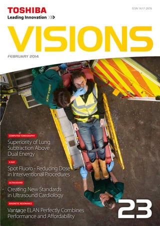
  • 1. Spot Fluoro - Reducing Dose in Interventional Procedures Superiority of Lung Subtraction Above Dual Energy Creating New Standards in Ultrasound Cardiology Vantage ELAN Perfectly Combines Performance and Affordability 23 X-RAY COMPUTED TOMOGRAPHY ULTRASOUND MAGNETIC RESONANCE ISSN 1617-2876 VISIONSFEBRUARY 2014 Caspar Berger Caspar Berger - Bone at Beelden aan Zee Vita Subsidiaria Calva/Self-portrait 31 (excerpt) Caspar Berger, 2014 Material: Print, argmented reality dimensions: 102 x 72 This artwork is about the question of whether we can rethink life, faced with the possibility of science cracking the code of life, of being able to make and fix life. If we were offered the choice, would we dare to be immortal? Download the ‘Caspar Berger Skeleton’ app, point your mobile device at the artwork and give the skull a check-up! About the Bone Exhibition For his project ‘Skeleton’, award-winning Dutch artist, Caspar Berger, made an exact copy of his own skeleton using a Toshiba Aquilion CT scanner and 3D-print technology, which he then incorporated into a wide variety of creative interpretations. The result is a lifelike ‘interior’ portrait of the artist, although none of the individual works features his fully intact skeleton. For ‘Bone’, Berger explored, for example, the concept of humans in fossil form. The hard parts of his body were effectively ‘preserved’ in stone objects. The piece entitled “Do Not be Afraid of Becoming a Bench/Self-Portrait 28” contains casts of all the bones of his skeleton. While seated on the stone benches, which can be interpreted as symbolizing the possible role of Man in the history of the World , the viewer can watch the video projection, entitled ‘Attraction/Self-Portrait 30’ which shows a reversed process of creation. In the video, silver- grey balloons with cast parts of Berger’s skeleton attached descend from a cloudy sky. Then the sun emerges, encouraging viewers to question if Man’s ‘place’ is actually between heaven and Earth. About Caspar Berger Sculptor Caspar Berger (1965) studied at the AKI, Enschede and at the Jan van Eyck Academy in Maastricht, the Netherlands. In his work Berger investigates the relationship between the interior and exterior, between reality and image. Berger gives shape to his ideas using silicone casts of models and of his own skin or even what’s underneath, leading to its end result in bronze, silver and sometimes gold; more recently, video installations, concrete and print. In 2013 he was awarded Singer prijs 2013 and recently he received the Sacha Tanja Penning 2014. www.casparberger.nl Admission Free Museum Beelden aan Zee Harteveltstraat 1 2586 EL Den Haag T: +31 70 358 58 57 www.beeldenaanzee.nl This voucher entitles free admission for one person to Museum Beelden aan Zee. Valid until June 1st 2014 Hidden in the dunes of Scheveningen, like a pearl in the sand, you will find museum Beelden aan Zee, a truly unique museum in the Netherlands that focuses on modern and con- temporary international sculpture. Man - the human image - is the leitmotiv of the collection of Beelden aan Zee. The collection holds work from sculptors as: Karel Appel, Atelier van Lieshout, Fernando Botero, Cesar, Tony Cragg, Igor Mitoraj, Jan Meefout and Ossip Zadkine. As from January 31st 2014 the following expo- sition can be seen at museum Beelden aan Zee: • Caspar Berger - Bone. • Yubi Kirindongo - Rebel in Art Soul. • George Minne - Voorbode van de Moderne Kunst Nick Ervinck - GNI-RI. www.beeldenaanzee.nl This artwork is made possible in part by: Toshiba Medical Systems Europe and Beyond Reality
  • 2. Imprint Publisher: TOSHIBA Medical Systems Europe B.V., Zilverstraat 1 NL-2718 RP Zoetermeer Tel.: +31 79 368 92 22 Fax: +31 79 368 94 44 Web: www.toshiba-medical.eu Email: info@tmse.nl Editor-in-chief: Jack Hoogendoorn (jhoogendoorn@tmse.nl) Modality coordinators: CT: Roy Irwan UL: Joerg Schlegel XR: Jaco Terlouw “Creating New Standards in Ultrasound Cardiology”“Customer Focus: Rigshospitalet, Copenhagen” by The Creative Practice (www.thecreativepractice.com) Design Layout: Boerma Reclame (www.boermareclame.com) Printing: Schefferdrukkerij (www.schefferdrukkerij.nl) Subscription Service: www.toshiba-medical.eu/visions © 2014 by TOSHIBA Medical Systems Europe All rights reserved Museum Beelden aan Zee Harteveltstraat 1 2586 EL Den Haag T: +31 70 358 58 57 www.beeldenaanzee.nl Photography: Erik en Petra Hesmerg
  • 3. VISIONS23 | 3 I grew up following the science fiction TV series,‘Star Trek’. The original series was broadcast in the Netherlands between 1966 and 1969 and at that time, I was already intrigued by the foresight of the series’ Writer and Producer, Gene Roddenberry, and his team, particularly on technology and medical science. Devices in the series, such asThe Communicator, Hypospray and MedicalTricorder, were presented as everyday utilitarian objects. Central characters, such as Captain James T. Kirk, used communicators to talk across galaxies with the starship, ‘The USS Enterprise’. And the ship’s Chief Medical Officer, Dr. McCoy, collected physiological information about patients and diagnosed diseases in just a couple of seconds. Further extensive clinical diag- noses were possible on board the ship using even more futuristic and astonishing systems and technologies. ‘Completely logical’, I imagine Science Officer, Spock, would say. Now, almost 50 years later, we consider mobile phones as an essential part of our lives, ourselves, and some- times, even our personality. While they are true commodities, promising new concepts, technologies and products are emerging on the healthcare horizon. For example, Google Glass1 that enables live transmission of surgeries to medical colleagues and students. Or the Google Smart Contact Lens2 that can help people with diabetes control blood sugar level by measuring glucose levels in their tears second-by-second. Last but not least, the Scanadu Scout3. A scanner packed with sensors that enable anyone to conduct sophisticated physical examinations in an instance. A first step in the realization of Dr. McCoy’s Medical Tricorder? These are all fascinating developments that might have immediate impact on medicine, or might become important in the very near future. We, at Toshiba, follow all kinds of medical developments with great interest, but nevertheless, prefer to focus on continuously creating leading innovations in our field of expertise: medical imaging and treatment. Every year, we file thousands of patents, making innovation a key part ofToshiba’s fabric. And with each new product and technology that we develop, we keep in mind that the best patient treatment starts with a fast, safe and accurate examination to enable reliable diagnosis. Our aim and dedication is to provide you with amazing, unprecedented, state-of-the-art imaging and treat- ment products, systems and technologies that you never thought to be true. Or, as Dr. McCoy would say: “It is medical imaging, Jim, but not as we know it”. Kind regards, Dear reader, EDITORIAL Jack Hoogendoorn Sr. Manager Marketing Communications Toshiba Medical Systems Europe BV 1 http://tinyurl.com/nfdfuwc 2 http://tinyurl.com/mfd8aua 3 http://www.scanadu.com
  • 4. ©2014 TOSHIBA MEDICAL SYSTEMS4 | VISIONS23 10 ECR 2014 abstracts 15 Time to ditch the chest X-ray? A comparison with CT 17 Usefulness of 320-row CT in the oil industry 20 Medical Imaging Enters the Next Generation 24 Planning a Hybrid Lab 29 Dual Energy CT in the PRIME Time 32 Dual Energy Raw Data Based Decomposition Analysis on Aquilion ONE 38 Spot Fluoro – A novel innovative approach to reduce the dose during interventional procedures. 40 Toshiba’s new Vantage ELAN Combines Strong Performance and Affordability 20 24 40 Further enhancement and technical innovation have taken Toshiba Medical Systems’Aquilion ONE™ onto a new level. There are many challenges that have to be met to achieve an environment that works well for all. The impressive qualities of high performance, affordability and enhanced patient comfort define Toshiba’s new MR system. CONTENT Chest, Radiation Dose Reduction, AIDR3D Research, environment, oil recovery Product Introduction, Aquilion ONE Next Generation Indications, planning, equipment Dual Energy Dual Energy Dose Reduction, Intervention, Spot Fluoroscopy Product Introduction, Vantage ELAN
  • 5. VISIONS23 | 5 43 Single energy metal artifact reduction algorithm for CT evaluation of periprosthetic soft tissues: Clinical applications 46 Creating New Standards in Ultrasound Cardiology 49 Superiority of Lung Subtracton Above Dual Energy 52 Violation of longitudinal strain of the left ventricular myocardium as a predictor of positive stress echocardiography results 55 CUSTOMER FOCUS: Rigshospitalet 67 Subtraction CTA of the brain detects cerebral aneurysms 68 Diagnosis and treatment of primary sarcoma of the liver 74 320-row CT as a single and effective platform for anatomical and functional evaluation of coronary artery disease: the CORE320 trial 76 Secondlife Mobile CT 46 55 68 New Guidelines Incorporating 2D Wall Motion Tracking to Assess Non-ST Elevation Acute Coronary Syndrome Could Save Lives The Radiology Department at Rigshospitalet Copenhagen in Denmark This case report confirms the requirement for accurate morphological verification of tumor process before the start of the antitumor therapy. 03 Editorial 06 News 09 Message from the President VISIONS SPECIAL Orthopedics, SEMAR, metallic artifacts 2D WMT, NSTE-ACS, Guidelines CTPA, Lungs, substraction 2D Speckle Tracking, Artida Cerebral Aneurysms, Subtraction CTA, AIDR 3D Histiocytic sarcoma, malignant histiocytosis, liver, differential diagnosis Myocardial Perfusion, Coronary Angiography Mobile CT, temporary imaging, Secondlife
  • 6. ©2014 TOSHIBA MEDICAL SYSTEMS6 | VISIONS23 The page on the right is part of the VISIONS Photo Page Series reflecting an eye for the beauty of our planet, the environment and the direct surroundings where Toshiba’s systems are installed by Toshiba and its customers. Not the actual imaging products but photos of sceneries, cities, countries or other cultural aspects are highlighted on this photo page. The Photo Page is based upon an idea of Prof. Edwin van Beek. Every reader of VISIONS can participate and get their picture published. The submitted content should include: high resolution (300dpi) image, photo of the hospital and a brief text, name of photographer and Toshiba system(s) installed. The complete result is shown on the opposite page. Send your pictures and texts to: jhoogendoorn@tmse.nl, Subject: Photo Page ► NEWS Edinburgh: Transforming Global Healthcare Edinburgh is home to world-leading medical researchers who work in partnership with the NHS and the private sector. This close cooperation helps to accelerate medical discoveries and transform them into treatments for patients. Toshiba is featured in a promotional clip from the inspiring captial of Scotland. Have a look and pay spe- cial attention to the frames from 1:23m-1:44m where Dr. Ken Sutherland explains about Toshiba’s activities in Edinburgh. http://tinyurl.com/punkwhp Self-portrait forms a red thread throughout the work of Caspar Berger. He became famous for self-portraits made from direct casts of his body, previously taken from the skin. This time the award-winning Dutch artist made an exact copy of his own skeleton using a Toshiba Aquilion CT scanner and 3D-print technology, which he then incorporated into a wide variety of creative interpreta- tions. The result is a lifelike ‘interior’ portrait of the artist, although none of the individual works features his fully intact skeleton. Curious how this is done? Have a look at: http://tinyurl.com/pk9rbhv In ‘Bone’, Berger explored the concept of humans in fossil form. The hard parts of his body were effectively ‘preserved’ in stone objects. The piece entitled ‘Do Not be Afraid of Becoming a Bench/Self-Portrait 28’contains casts of all the bones of his skeleton. The exhibition ‘Bone’ can be seen at museum Beelden aan Zee, the Netherlands from February till June 2014. The Bone Exhibition Vanitas/Self-portrait 35, 2014 (exerpt) Material: sealing wax, wood Dimensions: 140x85x80 Photography: Erik en Petra Hesmerg www.casparberger.nl www.beeldenaanzee.nl/en Follow us Admission Free with the voucher on the back of this magazine!
  • 7. VISIONS23 | 7 Kruger National Park is one of the largest game reserves in Africa. It covers an area of 19,633 km2 in the provinces of Limpopo and Mpumalanga in northeastern South Africa, and extends 360 km from north to south and 65 km from east to west. Areas of the park were first protected by the government of the South African Republic in 1898, and it became South Africa’s first national park in 1926. Text Source: Wikipedia – Photography: Jaco Terlouw Nelspruit Medi-Clinic offers a magnificent panoramic view of the Drakensberg Mountain range in South Africa, and is conven- iently situated within easy reach of major arterial traffic routes. The hospital services a community that spreads as far as Belfast to Hoedspruit to Swaziland to Maputo. The imaging facilities of Drs Van Rensburg and Partners provide a full radiological service to the patients and doctors of Nelspruit Medi-Clinic, offering all diagnostic modalities including a Toshiba Aquilion64 CT scan- ner and Toshiba Zexira/FPD medical diagnostic imaging system. Text sources: www.mediclinic.co.za http://vrprad.co.za/nelspruit-medi-clinic.html Photography: Jaco Terlouw
  • 8. www.toshiba-medical.eu ULTRASOUND CT MRI X-RAY SERVICES Our lives and social environment are subject to constant change and create ever-increasing needs and high demand for better medical solutions. We at Toshiba aim to maximize the quality, safety, and efficiency of medical care, supporting clinical practice with reliable quality products and innovative, cutting-edge technologies. The high image resolution and superior operability of our medical systems creates new clinical value. While our advanced applications, supported by highly reliable technologies, open the door to the next stage of medical care. We will continue to provide a wide variety of leading-edge solutions for the benefit of all people around the world, and seek to further development in the field of healthcare following our basic commitments: “Improving the quality of life”, “Lifelong commitment to innovation”, and “Achieving lifetime partnerships”. Toshiba: Made for Life! MADE FOR LIFE.
  • 9. VISIONS23 | 9 PRESIDENT’S MESSAGE Satoshi Tsunakawa President and Chief Executive Officer Toshiba Medical Systems Corporation Toshiba’s primary focus for FY2013 was to better understand our customer and the needs of their business in medical imaging – we always want to keep the customers first. In our CT business, we reached a very significant milestone with the manufacture of our 30,000th CT system in November 2013.We have continuously offered leading, innovative products based on the clinical needs of our customers since our first CT introduction. We believe that lowering CT dose and improving workflow are not choices that clinicians should have to make, so Toshiba CT is putting customers first by providing the industry’s best solutions to solve these challenges. We introduced Toshiba’s most advanced dose reduction technology, AIDR 3D, which enables our customers to achieve the best clinical results at the lowest dose. More recently we have introduced the suite of Adaptive Diagnostic CT technology, which makes complex examinations easier while simultaneously improving diagnostic accuracy and reproducibility to solve clinical challenges faced daily in routine practice. In our X-Ray business, we had the worldwide introduction of the DTS (Dose Tracking System) at RSNA 2013. This technology provides a real-time display of the cumulative skin dose distribution during interventional procedures. The push for developing this technology came from the strong desire from the doctors to be able to monitor and minimize patient radiation which had risen sharply in recent years during interventions. Our team listened to this VOC (Voice of Customers) from around the world and successfully developed this system – understanding our customers’ business needs to protect their patients. In the MRI business unit, we launched Vantage Elan™, which is providing excellent image quality, while achieving ease-of-use, in a quiet environment, and with a very compact footprint. The operation of Vantage Elan is simple for any level of user which means better images for every customer while increasing operational efficiency. Vantage Elan requires only 23m2, and 5 working days for the complete installation after system delivery which means real savings for the customer. Vantage Elan is the realization of the MRI team’s design goal of “No Compromises in Image Quality”, in spite of its compactness. I am confident that this system will become the new standard of the next generation of MRI systems. Our Ultrasound team launched a new technology called SMI (Superb Micro vascular Imaging). This new algorithm isolates and removes clutter while preserving the underlying hemodynamic flow information. This technology allows you to see the flow information that is behind the clutter and visualize extremely low velocity flows that are typically obscured with conventional Color Doppler. See the unseen – A giant step towards achieving our goal of “Picture Perfect Ultrasound” starts from here. Finally, last but not least, as of October 1st, healthcare has become one of the three business pillars of the Toshiba Corporation, andTMSC will be the core member of the new Healthcare Systems Services Group. We will be working closely with the new established Healthcare Business Development Division in Toshiba Corporation. The mission of the Healthcare Group is to extend the scope of business from medical diagnostics to new healthcare related business, including disease prevention and patient care. With this initiative, and carefully expanding our businesses, we are continuing in our corporate philosophy to keep the customers and more importantly their patients first. “We always keep our customers first.” By focusing on low dose, high-quality imaging technologies for accurate diagnosis and treatment, Toshiba continues to improve the quality of life for all people.
  • 10. ©2014 TOSHIBA MEDICAL SYSTEMS10 | VISIONS23 1) Prof. F. Barkhof A 3T MRI system brings major advantages in neuroradiology compared to the 1.5T systems. Our initial experience with a latest generation 3T system, the Vantage Titan, indicates that the system provides increased patient comfort while offering significant quality and diagnostic benefits for several clinical applications. In this presentation we will discuss our initial clinical experience with the Vantage Titan 3T system, including clinical cases of time-of-flight MRA for evalu- ation of the vasculature, isotropic 3D FLAIR in the assessment of glioma and multiple sclerosis. Moreover, flow-spoiled black blood (FSBB), a proprietary Toshiba technique, is explored for the evaluation of microvasculature (lenticulostriate arteries) in aging and dementia. 2) Prof. A. Blum Total Hip Arthroplasty (THA) is frequently performed for the treatment of advanced degenerative joint disease of the hip. Complication rates are low but the great number of hip prosthesis implanted renders radiologists prone to be confronted to post surgical peri prosthetic complications. Radiography remains the first step to assess a THA but CT-scans, MRI and sonography are now complementary tools that might be useful for the etiological diagnosis of a painful THA. Common indications for CT of THA are a painful hip prosthesis with normal radiographic findings, the assessment of osteolysis before surgical revision, the evaluation of soft tissues complications and the measurement of acetabular cup placement. Classic CT-scan is hampered by metal artifacts and is responsible for high radiation dose to the patient. Soft tissues anomalies are hardly seen with classic CT-scan. When optimized to reduce metal artifacts using suitable acquisition techniques and new algorithms, CT-scan is the most polyvalent technique to assess the components of the prosthesis, the cement and the bone interface, the bone stock and the soft tissues. These new algorithms: iterative reconstruction (AIDR 3D) and metal artifact reduction (SEMAR) software improve the image quality while reducing the dose. The objective of this presentation is to show how to improve prosthesis evaluation with CT scan. 3) Prof. M. Prokop Pulmonary embolism with CT is a great challenge because small thrombi can easily be missed (many small arteries), differentiation partial volume making real embolus difficult. Furthermore, reduced lung perfusion almost invisible on standard CT. In this lecture, we will look into Dual Energy and Subtraction imaging techniques to detect perfusion defects, to help localize small emboli, and to increase a prognostic value. Both advantages and down sides of each method will be analysed and presented. Satellite Symposia MR/CT Friday, March 7, 12:30 - 13:30 hrs, Room B Moderator: Prof. Dr. D. Hahn, Berlin, Germany ECR 2014 Abstracts Adaptive Diagnostics - Solving Clinical Challenges Speaker Time Slot Title Prof. Dr. F. Barkhof, Amsterdam, The Netherlands 12.30 - 12.50 Latest generation 3T MRI first experience in Neuroradiology 1) Prof. A. Blum, Nancy, France 12.50 - 13.10 A new reconstruction algorithm: Single Energy Metal Artefacts Reduction (SEMAR), applied for evaluation of hip protheses 2) Prof. M. Prokop, Nijmegen, The Netherlands 13.10 - 13.30 Lung subtraction versus Dual Energy 3) Prof. A. Blum Prof. F. Barkhof Prof M. Prokop
  • 11. VISIONS23 | 11 Satellite Symposia ULTRASOUND Saturday, March 8, 2014, 12.30 - 13.30 hrs, Room E1 Moderator: Prof. V. Mitkov, Russia 1) Dr. T. Fischer Prostate: Men with an elevated serum level of prostate-specific antigen (PSA) or suspicious findings on digital rectal examination (DRE) are examined by transrectal ultrasound (TRUS). For histological confirmation and therapeutic plan- ning, ultrasound guided systematic biopsy of the prostate is performed. However, in a subgroup of patients with elevated PSA levels, no malignancy is detected by biopsy or up to four biopsies are performed before prostate cancer is detected. A negative biopsy therefore does not exclude prostate cancer. In consequence, unnecessary biopsies with an increase of complications are performed in healthy men. Real-time MR/US image fusion may enhance cancer detection rates of TRUS-guided biopsies and contributes to lesion characterization by state-of-the-art US techniques. The study presented here for the first time compares state-of-the art US techniques after contrast medium administration with MRI and histology as the standard of reference. Scrotum: Ultrasound is the imaging modality of choice for the examination of the scrotum. The propblem of tumour / haematoma misinterpretation can result in unnecessary orchiectomy. CEUS for the testis can be currently recom- mended for the differentiation between hypovascular and avascular lesions (benign). Learning objects: - To investigate whether multiparametric magnetic resonance imaging (MRI) allows lesion localization in prostate can- cer in patients scheduled for MR/US fusion biopsy and whether the findings correlate with new ultrasound techniques - Prostate lesions were classified on the basis of MRI and US (B-mode scan, power Doppler, elastography/TDI, CEUS) - Targeted biopsies were performed in the MR/US fusionmode - Indications for the use of CEUS in focal testicular lesions, secmental infartion, after trauma and abscess formation in severe epididymoorchitis. 2) Prof. A. Lim This talk will encompass the latest technological developments of the Aplio 500 with particular focus on a few advanced applications for abdominal and small parts ultrasound.These newest developments include the improved visualisation of the microvasculature without the need for contrast and its potential clinical applications will be discussed. There have also been significant improvement in specialist applications such as elastography and also improved sensitivity in high frequency contrast technology. The latter now also allows reliable detection of the sentinel lymph node and their clinical impact will be outlined and illustrated. Expanding Clinical Boundries in Ultrasound Speaker Time Slot Title Dr. T. Fischer, Berlin, Germany 12.30 - 12.50 Genitourinary Ultrasound: Advanced Diagnostics in Prostate Cancer and Scrotal Lesions 1) Prof. A. Lim, London, United Kingdom 12.50 - 13.10 Abdominal and high-frequency Ultrasound imaging: Latest technologies for improved lesion detection and characterisation 2) Prof. J. Hata, Kawasaki, Japan 13.10 - 13.30 Seeing the Unseen - New Clinical Findings by Novel Imaging Techniques 3) Dr. T. Fischer Prof. A. Lim BRINGING INNOVATION TO LIFE
  • 12. ©2014 TOSHIBA MEDICAL SYSTEMS12 | VISIONS23 3) Prof. J. Hata In this session, the clinical experience of Superb microflow imaging (SMI), which is the novel application of Aplio 500 from Toshiba, is discussed. SMI is a unique ultrasound Doppler imaging employing multi-dimensional filter that makes it possible to depict minute vessels with slow velocity, without using contrast agents. In other words, the advantages of SMI are; 1.high frame rate, 2. high sensitivity, 3. high resolution, and 4. less motion artifact. Therefore, SMI is useful in various situations encountered in daily practice. The evaluation of the density and the shape of tumor vessels with SMI help us diagnose and differentiate tumorous lesions and also could be the non-invasive modality for the assessment of therapeutic effect of chemotherapy. Since SMI clearly visualizes minute vessels, it can also be used to evaluate the disease activity in inflammatory disorders, even in bowel inflammation such as ulcerative colitis, Crohn’s disease, and so forth. Furthermore, the diagnosis of organ ischemia can be done accurately with SMI, which is expressed as the focal defect of minute vessels in a real-time fashion, with high spatial resolution. In addition, SMI provides more sensitive view of minute vessels after the injection of contrast agent, which helps us to obtain the sustained dynamic image of microflow even after the first pass of the contrast agent. In conclusion, SMI, which provides the dynamic image of microvessels, is an essential tool for the diagnosis and the evaluation of various clinical conditions. Toshiba Workshop Sessions will be offered Friday, March 7 thru Sunday, March 9. Times as listed below in room Lounge 6 - Level 01 (max 20 seats). Workshops are open to any ECR 2014 attendees. On-line registration in the “myuserArea”of the ECR web page. Toshiba is offering a series of educational sessions for radiologists throughout ECR 2014. Each mentored workshop is preceded by a short lecture providing an overview on data acquisition, clinical use and image interpretation. Clinical cases will be analyzed and interpreted by participants on the workstations provided. The maximum number of participants per session is 20 (2 participants per workstation). CME credits will be provided, and the workshop language is English. Date Time Slot Title March 7, 2013 1 10:00 - 11:00 Ultrasound - Elastography Quantification, guest speaker: Dr. V. Cantisani, Rome, Italy March 7, 2013 2 11:15 - 12:15 Ultrasound - Fly Thru Acquisition and Reconstructions, guest speaker: Dr. B. Smith, London, UK March 7, 2013 3 12:30 - 13:30 Ultrasound - DCEUS in a multicenter study, guest speaker: Dr. N. Lassau, Paris, France March 7, 2013 4 13:45 - 14:45 Ultrasound - Elastography Quantification, guest speaker: Dr. V. Cantisani, Rome, Italy March 7, 2013 5 15:00 - 16:00 Ultrasound - Fly Thru Acquisition and Reconstructions, guest speaker: Dr. B. Smith, London, UK March 7, 2013 6 16:15 - 17:15 Ultrasound - DCEUS in a multicenter study, guest speaker: Dr. N. Lassau, Paris, France March 8, 2013 7 10:00 - 11:00 CT - Ultra low dose brain perfusion, guest speaker: Dr. E. Smit, Nijmegen, The Netherlands March 8, 2013 8 11:15 - 12:15 ViTAL - Lung Density Analysis, guest speaker: Dr. S. Niehues, Berlin, Germany March 8, 2013 9 13:45 - 14:45 CT - Myocardial perfusion, guest speaker: Dr. K. Kofoed, Copenhagen, Denmark March 8, 2013 10 15:00 - 16:00 CT - Liver perfusion, guest speaker: Dr. H. Schöllnast, Graz, Austria March 8, 2013 11 16:15 - 17:15 CT - Ultra Low Cardiac CTA, guest speaker: Dr. R. Bull, Bournemouth, UK March 9, 2013 12 10:00 - 11:00 ViTAL - TAVR planning, guest speaker: Dr. S. Niehues, Berlin, Germany March 9, 2013 13 11:15 - 12:15 CT - Ultra Low Cardiac CTA, guest speaker: Dr. R. Bull, Bournemouth, UK March 9, 2013 14 12:30 - 13:30 ViTAL - Lung Density Analysis, guest speakers: Dr. S. Niehues, Berlin, Germany March 9, 2013 15 13:45 - 14:45 CT - Liver perfusion, guest speaker: Dr. H. Schöllnast, Graz, Austria March 9, 2013 16 15:00 - 16:00 CT - Myocardial perfusion, guest speaker: Dr. K. Kofoed, Copenhagen, Denmark March 9, 2013 17 16:15 - 17:15 CT - Ultra low dose brain perfusion, guest speaker: Dr. E. Smit, Nijmegen, The Netherlands Prof. J. Hata
  • 13. VISIONS23 | 13 BRINGING INNOVATION TO LIFE DCEUS Quantification Workshop Bracco/Toshiba Saturday, March 8, 2014, 12.30 - 13.20 hrs, Room Lounge 1 - Level 01 (max 30 seats) Moderator: Prof. Jean-Michel Correas, Paris, France Quantification of Renal Perfusion Bracco and Toshiba offer a 50 minutes educational session for radiologists on quantification of Dynamic Contrast Enhanced Ultrasound (DCEUS) based on the use of the Bracco quantification softwareVueBox™ and contrast-enhanced ultrasound sequences acquired with the Toshiba Aplio 500. Brief introductionary lectures will provide an overview of concepts in DCEUS quantification and in renal perfusion. In the hands on session, the participants will perform their own analysis ofToshiba Aplio 500 clips under the supervision of Prof. Jean-Michel Correas using laptops equipped with the VueBox software. Floor plan is an indication. Please follow the signage TOSHIBA IWS.
  • 14. Want to be up-to-date on Diagnostic Imaging Innovations? www.toshiba-medical.eu ULTRASOUND CT MRI X-RAY SERVICES Adaptive Diagnostics - Solving Clinical Challenges Date: Friday, March 7, 2014 Time: 12.30 - 13.30 hrs Room: Room B Quantification of Renal Perfusion (Toshiba Bracco) Date: Saturday, March 8, 2014 Time: 10.30 - 11.30 hrs Room: Room Lounge 1 - Level 01 Expanding Clinical Boundries in Ultrasound Date: Saturday, March 8, 2014 Time: 12.30 - 13.30 hrs Room: Room E1 Toshiba Workshops for the duration of ECR Room: Software Demonstration Room, Lounge 6 - Level 01 Sessions open to any ECR 2014 attendees. On-line registration in the “myuserArea” of the ECR web page. Join the Toshiba Sessions CME credited! Earn 1 CME credit! Earn 1 CME credit!
  • 15. VISIONS23 | 15 MATERIALS AND METHODS We examined 13 patients with suspected pulmonary lesions, following chest radiography with ultralow-dose CT acquired on an Aquilion ONE scanner. Images were acquired at 135 kV and 10 mA, with 0.5-mm slice thick- ness and rotation time of 350 msec. Three experienced chest radiologists rated both sets of images for pathological findings on a three-point scale, and image quality was rated according to nine criteria of the European guidelines for chest CT and chest radiographs. RESULTS Both image quality and the ability to detect and exclude pathology was strikingly better for ultralow-dose CT than for radiography. The radiation dose for CT and conven- tional radiographs was comparable and the laboratory time only marginally longer for CT. In summary, we found the following: • 33 relevant findings including 19 masses, nodules, and micronodules. • Detection sensitivity for all observers and all findings was 18% ± 3% for radiography and 89% ± 2% for ultralow-dose CT. • The positive predictive value for radiography was 37%, including 31 false-positive findings, while the positive predictive value for CT was 98% with two false-positive findings. • Image quality for all readers was significantly higher for the CT images (p 0.05). • The average effective dose was 0.05 mSv for chest radi- ography and 0.105 mSv for CT. • The average laboratory time was five minutes for chest radiography versus seven minutes for CT. Time to ditch the chest X-ray? A comparison with CT It is widely accepted that Computer Tomography (CT) is both more time consuming and expensive than chest radiography. Furthermore, CT incurs a considerably higher radiation dose than the conventional chest X-ray. Ideally we wish to combine the low- dose of the chest X-ray with the high image quality of CT, but this ambitious aim has, untilnow,notbeenpossible.Inthisstudywecomparedconventionalchestradiography to 320-detector-row CT Aquilion ONE™ using advanced iterative reconstruction (AIDR 3D) in terms of image quality, radiation dose and laboratory time. Dr. T.M. Aaløkken J.F. Kristiansen MSc Dr. A. Günther A.C.T. Martinsen PhD 1-4) Oslo University Hospital, Norway CTEU130074 CLINICAL CASE COMPUTED TOMOGRAPHY Chest, Radiation Dose Reduction, AIDR3D Figure 1: Above is a false-negative chest X-ray. Figure 2: an ultralow-dose 3-mm axial chest CT of the same patient acquired on a 320-detector-row scanner (Aquilion ONE) and reconstructed with AIDR 3D shows a 7-mm nodule. Dr. T.M.Aaløkken 1), J.F. Kristiansen MSc 2), Dr. A. Günther 3), A.C.T. Martinsen PhD 4)
  • 16. ©2014 TOSHIBA MEDICAL SYSTEMS16 | VISIONS23 CONCLUSION The study demonstrated that the diagnostic yield from ultralowdoseCTisfarsuperiortothatofchestradiographs. Chest CT has traditionally incurred an effective radia- tion dose of approximately 4 mSv. Iterative reconstruction techniques now enable CT imaging at much lower doses and CT has become a fast and accessible modality. In this light the only remaining advantage of chest radiography is the low expense. This work was presented at RSNA 2012. Moderator Dr. Narinder Paul from the University of Toronto rose the issue of an increase in findings with CT potentially leading to additional tests, costs, and risks. Figure 3: Axial (previous page) and Coronal (above) maximum-intensity projection (MIP) show a 7-mm nodule in the left lung and some atelectasis in the right lung. AIDR 3D is an advanced iterative reconstruction algorithm that reduces noise in both the three dimensional reconstruction data and raw data domains. With the integration of AIDR 3D into SUREExposure controls, radiation exposure is automatically reduced before the scan, ensuring that the lowest possible dose is employed for the specific diagnostic objective irrespective of the size or shape of the patient. AIDR 3D integrated in SUREExposure 3D Adaptive can be applied to all acquisition modes for routine clinical use and is able to remove up to 50% of image noise resulting, which corresponds to a dose reduction of up to 75% by the same Standard Deviation (SD) for noise. Clinical results show that AIDR 3D substantially reduces noise, while preserving sharp details, providing “natural looking” images. As well this new technology is especially good in artifact suppression (streaks, beam-hardening, etc.). Reconstruction performed with AIDR 3D per- mits substantial dose reduction when compared to scans performed with traditional Filtered Back Projection (FBP) techniques. The AIDR 3D algorithmisdesignedtoworkinboththerawdataandreconstruction domainsandoptimizesimagequalityforeachparticularbodyregion. AIDR 3D - ADAPTIVE ITERATIVE DOSE REDUCTION 3D Key features of AIDR 3D - Full integration in scan protocols for improved workflow - Dose reduction in clinical setting by up to 75% - Minimal penalty in reconstruction times - Noise reduction and improved Spatial Resolution - Superb artifact suppression
  • 17. VISIONS23 | 17 As a world leader in energy technology and innovation, HOOS works with companies to enhance the recovery of unconventional oil. The goal is to help oil producers engage in sustainable development of Alberta’s resources through technologies that reduce costs, improve recovery, and decrease environmental impact. Research in this area is facilitated by the Aquilion ONE, which was purchased primarily as support for applied RD in petroleum recovery and advanced materials. This article describes the use of CT with Aquilion ONE for research at Tech Futures. COMPUTED TOMOGRAPHY AT TECH FUTURES Through computed tomography, non-destructive exami- nation of internal structure is accomplished and high-res- olution 3D images of items like geological core samples and sand packs are created. A wide variety of reconstruc- tion algorithms and post-processing options are used at Tech Futures to suit the material being scanned and the imaging needs of the customer. Accurate extrac- tion of quantitative information from advanced image processing, along with 2D and 3D reconstructions, is often paramount. Quantitative information is delivered to clients by way of customized calibrations for each object or experiment scanned. Targeted noise-reduction techniques provide methods to acquire the best reso- lution possible. Specifically, this involves repeat scans and averaging data whenever possible to reduce noise. Further discrimination of materials can be done through dual energy scanning, which exploits subtle differences in attenuation spectra. Tech Futures’ current strategy is to design CT experi- ments conducive to the energy level and geometry of the Aquilion ONE. Low-attenuation vessels and materials suitable for X-ray use are considered in the experimen- tal design stage. The speed of the scanner, for both the acquisition and reconstruction of data, makes it possible to track processes that change on a scale of minutes. The usefulness of a 320-row CT scanner in the oil industry Alberta Innovates -Technology Futures (Tech Futures) is part of an integrated provincial system that works to further research and innovation in Alberta, Canada.The priority of the corporation is to accelerate the development and growth of new and existing industries in agriculture, forestry, energy, environment, and health. To help meet research needs in Alberta, the Heavy Oil Oil Sands (HOOS) group at Tech Futures acquired an Aquilion ONE™ CT scanner. CTEU130078 Shauna Cameron, P.Ag, Imaging Specialist 1) Alberta Innovates – Technology Futures. Alberta Innovates - Technology Futures, Edmonton, Canada Shauna Cameron, P.Ag 1) This is invaluable for fluid flow experiments in cores and sand packs. Efforts are also made to carefully register experiments when repeat scans are taken in order to successfully subtract images and uncover small changes in density. Orbital sync has been a useful tool in securing registra- tion. This function ensures that the source and detector are located in precisely the same starting position for each repeat scan allowing the images to be either subtracted for discrimination of tiny density changes or averaged for noise reduction. Radiation dose reduction is not critical to scans carried out at Tech Futures since subjects are not living. In fact, objects or experiments are scanned repeatedly at the highest available settings in order to extract the best quantitative information. Often, protocols are designed so objects are scanned in short lengths, with multiple volumes necessary to cover the desired length. This technique permits the highest allowable dose to penetrate objects much denser than the human body. OIL RECOVERY FROM ROCK: DIGITAL ROCKS One application for advanced imaging with Tech Futures’ scanner is digital rocks, which is an emerging concept in characterizing reservoirs by obtaining physical properties directly from images of rocks. The technology combines OIL INDUSTRY COMPUTED TOMOGRAPHY Research, environment, oil recovery
  • 18. data can be very useful in studying pore architecture and physics of the processes happening during enhanced oil recovery (EOR). It is especially important for unconven- tional resources like shale, carbonate, tight gas, and coal seam gas. One of the most interesting aspects at this time is characterizing bitumen-bearing carbonates, especially fracture characterization. Digital rocks enables Tech Futures to engage in a unique up-scaling effort of merging micro and macro imaging to observe a wide range of fluids, conditions, and detailed behaviour in EOR processes. The advantage of the technique is the considerable cost savings over securing individual laboratory analysis for each desired property. The procedure involves first obtaining full core mm-scale resolution scans with the Aquilion ONE scan- ner to identify sites representative of a range of image properties. This is followed by micro CT scans (approxi- mately 10 micron resolution), available from various out- sourced labs, which are suitable for sand packs or other very small samples. Imaging with sufficient resolution to resolve individual pores in a sand pack or a core is inevita- bly restricted to a very small volume. Simulation software is used to upscale the results of pore scale multi-phase flow simulations to obtain flow properties at each site and look for correlations between flow properties and CT at different scales (mm to micron resolution) with X-ray and scanning-electron microscopy (SEM) to create images of the internal structure of the pore spaces as well as minerals and organic matter within them. Such Figure 2: Axial image of Alberta Carbonate Core Dinosaurs first appeared 225 million years ago and died out 65 million years ago. Today most dinosaur fossils are found by doing fieldwork, but also during excavations for tunnels, buildings and drilling for oil wells. The Aquilion ONE at Alberta Innovates - Technology Futures scans these fossils to provide detailed information to paleontologists on the structures of the animals. Below are two examples of dinosaur fossils scanned with Aquilion ONE. 3D Image of a Juvenile Ceratopsian 3D image of the Snout (Antorbital Region) of a Tyrannosaur 225 TO 65 MILLION YEARS IN THE MAKING Specimens are from the collections of the University of Alberta, Laboratory of Vertebrate Paleontology AQUILION ONE ASSISTS IN MEASURING PERMAFROST Water is trapped as ice in the permafrost of the world’s artic regions. If the Earth is warming, the amount of water in the permafrost should decrease as the ice melts. How can we measure the ice in permafrost? The Aquilion ONE at Alberta Innovates - Technology Futures, is doing just such measurements. Permafrost cores are scanned and 3D volume rendered images are produced depicting ice and sediment. Quantitative measurements can also be made to determine the percentage of ice in each core. By imaging cores from the same region over many years, the change in the amount of ice can be measured. Three images from CT data. (A) Axial slice; (B) 3D reconstruction; (C) Longitudinal slice. Soil is white, ice is gray and gas is black. Calmels, F., Froese D.G., and Clavano, W. Cryostratigraphic record of permafrost degradation and recovery following historic (1898-1992) surface disturbances in the Klondike region, central Yukon Territory. Can. J. Earth Sci. 49: 938–952 18 | VISIONS23 ©2014 TOSHIBA MEDICAL SYSTEMS
  • 19. VISIONS23 | 19 image properties. Properties are then mapped back onto the macro CT scan using the correlations and core scale flow simulations are performed with the results to make predictions for recovery from the oil reservoir. OIL RECOVERY FROM SAND: IMAGING SAND PACKS Other HOOS applications of the Tech Futures scanner include calculating porosities and saturations in sand packs, tracking solute in sand packs, testing solute uptake in oil, studying turbulent flow in water as well as laminar flow in oil, monitoring sand failure or production, and investigating the waterflood process used in oil recovery at the scale of sand grain pores. CONCLUSION The use of CT to image and quantify materials greatly assists scientists in determining potential sites for com- mercial oil extraction in unconventional materials such as carbonates and oil sands. This is a very interesting and different application of CT from its usual medical uses. Figure 3: Merging macro and micro scanning to observe a range of conditions to determine the behaviour of enhanced oil recovery. Figure 4: A sagittal section through a waterflood experiment in a cylindrical sand pack. Flow is from left to right. Blue indi- cates “unswept” regions with oil saturations of about 90%. Red indicates where water has displaced about 25% of the oil. This “synthetic radiograph” was obtained by averaging a number of sagittal slices near the axis of a cylindrical pack, to reduce noise and improve the spatial resolution. Research is facilitated by the Aquilion ONE, which was purchased in 2013, primarily as support for applied RD in petroleum recovery and advanced materials. This CT system was an upgrade to an Aquilion 64, which had been installed in the Edmonton facility in 2007. The decision to purchase the original Aquilion 64 in 2007 was based partly on its ability to monitor dynamic processes. Another deciding factor was the presence of a local service base, which enables Tech Futures to obtain same-day service calls to address instrument problems. While the energy range of the medical scanner is lower than that of the industrial CT scanner HOOS used previously, the trade-off of higher energies and longer scan times for greater speed and local service was deemed favourable. The speed of the scanner, for both the acquisition and reconstruction of data, makes it possible to track processes that change on a scale of minutes. This is invaluable to fluid flow experiments in cores and sand packs. The wide area coverage allows cores to be scanned in fewer sections and enhances the measurements of dynamic processes. LOCAL SERVICE SEALS THE DEAL Core experiments on the Aquilion ONE Flow Experiments on the Aquilion 64 CTEU130078
  • 20. ©2014 TOSHIBA MEDICAL SYSTEMS20 | VISIONS23 AQUILION ONE NEXT GENERATION The latest evolution of the leading dynamic volume CT system sees increased ease of use for radiologists and radiographers, better patient safety and comfort as well as outstanding image clarity. Like its predecessor, the next generation of the Aquilion ONE system has the ability to scan entire organs in a single gantry rota- tion. The 16 cm detector makes it possible to capture morphology at a single moment in time - be it a heart, foot, or an infant’s chest - and eliminates movement artefacts. Patient comfort and safety are optimized by a larger gantry aperture and a newly developed Quantum VI detector, providing higher light output for optimized dose reduction. The new CT is entering the market at a time when Toshiba Medical Systems is set to become an ever more important‘player’within theToshiba Corporation and the corporation’s healthcare sector already has plans for fur- ther expansion. Satoshi Tsunakawa, President and CEO of Toshiba Medical Systems Corp., Japan, emphasizes that the main aim of the business is to further extend the scope from medical diagnostics to new business to include disease prevention and patient care. Toshiba is already a key player in diagnostic imaging and has already sold 30,000 CT machines worldwide but believes there are further opportunities within the sector. “In order to realise this vision, our target for revenue in this year is 10% growth over last year,” Tsunakawa adds. Medical Imaging Enters the Next Generation The world’s best scanner just got even better. The performance of Toshiba Medical Systems’Aquilion ONE™ has impressed radiologists in recent years but further enhancement and technical innovation have taken it onto a new level and added yet another dimension to CT imaging. NEW COMPUTED TOMOGRAPHY Product Introduction, Aquilion ONE Next Generation The next generation of Aquilion ONE offers enhanced workflow and patient comfort along with innovative new imaging tools. Ground-breaking Aquilion ONE Technology
  • 21. VISIONS23 | 21CTEU140086 At the heart of that growth – and a critical component in the evolution of the new Aquilion ONE – is the strong ethos of innovation within Toshiba. Henk Zomer, Senior Manager of the CT Business Unit at Toshiba Medical Systems Europe, says that innovation is a strong theme running through the development of CT within Toshiba which has seen the company become the CT market leader in Japan. At present, it is in third place on the global stage but has clearly-defined aims to become the number one CT manufacturer in the world. “Innovation never stops,” Zomer points out, and adds “it is a never- ending challenge between highly professional creative users and our engineers.” ADAPTIVE DIAGNOSTICS What sets the next generation of Aquilion ONE apart is its flexibility and performance, making use of new innovations like Adaptive Diagnostics, including Dual Energy raw data analysis, Variable Helical Pitch, SEMAR (Single-Energy Metal Artifact Reduction) and new SURESubtraction applications. Adaptive Diagnostics is Toshiba’s patient-centric suite of unique imaging solu- tions to simplify complex protocols and ensure consist- ent quality of results and simplifying workflows. Meanwhile Dual Energy scanning is helping to bring greater consistency to clinical results. While anatomical structures attenuate X-rays differently, Dual Energy raw data analysis increases the amount of information avail- able from CT imaging. In addition the SURESubtraction applications provide clinical solutions to the challenges faced in everyday clinical practice: the brain subtraction algorithm enables accurate subtraction of the skull and medical implants; the neck subtraction deformable registration algorithm creates high-resolution images freed of bone structures; the lung subtraction provides iodine maps of the lung parenchyma with exceptional high contrast-to-noise ratio; and ortho subtraction ensures accurate subtraction of skeletal structures and calcified plaques. With a constant focus on radiation dose, patient and staff safety, Toshiba developed fully integrated AIDR 3D (Adaptive Iterative Dose Reduction). AIDR 3D assists the radiologist in automatically saving dose on every exami- nation while maintaining excellent diagnostic image quality at a radiation dose suitable for each patient. A major challenge in CT remains the interpretation of scans from patients with metallic implants. This is where SEMAR technology plays a role by employing a sophis- ticated reconstruction algorithm to eliminate artefacts caused by metal while still improving visualisation of the implant. SEMAR can be used in routine low dose standard volume acquisitions and the combination with AIDR 3D provides excellent image quality. INITIAL CLINICAL EXPERIENCE WITH THE NEW AQUILION ONE Radiologists who have experienced the Aquilion ONE in recent years have been impressed with the next genera- tion of this dynamic volume CT system. ImageQualityandWorkflow Consultant radiologist Dr. Russell Bull from the Royal Bournemouth Hospital in the south of England has been using an Aquilion ONE since 2009. At that time, he recalls, the facility to cover a whole organ for the first time was a major leap forward in scanning for his department. But as he points out: “None of these advances matter unless they change the diagnosis or experience of the patient, the throughput or cost of procedure. If it is just clever that does not matter, but the Aquilion ONE completely Satoshi Tsunakawa, President and CEO, Toshiba Medical System Corporation, Japan Dr. Russell Bull is particularly impressed by the ease of use, image quality and integrated dose reduction of the new Aquilion ONE.
  • 22. ©2014 TOSHIBA MEDICAL SYSTEMS22 | VISIONS23 revolutionised our practice.” He saw improved efficiency and throughput. “It increased scan efficiency and the volumetric scanning allowed use of much lower doses of contrast agent.” “We wondered whether it could ever get any better,” he says, “but it did, we have now the Aquilion ONE Next Generation.” He is particularly pleased with the new Quantum Vi Detector which he says has improved detec- tor efficiency.“The improvements we have seen are large; much better patient experience from the large bore, very large patients receive low radiation dose and image quality is again better. There are fewer artefacts and less IV contrast is needed. Furthermore we have the ability to scan a wider range of patients as well seeing much faster reporting. As a result, the workflow has improved enormously.” “We always thought that our Aquilion ONE was the ‘world’s best scanner’but it is clear that the Aquilion ONE Next Generation is now the ‘world’s best scanner’. It is unbeatable for ease of use, image quality, low radiation and is completely revolutionising what we do.” Matthew Benbow, who is a SuperintendentTechnologist at Royal Bournemouth Hospital, stated that the new Aquilion ONE has made important differences to his team. The wider bore has made it more comfortable for patients, especially for those whose movements are restricted and those who are unable to place their arms above their heads. Aquilion ONE has been ergonomi- cally designed for fast patient throughput and increased patient comfort, with the technology aligned in such a way to ensure that examinations are conducted with minimal steps. Benbow said: “Our Aquilion ONE with motorized lateral table slide function allows the patient to be positioned without physical strain. This is an invaluable feature safeguarding the health, safety and wellbeing of radiographers from a lifting perspective”. SEMARMetalArtefactReduction The Aquilion ONE has clear benefits across a whole range of innovative applications ranging from muscular-skeletal to the lungs. Prof. Alain Blum, Head of the Radiology Department at Service d’Imagerie Guilloz – C.H.U. Hôpital Central Nancy, France, believes that the new system offers “a huge step forward for the evaluation of soft tissues.” Especially in the postoperative evaluation of patients with prosthesis, CT will play an increasing role. Since the classic CT scan is hampered by metal artefacts, new algorithms such as iterative reconstruction and SEMAR will definitely help to minimize those artefacts while presenting a better image quality – at a much lower dose than ever before. A preliminary study of 68 patients with hip prosthe- sis recently conducted by Prof. Blum demonstrates that newly released Adaptive Diagnostics software for the Aquilion ONE significantly improves image quality, espe- cially in those patients with bilateral hip prosthesis. In addition, he states a 30 percent increase in the detection rate of peri-articular pseudo-tumours. Summarizing the advantages of the new developments Prof. Blum says: “CT is the only technique providing detailed information on the implants, bone and the soft tissue. By using iterative reconstruction and SEMAR, Superintendent Technologist Matthew Benbow values the enhanced comfort Aquilion ONE offers for both his patients and his staff. Prof. Alain Blum sees a significant increase in image quality with the new metal artefact reduction technology SEMAR, especially in patients with bilateral hip prosthesis.
  • 23. VISIONS23 | 23 artefacts are significantly reduced and diagnostic image quality is dramatically improved.This will surely lead to an increase of CT-examinations for patients with hip pros- thesis, so far difficult to examine.” SURESUBTRACTION LUNG IODINE MAPPING Prof. Mathias Prokop, Chairman of the Department of Radiology at Radboud University Medical Center in Nijmegen in The Netherlands, has evaluated SURESubtraction Lung. This newly developed Adaptive Diagnostics software creates iodine maps by subtracting a low-dose pre- contrast CT from a CT angiogram of the lung.“The iodine maps created by SURESubtraction on the Aquilion ONE,” Prof. Prokop points out, “have greatly improved signal and higher spatial resolution compared to iodine maps derived from dual energy acquisitions. SURESubtraction Lung shows perfusion defects and thus provides direct insight in the effect of embolism on lung perfusion. By providing a perfusion map of the lung it can help SURESubtraction Lung provides iodine maps of the lung parenchyma with a high contrast/noise ratio. The color overlay allows easy identification of hypo-perfused areas. SEMAR removes artefacts caused by metal and improves visualization of the implant, supporting bone and adjacent soft tissues for clearer and more confident diagnosis. Prof. Matthias Prokop points out that SURESubtraction Lung creates excellent iodine maps without increase of radiation dose. clinicians find small emboli they could have missed oth- erwise. It also has enormous unexplored potential, for example with regard to diagnosing the activity of inter- stitial lung disease, or evaluating the interaction between aeration and perfusion in diseases like emphysema or small airway disease.” In acute and chronic pulmonary embolism stud- ies SURESubtraction Lung creates excellent iodine maps without increase of radiation dose.“SURESubtraction Lung,” Prof. Prokop underlines,“is an easy to learn technique with simple scanning procedures and fast processing times that do not burden the workflow. In short, a very powerful technique with outstanding image quality.” SUMMARY The next generation of Aquilion ONE marks a major step forward in scanning technology enhancing the whole CT experience for the patient and the health professional. With reduced radiation dose, less contrast agent, quicker delivery of results, improved imaging and increased versatility it offers a major advantage in technology and helps lead to better patient diagnosis and outcomes. CTEU140086
  • 24. ©2014 TOSHIBA MEDICAL SYSTEMS24 | VISIONS23 Whilst it may seem a simple task to combine an imaging suite with a surgical one, there are many challenges that have to be met to achieve an environment that works well for all. Angela Nightingale 1) Angela Nightingale 1) Toshiba Medical Systems Ltd, Crawley, United Kingdom PRACTICAL X-RAY Indications, planning, equipment Advanced technology is changing the way surgery is being performed, with minimally invasive techniques being applied to many areas. To undertake these com- plex procedures a new ‘Hybrid’ operating and imaging environment has been developed which combines a full operating theatre with high level surgical facilities and advanced imaging equipment. There needs to be a great deal of thought given to whether it is even appropriate, so there should be detailed analysis of what procedures will be undertaken, in what numbers and by whom, to ensure that the maxi- mum use will be made of such a major financial invest- ment. In some cases it may be more appropriate and financially prudent to install separate facilities. If the clinical and financial indications are positive, then all the stakeholders must be identified at a very early stage, their input sought and a room usage plan devel- oped. These high technology rooms require too large an investment to be used on a part time basis, so care should be taken to maximise use. Planning a Hybrid Lab
  • 25. VISIONS23 | 25 CLINICAL INDICATIONS FOR A HYBRID LAB Cardiac Initially developed for Paediatric Cardiology, use of hybrid labs has expanded into adult surgery with coronary revas- cularisation, trans catheter valve replacement (TAVI) and repair,leftventricularassistdevices(LVAD’s)andaorticstent placement ideally performed in such an environment. Tavi Trans catheter replacement of aortic valves is still limited to patients at high risk during conventional surgical tech- niques. The European Societies of Cardiac Surgery and Cardiology have recommended the hybrid environment as the ideal for these new less invasive techniques. Figure 1: Tavi procedure picture Congenital heart disease In certain groups of patients and conditions the combi- nation of imaging and a percutaneous approach reduces the challenge of navigating complex anatomy, bypass time, overall risk and therefore improves outcomes. Coronary artery disease Primary diagnosis will always be via CT or a conventional Cath, but in cases of graft failures research suggests that 13-20% could be diagnosed and then immediately repaired, but currently the two procedures are generally regarded as separate options. A hybrid approach can decrease morbidity and mortality when compared with conventional surgery. Endovascular aortic repair Endovascular repair of the descending aorta (EVAR) is a well-established technique with a higher survival rate than open surgery, but only recently has the same technique been applied to the rest of the aorta. This is often com- bined with open surgery, a situation for which a hybrid lab is ideally suited and minimises risk for the patient. XREU130018 Figure 2: Evar procedure image Pacemaker and icd implantation The hybrid lab offers better imaging and superior angula- tion than a mobile unit and higher infection control than a conventional lab, minimising risk. EP Theoretically the combined use of surgical epicardial and interventional endocardial approaches for atrial fibrillation can offer advantages over conventional EP treatment. Non Cardiac The last two decades have seen a paradigm shift in the treatment of vascular diseases, from traditional open surgical repairs (OSR) to percutaneous interventions. Neither the classic operating room nor the conventional angiography suite is optimal for both. Thoracic aortic aneurysm Diseases of the thoracic aorta are currently commonly being treated by transfemoral endovascular procedures, avoiding the inherent morbidity of other more invasive procedures and involving a considerably shorter recovery period. Abdominal aortic aneurysm When aortic repairs are combined with revascularisation or embolisation of other vessels the need for a combined imaging and surgical facility becomes essential. Limb ischaemia Endovascular treatment has also gaining in acceptance for the treatment of chronic limb ischemia. Many of these patients have multilevel disease so that iliac and femoral revascularisation is often needed, sometimes in conjunc- tion with the popliteal segment.
  • 26. ©2014 TOSHIBA MEDICAL SYSTEMS26 | VISIONS23 Neuro-interventional For Neuro-interventional procedures, a hybrid theatre can provide clinical benefits in stroke, aneurysm, trauma, paediatric and more complex cases. New procedures, such as endovascular neurosurgery, are evolving as the latest imaging equipment can provide advanced image guidance to allow more accurate positioning of Catheters, stents, coils and guide wires. PLANNING A HYBRID THEATRE Addressing all clinical needs For the successful development of a hybrid lab the roles of each of the stake holding groups need to be under- stood and taken into account. The key internal people who should be involved on hybrid suite planning com- mittees include the surgeons and cardiologists who will use the facility, the theatre and imaging lab staff, surgical nurses, IT staff, facilities/estates manager, infection con- trol personnel, hospital engineers and directorate man- agement. External stakeholders are major equipment manufacturers, the architect, M and E consultants, struc- tural engineers, electricians and IT. Location, location, location It is generally simpler to put a hybrid room close to or in an Operating Theatre environment as the infrastructure to support its use is close at hand, rather than try and reproduce a full surgical environment in an open depart- ment. In a theatre, positive pressure ventilation, sterilisa- tion equipment, scrubs, etc., are all available so do not need to be duplicated. Size does matter A hybrid lab needs to be substantially larger than either a conventional surgical theatre or an interventional imag- ing room as, unlike an imaging only room, there needs to be space to park the imaging equipment out of the way of the surgical area. Physical room considerations Interventional imaging equipment may suspended from the ceiling, located on the floor, or both, so the floor and ceiling strength needs to be determined, before a choice of equipment is made. Ceiling mounted systems require a minimum and maximum height for installation which may be difficult to achieve in an existing space. Ancillary rooms These should include patient preparation and recovery areas, clean and dirty utility, staff areas and offices. If the Hybrid room is located in the same area as other operat- ing theatres, this will allow sharing of some of these facili- ties, reducing cost and increasing usage rates. Construction access Typically the imaging equipment in a hybrid room is delivered in one or two very large pieces which require a delivery route with minimum door and ceiling heights and widths, which should be taken into account when looking at potential locations. IN-ROOM PLANNING Radiation protection A radiation protection advisor should be consulted when planning the layout of the area so that they can advise on the level of protection required, which may include lead shielding of wall/doors, radiation warning lights and local rules. When selecting the imaging equipment for the room, input should be sought from radiology or cardiology staff to look at clinical examination dose levels v image quality and potential dose saving features of each system. Figure 3: Aortic aneurysm repair Figure 4: Avm embolisation image
  • 27. VISIONS23 | 27XREU130018 Lighting There are many different types of lighting that need to be planned in this type of suite – general/ambient, general surgical and procedure specific. The various users of the room should suggest the lighting they need and the pro- ject manager and building works contractor will plan a scheme in conjunction with specialists if needed. Anaesthetic considerations In an ideal situation the positioning and type of these facilities would suit every surgeon’s/anaesthetist’s preference and procedure type, but this is rarely possi- ble. Ceiling mounted units will provide the maximum flexibility whilst keeping the floor clear of obstructions, facilitating high levels of infection control and the best access to the patient. Air conditioning Full compliance with current codes of practice and guidelines for a surgical theatre will be required. Imaging equipment has an temperature range for optimum reli- ability and image quality and its heat output and that of other equipment in the room will need to be included in all air conditioning calculations. Specific surgical proce- dures may also require specialist environmental condi- tions and these should be taken into account. It considerations Hybrid surgical suites are often one of the most techno- logically advanced rooms in a hospital, so the suite will require multiple network points to access patient records, previous studies and other data, which may also require additional display monitors. External AV conferencing is also frequently required which will need to be planned for – microphone and camera positions, network points, bandwidth etc. all have to be part of the suite specification. Infection control It is important that the site’s IC department is consulted at an early stage to ensure that their recommendations are incorporated into the design of the suite and possibly the choice of equipment within the lab. Storage/catheters Opinion on storage for Catheters, devices, consumables and other theatre items varies widely and will largely depend on local opinion and the predominant use of the room. For imaging there may be a need to store Catheters within the room and appropriate cabinets will need to be installed to comply with local infection controls regulations. If space permits it may be better to have a storage area adjacent to and accessible from the theatre itself. ADDITIONAL EQUIPMENT Specialized Equipment The list of additional equipment that may be used in this type of environment is almost infinite and will change over time as new procedures are developed. Imaging equipment choice C-Arm For the successful implementation of a hybrid suite the choice of imaging equipment is of vital importance. Increasingly complex procedures require high quality imaging with a choice of acquisition and display modes such as rotational and 3D angiography, CT like images and stent enhancement as well as integration with other diagnostic and treatment technologies. The first choice to be made is between a mobile C-arm and a fixed system. Mobile systems This type of C-arm is in common use in Theatres and is used for many surgical applications. However the power output and image quality are insufficient to show the fine vessels and guide wires that are used in hybrid procedures. Fixed systems A fixed system has the ability to move quickly and eas- ily to a park position and offers flexibility in positioning, without compromising clinical working. Lateral move- ment is desirable to allow easy access to both sides of the patient with minimal table movement. The size of the flat panel detector (FPD) and its housing should also to be considered, as there is wide variance which will impact positioning and dose levels. Mono or biplane system Any hybrid suite is a very crowded area and a biplane system makes use of the theatre more complex, with reduced patient access. Single plane systems
  • 28. ©2014 TOSHIBA MEDICAL SYSTEMS28 | VISIONS23 are generally the system of choice for most vascular, cardiac, GI and orthopaedic suites. A biplane system may be considered for dedicated neuroradiology, paediatric cardiology or electrophysiology hybrid theatres, but the actual use should be clarified with all stakeholders to ensure that the need for Biplane imaging outweighs the drawbacks of the room complexity. Floor or ceiling mount Ceiling mounted systems generally have a greater range of movement, take up no floor space and are easier to park away from the operating table. Floor-mounted sys- tems have no overhead cabling or ceiling tracks above the operative field, reducing the infection control burden. Reconciling these two aspects is an individual choice, but the majority of hospitals decide on ceiling mount as they provide best access to the patient without the need to move the table and the most flexible use of the theatre. Table The challenge in selecting a table for a hybrid theatre is the compromise between imaging and surgical needs. Surgery needs a breakable table that can be customised with different tops and accessories to cater for the varying types of procedures that will be undertaken, but imaging requires a radiolucent floating table top which is not available with surgical tables. For 3D and CT like imaging the table needs to be integrated into the imaging system and so the choice of tables is normally limited to the one that is standard on the system and one or two theatre type tables. SUMMARY Implementation of a hybrid surgical/imaging suite is a complex process involving many different clinical and non-clinical disciplines with different requirements and priorities. Keys to a successful and economically viable outcome are consultation, accurate understanding of the purpose of the suite and detailed planning. Procedure Mix Equipment Recommendations Procedure Majority Endovascular Primary Pediatric Endovascular Primary Neuro Endovascular Primary Adult Endovascular Greater than 70% Wide Mix of Endovascular, Open Vascular, General Surgeries Majority Open Surgical Hybrid Cath Table/Conventional OR Table with Vascular and Universal Tabletops Hybrid Cath Table/ Tilting Angio Table/ Conventional Angio Table Conventional OR Table with Vascular and Universal Tabletops Table Type Cardiovascular Systems Single Plane Ceiling/ Single Plane Floor/ Biplane Biplane Biplane Single Plane Ceiling/ Single Plane Floor/ Single Plane Ceiling/ Single Plane Floor/ Single Plane Ceiling/ Single Plane Floor/ Figure 5: Operation view (tumour arrowed)
  • 29. VISIONS23 | 29 By virtue of the interaction of emitted xrays from the CT tube with the k-edge of matter, CT has not had difficulty differentiating anatomic structures with significantly dif- ferent composition or atomic number. The attenuation coefficient of photons varies significantly with differing k-edge binding energies. Fat, bone or air for example will not be mistaken on CT. The challenge arises for matter with similar k-edge binding energies. Two materials will behave differently at differing photon energies, depend- ing on the elements they are composed. Matter with a high atomic number such as calcium has a higher change in attenuation at differing kVp energies than uric acid for example, which has a lower atomic number. By emitting two different photon energies, 135 and 80 kVp, matter with close k-edge binding energies can be differentiated. Aquilion ONE and Aquilion PRIME (Toshiba), are amongst the scanners that are leading the way in this new era of CT technology. PRACTICAL CONSIDERATIONS FOR DUAL ENERGY CT 1. Noise and image quality: How to prevent noise on the low dose scan? Although two separate volumes with separate energies are acquired, the Toshiba’s DE technology automatically adjusts the mA settings for each kVp to provide image volumes with similar signal to noise ratios. 2. Coverage: Complete coverage of a region is acquired at both energies by using a very low pitch. Scan start times are synchronized. With the z axis resolution of 16cm in the Aquilion ONE, volumetric scanning of a solid organ can be acquired with two temporally uniform data sets provided. Any potential for motion artifact is eliminated Dual Energy CT in the PRIME Time The storms of the ‘slice war’, ‘gantry rotation war’ and ‘dose war’ have calmed, for now. Significant technologic advances are now available such as 320 detector row panels with 16cm volume coverage and remarkable gantry rotation times (275ms) on the Aquilion ONE™ (Toshiba). Toshiba AIDR 3D technology has shown the effective dose exposures can be reduced by up to 75%. What is next for CT, what battle to choose now? Huge advances in spatial and temporal resolution aren’t enough? CT now leaps further into the realm of tissue characterisation like its rival MRI? Dr. Orla Buckley 1) Dr. Orla Buckley 1) Tallaght Hospital Ireland CTEU140081 TECHNOLOGY COMPUTED TOMOGRAPHY Dual Energy with this capability. Scanners with less detector rows take advantage of a very low pitch to ensure coverage of the required volume. 3. Radiation exposure: Low pitch, dual energy exposures… Is this recipe for high radiation exposure? Depending on the body mass of the patient and the volume of the target organ, the mA can be manually set to a higher or lower on the console prior to scanning. Secondly, exposure can be manually turned OFF in the upper part of gantry rotation that would expose the ventral side of the patient and potentially radiation sensitive areas such as breast tissue in females (Fig. 1). Figure 1: Tube exposure can be manually turned OFF in the upper 180° of gantry rotation that would expose the ventral side of the patient and potentially more radiation sensitive areas such as breast tissue in females.
  • 30. ©2014 TOSHIBA MEDICAL SYSTEMS30 | VISIONS23 Figure 2a: Coronal CT images of the kidneys at low kV, high mA (left), high kV, low mA (right) Figure 2b Although noise is reduced by automatic adjustment of the mA settings on the low dose scan, the DE images will often appear‘noisier’than the diagnostic standard expo- sure. Radiologists need to recognize that the dataset will provide images with low noise for general diagnosis in addition to lower kVp data, which is used for the tissue decomposition algorithm. 4. Post processing Post processing of the image datasets produces not only color-coded cross-sectional images but three-dimen- sional, 360° rotational volume-rendered models. This is a relatively automated process. As an example, in the set- ting of analysis of renal stone composition, a two mate- rial composition algorithm is utilized to separate stones composed of uric acid from other compositions such as calcium or struvite, Colour coding of the post processed data set readily identifies the composition of the stone. 5. What can Dual Energy CT do? By virtue of its ability to differentiate tissue composition and behavior of matter at different energies, DECT can characterise matter, can be used to generate virtual non contrast data sets and can enhance depiction of iodine containing structures at low energy exposures. CLINICAL APPLICATIONS Case 1 A 62 year old male patient presented to the emer- gency department with renal colic. Background history includes obesity, hypercholesterolaemia and a personal history of renal stones. Dipstick urinalysis demonstrates microscopic haematuria. Review of prior imaging dem- onstrates a longstanding left lower pole renal stone measuring 1cm. Discussion with the urology service indicates prior failed lithotripsy. CT KUB was ordered (Fig. 2). A dual energy proto- col was performed to identify the composition of the known stone in light of prior failed lithotripsy. Fig. 2a and 2b are select coronal images from the DECT acquisition. Fig. 2a illustrates the coronal CT images of the left kidney at the high and low kVp with adjusted mA. The 1 cm high attenuating renal calculus is seen at the lower pole of the left kidney. Application of the colour coded decomposition algorithm indicates this stone is made of mixed composition. 10 % of renal stones are composed of uric acid. Lithotripsy is of reduced therapeutic benefit in these patients compared to other stone compositions and urine alkalinisation can be a better therapeutic option. At our institution, a National Urology centre, renal stone characterization has had a big impact on stone man- agement. Alternate pathways of treatment are suited to different stones and DECT permits this characteriza- tion. Some are suitable for lithotripsy, uric acid stones response to urine alkalinisation and very high density stones such as brushite stones may be refractory to all non-invasive treatment and percutaneous nephrosto- lithotomy is the best therapeutic option. Recognition of the stone composition and selection of appropriate therapy results in better clinical outcome, better patient satisfaction as a result and also economic benefit. Case 2 A 64 year old gentleman presented to the Rheumatology clinic in August 2012 with a 20-year history of joint pain and swelling. His symptoms had started in the right second metatarsophalangeal joint (MTP). Subsequently over years this progressed to involve his elbows, ankles, knees and fingers and toes. At presentation he was tak- ing NSAIDs, allopurinol and colchicine as prescribed by his general practitioner. Joint aspirate was non diagnostic. Ultrasound of the small joints of the hands was suggestive of gout.
  • 31. VISIONS23 | 31 Although the clinical picture was highly suggestive of gout, prior to initiation of uric oxidase infusions, DECT was performed to confirm the presence of uric acid and quantitatively assess tophus burden so treatment efficacy could be monitored on serial scanning. DECT of the hands and feet was acquired as shown in Fig. 3 and Fig. 4. DECT allows earlier detection of gout before erosive changes have taken place. Clinically challenging cases where the cause of the inflamed joint is unclear and serum biochemistry and ultrasound are equivocal often occur and DECT can provide a definitive answer as the presence of uric acid crystals or other causes. With the advent of new costly uric acid pharmacologic agents such as the new recombinant uric oxidase agents, there is greater pressure to determine tophus burden before, during and after treatment to try and prove efficacy of treatment. The ability to perform volumetric analysis of the tophus burden is of particular utility in quantifying response to treatment. References 1. Nicolaou S, Yong-Hing C, Glea-Soler S, Hou DJ, Louis L, Munk P. Dual Energy CT as a Potential New Diagnostic Tool in the Management of Gout in the Acute Setting. AJR 2010 194: 1072-1078. 2. Desai MA, Peterson JJ, Warren Garner H, Kansdorf MJ. Clinical Utility of Dual Energy CT for Evaluation of Tophaceous Gout. Radiographics 2001; 31: 1365-1375. 3. Coursey CA, Nelson RC, Boll DT, Paulson EK, Neville AM, Marin D, Gupta RT, Schindera ST. Radiographics. 2010 Jul-Aug;30 (4):1037-55 Figure 3: Extensive uric acid deposits are demonstrated in these volume rendered images and Multi Planar Reformat images. In these images acquired using dual energy with application of a gout algorithm, uric acid crystals are coded in red. Gout is present at the second MCP in particular where a volume of 0.22cm3 of crystal deposit is present. Figure 4: Uric acid deposits colour coded in red at the first MTP joint associated with underlying bone erosions (osseus erosions better depicted on bone algorithm). Volumetric quantitation of the uric acid deposits provided, 0.37 cm³ in this example. CTEU140081
  • 32. ©2014 TOSHIBA MEDICAL SYSTEMS32 | VISIONS23 Dual energy CT, in which the subject is scanned at two dif- ferent energies (tube voltages), can be used to perform material decomposition based on the differences in material absorption coefficients at dif- ferent energies and also to create refer- ence material images for various types of analysis. Specifically, it supports material decomposition, the creation of iodine maps and virtual non-contrast images, effective atomic number analysis and electron density analysis. The basic requirements for dual energy analysis are that the datasets must be temporally and spatially matched. Specifically, there must be no temporal differ- ence between the two scans and the tube orbits must match. It is also important that there should be a large dif- ference between the two energies and the exposure dose should be adjusted so that the noise levels are the same. Aquilion ONE supports dual energy imaging and analysis using both volume scanning and helical scan- ning. In volume scanning, it is possible to switch the tube voltage between 135 kV and 80 kV in as little as 0.2 s. In helical scanning, a mode in which exposure is performed only from the patient’s back is provided to minimize X-ray exposure to the breasts. Dual Energy analysis methods can be broadly classi- fied into two types: image-based analysis and raw data- based analysis. In Aquilion ONE, it is possible to perform Dual Energy Raw Data Based Decomposition Analysis on Aquilion ONE Dual energy raw data based decomposition analysis has recently become available on the Aquilion ONE™ CT scanner. Monochromatic images are generated using this tech- nique. Research performed at other sites has indicated that monochromatic images may have clinical value. In this article we outline our investigations into the use of this technology in both phantom experiments and in two clinical cases. Dr. Fuminari Tatsugami 1), Dr. Toru Higaki 2), Dr. Kazuo Awai 3) Dr. Fuminari Tatsugami Dr. Toru Higaki Dr. Kazuo Awai 1) Department of Diagnostic Radiology, Hiroshima University Hospital 2-3) Department of Diagnostic Radiology, Institute of Biomedical Sciences, Hiroshima University TECHNOLOGY COMPUTED TOMOGRAPHY Dual Energy raw data-based analysis using volume scan data. Analysis using raw data has been shown to produce more accu- rate dual energy results than image based analysis. RAW DATA-BASED ANALYSIS In raw data-based analysis, the count values (raw data- sets) acquired at two different energies are used for calculation. Specifically, the human body is assumed to be a mixture of two different materials such as water and bone or water and iodine, and the content of each mate- rial is calculated from the two count values (raw datasets) obtained by dual energy scanning (Fig. 1). Since the Liner attenuation coefficient of each material is known, the CT Figure 1: Raw data-based dual energy analysis uses projec- tion data from two different energies. Two raw datasets based on water and bone densities are created and recon- structed into volumes. These volumes are used to generate monochromatic images.
  • 33. VISIONS23 | 33CTEU130080 Figure 2: Beam hardening artifacts seen in a single energy 120kV image, image based dual energy image and monochromatic raw data based dual energy image. number at a given energy can be determined by combi- nation with the calculated content, making it possible to create monochromatic images (keV images). Novel analysis software which utilizes raw data based dual energy data available on the Aquilion ONE includes reduction of beam hardening artifacts, automatic generation of the best CNR images, virtual non-contrast image, iodine map, effective atomic num- ber and electron density imaging. Among these new technologies, electron density images has gained atten- tion of radiologists and radiation oncologists because it is a promising method for generating more accurate electron density images than conventional methods in radiotherapy planning. We performed phantom experiments to verify the accuracy of this method. PHANTOM EXPERIMENTS Improvement of beam hardening artifacts When two syringes containing a high-absorption mate- rial (iodine with a CT number of approximately 1000 HU) are placed in a phantom filled with water and scanned at a single energy, severe beam hardening artifacts are observed between the two syringes. The beam hard- ening artifacts are still seen in the Virtual 120kV image generated using image based dual energy analysis. In a monochromatic image created by raw data-based dual energy analysis, the beam hardening artifacts are signifi- cantly reduced (Fig. 2). Best CNR image The use of monochromatic images makes it possible to obtain an image with the highest possible contrast-to- noise ratio in the region of interest (called the Best CNR image). The results of comparison between image-based analysis and raw data-based analysis showed that the CNR was approximately 5% higher for raw data-based analysis. It is considered that beam hardening artifacts in high-absorption materials are reduced, resulting in more accurate CT number measurement in the subject. Virtual non-contrast image Investigations using a water phantom containing syringes filled with fat and with iodinated contrast medium at seven different concentrations showed that both the iodine map and virtual non-contrast image obtained using the raw data-based analysis tool were superior. In the raw data-based analysis, the influence of beam hardening artifacts was reduced and the CT number accuracy in the virtual non-contrast image was improved. Effective atomic number and electron density It is possible to determine the effective atomic number and the electron density of an object using the raw Aquilion ONE
  • 34. ©2014 TOSHIBA MEDICAL SYSTEMS34 | VISIONS23 data-based analysis application. The fat and water in the phantom were measured to determine their effective atomic numbers.The measured values were 5.45 and 7.31, respectively, which were quite close to the typical values of 5.92 for fat and 7.42 for water (Fig. 3).The higher the iodine concentration, the higher the effective atomic number. The measured electron density values for fat and water were 3.11 and 3.35, respectively, which were also quite close to the typical values of 3.09 for fat and 3.34 for water (Fig. 4). CLINICAL CASES Raw Data based dual energy analysis methods were then applied to two clinical cases to understand the clinical value of this application. Improvement of beam hardening artifacts This 52-year-old man visited a local physician complain- ing of narrowing of the visual fields. Physical examination showed papilledema. A CT was requested to investigate the cause of the visual disturbances. In Figure 5, a tumor Figure 3: The measured values for water and fat were close to the typical values for these materials. For iodine, the higher the concentration, the higher the effective Z value, the lower the concentration the closer the effective Z value is to water. Figure 4: The measured electron density values for water and fat were close to the typical values for these materials. The iodine measurements were equivalent to water, regardless of concentration.
  • 35. VISIONS23 | 35 Figure 5: Comparison of beam hardening artifacts in a dual energy scan of the brain. The artifacts are reduced in the 70keV monochromatic image. CTEU130080 Figure 6: The best CNR image was selected from a series of monochromatic images to demonstrate the tumor and parenchyma.
  • 36. ©2014 TOSHIBA MEDICAL SYSTEMS36 | VISIONS23 Figure 7: Comparison of virtual non-contrast images indicates that the SD and CNR are improved with raw data based analysis. is seen at the left cerebellopontine angle, but band-like beam hardening artifacts are superimposed on the tumor at the skull base. These artifacts could be reduced by creating a monochromatic image at 70 keV while maintaining good image quality. A tumor at the left cerebellopontine angle and hydro- cephalus associated with the tumor were confirmed by MRI.The patient was referred to the neurosurgery depart- ment of our hospital. Best CNR image This 75-year-old man visited a local physician complain- ing of speech difficulties, headache, and vomiting. CT showed a metastatic tumor in the left parietal region surrounded by hemorrhage in the left parietal lobe. Monochromatic images in the range from 35 to 135 keV were created, and the image with the optimal CNR for evaluating the tumor and cerebral parenchyma was gen- erated using the Best CNR tool. The CNR values for each keV are displayed as a graph. The Best CNR image for this patient was 63 keV (Fig 6).The patient was referred to the neurosurgery department of our hospital. Virtual non-contrast image The data from the same patient was then used to investigate virtual non contrast images. Figure 7 shows the comparison of image-based and raw data-based virtual non-contrast images. The noise level in the raw data-based virtual non-contrast image was lower than that in the image-based virtual non-contrast image. The SD of the raw data-based virtual non-contrast image was 8.4, which was superior to the SD of 11.6 for the image- based virtual non-contrast image. The CNR was also improved to 3.5 from 2.0. Effective atomic number and electron density The data from the same patient was then used to investi- gate effective atomic number and electron density. ROIs were placed in the bone, hemorrhage, and tumor to cre- ate effective atomic number images (Fig. 8). The measured value for bone was 12.67, which was close to the typical value of 13.8. The measured value for hemorrhage was 6.75, which was lower than that of water (7.42). Although the measured value for the tumor was 8.69, such measurements are affected by the iodi- nated contrast medium and may therefore vary depend- ing on the primary lesion or the malignancy grade of the tumor. The measured electron density value for bone was 5.12, which was higher than that of hemorrhage (3.7) or the tumor (3.58) (Fig. 9). The electron density values for hemorrhage and the tumor were close to that of water (3.34). Electron density images can be used to determine electron density values directly from clinical images, and it may therefore be possible to create more accurate dose distribution maps for radiotherapy planning.
  • 37. VISIONS23 | 37 Figure 8: Effective atomic number indicates that the hemorrhage has a value lower than water (typical value:7.42). The value for tumor could indicate grade of malignancy. Figure 9: Electron density image could potentially be used for radiotherapy planning as the electron density values can be calculated from the clinical images. CONCLUSION The main advantages of dual energy technology using Aquilion ONE are that volume data can be acquired over a range of 16 cm in a single rotation, the tube current can be adjusted for each tube voltage, and AIDR 3D can be used in combination. We have shown that monochromatic images can be used to reduce beam hardening artifacts. In addition, it is possible to use monochromatic images to measure effective atomic number and electron density which are expected to be employed in clinical practice in the near future. Effective atomic number images have the potential to be used to differentiate tumors, while electron density measurements could be used for more accurate radiotherapy planning. We look forward to further investigations into the clinical value of raw data based dual energy analysis.
  • 38. ©2014 TOSHIBA MEDICAL SYSTEMS38 | VISIONS23 The machine was designed to be very robust, but highly ergonomical and user-friendly. This system is character- ized by superior mobility of the C-arms and unique, flexible lateral isocenter, which enables an optimal com- bination of biplanar working projections, regardless of the size and position of the region of interest. The ‘jewel in the crown’ of the Infinix is, however, the Spot Fluoroscopy function, which is, according to Toshiba’s Development Team, an innovative system, designed to save dose in interventional procedures. Interventional procedures have become increasingly complex, which results in a significant prolongation of the fluoroscopic time during endovascular procedures. That is why, dose-saving techniques have gained more significance than ever before for patients and medical staff. Sport Fluoroscopy is a Toshiba-patented function that is based on: 1. Asymmetric Virtual Collimation that allows a free definition of any desired collimation based on Last Image Hold. 2. Superimposition of Last Image Hold information to keep anatomical or device relevant reference informa- tion visible during Fluoroscopy. 3. Novel Automatic Brightness Control (ABC) technique that avoids a dose increase, regardless of collimation. The core of the new function is the flexible sensing area of the ABC that adapts instantly to the predefined collimated field of view. The activation of the Spot Fluoroscopy by a simple, double mouse-click for each projection is enough to define the area of interest, required for exposure during Fluoroscopy. The locali- zation, shape and size of this area can be redefined as The Infinix™ VF-i/BP was installed in November 2011 at the Department of Radiology, University Hospital in Uppsala, Sweden. Since then, more than 480 interventions in the cerebrospinal blood vessels and more than 1000 cerebral angiographies have been performed using the system. Additionally, more than 120 peripheral AVM were treated under the same period. The system has also been used for neurosurgical interventions,nerve root blockades and spinal myelographies. 1) Unit of Neurointervention, Center for Imaging, University Hospital, Uppsala, Sweden 2) Toshiba Medical Systems Europe BV, Zoetermeer, The Netherlands 3) Toshiba Medical Systems Corporation, Otawara, Japan TECHNOLOGY X-RAY Dose Reduction, Intervention, Spot Fluoroscopy Dr. Ljubisa Borota MD PhD 1), Andreas Patz 2), Takuya Sakaguchi 3) many times as necessary using LIH (Last Image Hold). Since the last image hold is superimposed over the collimator blades, important anatomic landmarks are not lost during Fluoroscopy. This easy-to-use function reduces both the dose directly delivered to the patient and the scattered radiation that the staff is exposed to. Spot Fluoroscopy, coupled with the live zoom function, enable superior visualization of the target vascular struc- tures, without requiring an increased dose. During the summer of 2013, the Neurointerventional Team of the Uppsala University Hospital, together with Toshiba’s engineers, performed large-scale measure- ments of various dose parameters, analyzed these and explored their impact on daily work. Spot Fluoro - A Novel, Promising Approach to Reduce the Dose in Interventional Procedures Dr. Ljubisa Borota No radiation. Reference image based on LIH. No loss of information Only radiation in selected region of interest. No increased Entrance Dose SpotFluoroadvantages
  • 39. VISIONS23 | 39XREU140019 The results of the analysis showed statistically highly significant reduction of the Dose Area Product (DAP) and Dose Area Product Rate (DAP Rate) 1. The analysis also showed that activation of the Spot Fluoro function did not lead to any prolongation of the total Fluoroscopy time or, in other words, that activation of the Spot Fluoro did not have any negative impact on daily work 1. The image quality was assessed by two experienced neuro- radiologists. It was concluded that the steep angles of working projections and massive bony structures of the skull base surrounding the target might cause marginal degradation of image quality. Our results have clearly shown that reasonable combination of conventional Fluoroscopy and Spot Fluoroscopy can provide satisfactory image quality during neurointerventional procedure with the lowest possible dose to the patient and staff. When I became Head of the Centers in 2010, I put a focus on developing our Neuro-section. We changed our way of working and I have been lucky enough to be able to recruit expert competence. Our vision is to contribute to good health and good quality of life, and involves making the patients an integral part of the process. Since 2011, we have seen extensive development of Neurointerventional Radiology at the Centre for Medical Imaging. The number of interventions performed by the Neurointerventional Team has increased tremendously. This increase corresponds with the installation of Toshiba’s Infinix i-BP sys- tem. During the first phase of development in this specialist department, the Neurointerventional Team focused on acquiring the knowledge and skills nec- essary for treating a wide range of cerebrospinal vascular diseases. Despite the factthattheTeamwasunderstaffed,theyachievedamazingresults.Thenum- ber of interventions increased by 300% compared to before 2010. Toshiba’s teamofengineerscontributedsignificantlytofastdevelopmentofTheCenter’sneurointerventionalservice.Itcould be said that the Neurointervention Team and Toshiba’s engineers ‘grew up together’ during this time. Since the beginning of the 2013, the Neurointervention Team and Toshiba’s Development Team worked hard on the clinical implementation of Spot Fluoroscopy - a new innovative approach for reducing dosage in inter- ventional procedures. This represented a new phase in the development of the neurointervention at The Center. During the phase, Toshiba’s Development Team played a very important role. I am proud that the first results of this common project have already been successfully demonstrated at the ABC/WIN (Anatomy Biology Clinical Correlations Working Group in Interventional Neuroradiology) Congress 2014, in Val d’Isere, France, and now, in extended form, at the ECR (European Congress of Radiology) in Vienna, Austria. I really hope that the scientific collaboration between my department and Toshiba Medical Systems will continue in the same vein and with even better scientific results. Throughout 2013, the Neurointerventional Team has performed numerous demonstrations of the equipment for other potential customers by performing neurointerventions and complex examinations of the cerebral blood vessels, along with presentation of detailed descriptions of the system. I have received very positive feedback from this. I also hope that the future relations will continue to be based on mutual benefit and be as pleasurable as they have so far proved to be. Dr. Shalabi - Head of department Akademiska, Uppsala University, Sweden. DR. SHALABI LEADS THE IMAGING AND FUNCTIONAL MEDICAL CENTERS (BFC) AT AKADEMISKA UNIVERSITY HOSPITAL IN UPPSALA, SWEDEN. 1. Borota L., (Uppsala, SWEDEN); Patz A. (Zoetermeer, NETHERLANDS); Saka- guchiT. (Otawara, JAPAN): Spot Fluoro – A novel innovative approach to reduce the dose in interventional Procedures, Advantages and Draw- backs, ECR, Vienna, 7 – 10 March, 2014