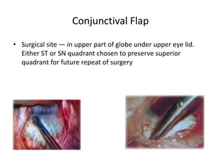
Trabeculectomy
Introduction
• A surgical procedure featuring a partial thickness scleral flap
that creates a fistula between anterior chamber and
subconjunctival space for filtration of aqueous and creation of
conjunctival bleb in an effort to lower lOP.
• TRABECULECTOMY+ANTIMETABOLITES = GOLD STANDARD
FOR SURGICAL MANAGEMENT OF GLAUCOMA
Mechanism
• Allows aqueous outflow from the anterior chamber to the
sub-tenon space, through the fistula & scleral flap borders
and finally collected in to episcleral & conjunctival veins
• A functioning flitering bleb forms over the sclerostomy site
which appears as a blister like elevation of the conjunctiva.
Indications
• ABSOLUTE INDICATIONS.
• Failure of conservative therapy
• Deterioration despite seemingly adequate lOP control
• Poor compliance
• Primary therapy — in advanced disease requiring a very low
target pressure, particularly in younger patients
• Patient preference
• RELATIVE INDICATIONS
• Economic considerations: In developing countries like India.
• Ocular or systemic side effects of antiglaucoma medications.
Relative Contraindications
• Blind eye
• Active inflammation
• Active anterior segment neovascularization
• Extensive conjunctival scarring /thin sclera(prior surgery ,
chemical trauma
Pre Op Evaluation
• Thorough ophthalmic evaluation including lOP, visual fields and
ONH evaluation
• Complete systemic evaluation with respect to diabetes,
hypertension, cardiovascular diseases
• Stop anticoagulants & antiplatelet agents
• Prophylactic peripheral iridotomy in angle closure disease.
• Conjunctival mobility should be checked pre operatively to plan
the site of surgery.
• Topical pilocarpine may be used preoperatively to constrict
the pupil
• Preoperative topical sympathetic agonists (e.g.
apraclonidine, adrenaline 0.01% or 0.1%) cause anterior
segment vasoconstriction and hence reduce intraoperative
bleeding
• Povidone-iodine is used to prepare the periorbital skin, the
eyelids and the ocular surface
• In uveitic glaucoma, the use of preoperative topical and/or
systemic steroids may be required to ensure optimal control
of ocular inflammation prior to surgery
Anaesthesia
• PERI/RETROBULBAR — don’t inject more than 5ml
• Topical anesthesia with intracameral anesthesia avoids
conjunctival damage, chemosis, SCH
• Subconjunctival anesthesia is less preferred
• General anaesthesia - in pediatric age group, highly anxious
patients or with suboptimal mental status. Allows maximal
control over systemic blood pressure and also lOP
intraoperatively.
• Eye painted & draped
• Eye exposed with lid speculum
• TRACTION SUTURE - To keep eye in inferior position
• 1. Clear corneal suture. Better exposure, less complications•
7-0 spatulated vicryl/silk at half thickness, 2mm anterior to
limbus
• SR bridle suture
• Associated with many complications
Conjunctival Flap
• Surgical site — in upper part of globe under upper eye lid.
Either ST or SN quadrant chosen to preserve superior
quadrant for future repeat of surgery
‘ Green Dots – restricted posterior aqueous
flow with a ring of scar tissue - RING OF STEEL•
Anteriorly directed aqueous flow (arrows)
Antimetabolites
• Polyvinyl alcohol sponges soaked in antifibrotic agent.
• Conjunctival edges kept away with T clamps. After usage
soaked pieces are removed & discarded, eye irrigated with 20-
6Oml BSS Larger area of antifibrotic treatment- diffuse non
cystic blebs
Complications
• Increased duration & concentraion — thin avascular cystic
blebs, hypotony
• Epithelial erosions — mainly 5-FU
• Intraocular penetration & damage – endothelial damage,
ciliary body destruction
• Infections - blebitis, endophthalmities and leakage.
• Scleritis, scleromalacia, necrosis
Scleral Flap
• To provide resistance to aqueous outflow & prevent
hypotony.
• Rectangular, triangular, trapezoids
Paracentesis
• Done to enable the surgeon to maintain the AC
• Infusion for continuous lOP maintenance by AC maintainer-
• To test for patency of filtration site by injecting fluid into AC.
• To prevent intra-op flat AC
Sclerostomy
• Fistula created by hand cut or KELLY DESCEMETS PUNCH
• Internal block excision
• Sclerostomy punch — preferred. An anterior corneoscleral
incision is made. The punch is then inserted to engage the
full-thickness of the limbus. It should be aligned
perpendicular to the eye to ensure a clean and nonshelved
sclerostomy
• Anterior corneoscleral entry into AC- reduces risk of iris
incarceration and bleeding from iris root and ciliary body
• Posterior extension — damage to ciliary body with
hemorrhage & ostium blockage by uveal tissue.
• 0.5—1.0 mm — adequate ostium size
Peripheral Iridectomy
• To prevent iris incarceration & ostium blockage performed
through the sclerostomy using Vannas scissors
• Base of the iridectomy should be little wider than sclerostomy
opening
• Complications: Hyphaema, inflammation, iridodialysis
Suture position determines control of tension
resistance to outflow‘ 10-0 Nylon suture‘ Suture knots
to be buried Types of sutures• Fixed, interrupted
sutures• Adjustable sutures• Releasable sutures
Scleral flap closure
Conjunctival closure
• Closure must be water-tight 10-O nylon or 10-0/9-O vicryl can
be used
• Single interrupted sutures
• Edge purse-string sutures
• Interrupted horizontal mattress
• Creation of corneal grooves for conjunctival closure of fornix-
base flap to minimize wound leakage and suture discomfort
• Anterior chamber is reformed with BSS through the
paracentesis
• Test leakage with Seidel technique
• At the end of surgery, cycloplegics/mydriatics can be used
• ATROPINE 1%
• Relaxation of ciliary muscle & pain relief Prevention of central
posterior synechae
• Less AC shallowing
Post Op management
• Follow-up closely
• Prednisolone acetate (1%) 2 hourly for 2 weeks and tapered
over 8 weeks
• Topical antibiotics: 4 weeks post operatively
• Topical mydriatic/cycloplegic agent : Atropine I % prevents AC
shallowing and risk of malignant glaucoma
• Oral or IV steroids: not routinely used , in severe uveitic
glaucoma
• Adjuvant subconjunctival 5-FU after first postoperative week
for up to several months to modulate wound healing• 5mg
(0.1 ml of 5Omgfml) 5-FU deep in superior fornix Indications
• As a part of planned postop regimen in cases high risk of
failure
• Signs of imminent bleb failure
• Adjuvant therapy after needling or re-exploration
• After several months for persistent healing response & rising
lop
Post op Assessment
• First 10 days are crucial
• Bleb
Migdel and Hitchings Classification
• TYPE I BLEB (very low lOP, elevated bleb) Thin,
transconjunctival flow of aqueous good filtration
• TYPE 2 BLEB (low lOP, elevated bleb) IDEAL BLEB
thin, diffuse, relatively avascular microcysts, good
filtration
• TYPE 3 BLEB (high lOP, low localized bleb) flat, no
microcystes engorged vessels, non filtering
• TYPE 4 BLEB (high lOP, encapsulated localised,
high elevated, engorged vessels. Cyst like cavity of
hypertrophied Tenon’s
• ANTERIOR CHAMBER
• Hyphema, hypopyon
• Shallow post-op AC
• Grade 1 : Peripheral corneal iris touch
• Grade 2: Corneal iris touch upto pupillary margin‘
• Grade 3: Lens cornea apposition
• CORNEA: look for epithelial erosion, edema 4.
• Iop
• FUNDUS : choroidal detachment, suprachoroidal hemorrhage,
decompression retinopathy
Argon suture Lysis
• Facilitated by compressing conjunctiva to visualize scleral
suture or high magnification suture lysis contact lens (Hoskins
or Blumenthal lens)
• Argon laser: 50-lOOpm, 0.05-0.1 sec duration, 200-400mW
power
• Within first 2 weeks: enhance filtration before scarring occurs
• Delayed (upto 6 weeks) if intraoperative antimetabolite used
Bleb needling
• Aim - to increase the size of the sub-Tenons aqueous lake
while avoiding overdrainage and hypotony
• Puncturing & loosening the scar tissue of filtration bleb to
increase sub-tenon’s aqueous lake
• Two types - Sub-Tenon’s Needling, Subscleral flap Needling
Complications
Conjunctival flap Related
• Tears and button holes
• Shrinkage
• Treatment -large button hole during early stage - select new
site in centre of flap - purse string suture
• Near limbus - Oversewn with adjacent conjunctiva or sutured
directly to cornea tenon’s capsule should be incorporated to
increase strength
Scleral flap Related
• SHALLOW AC
• Viscoelastic injection
• Preplaced scleral flap sutures
• HYPHEMA
• During Pl, conjunctival dissection, episcleral &perforating
vessels, sclerostomy site
• Stop antiplatelet, anticoagulants
• Gentle handing, adequate cautery
• Punch till blue-white junction & not beyond it
• Rx Light compression
• Keep scIeraI flap open to allow blood to exit along with gentle
irrigation
• Persistent bleed — visco/air tamponade
Suprachoroidal hemorrhage
• Can occur at any time intra-op & post-op
• Delayed
• Precautions
• Avoid prolonged hypotony
• Preplaced flap sutures
• Tighter flap closure & postop suture lysis
• Controlled decompression of globe
• Use of punch instead of block excision
• Signs — shallowing of AC, dark expansion of choroid.
• Rx — wound closure immediately — IV Mannitol
• Posterior sclerotomies — to drain hemorrahage
• Pl related
• Large Pl iridodialysis
• Vitreous loss & lens injury
• Zonular-lens complex damaged during Pl
• Sudden decompression of globe with forward shift of iris-lens
diaphragm
• Rx‘ Anterior vitrectomy to be done to avoid ostium blockage
Wipe out phenomenon
• 1-2% risk in all glaucoma surgeries.
• Early undetected visual field loss/central fixation loss
• Typically occurs in advanced glaucoma with split fixation
or VF loss within 5 degrees of fixation. Precautions
• Sub Tenons anesthesia
• Avoid Adrenaline use
• Avoid post-op lOP spike
• Prompt management of post-op lOP spike
Shallow AC with low IOP
• Causes
• Overfiltration
• Choroidal detachment with decreased aqueous production
• Wound leak Treatment
• Grade 1 — reforms spontaneously
• Grade 2 — observation Reform AC with visco/air
• Grade 3 — immediate correction choroidal drainage
Shallow AC with high IOP
Deep AC with high IOP
• Causes
• Obstruction of ostium
• Tight flap sutures
• Failing bleb
• Steroid induced lOP response
• Bleb is flat/low
• Rx Gonioscopy- to look for patency of ostium
• Nd YAG laser- to disrupt fibrin, vitreous, iris.
• Ocular massage/suture release.
• Failing bleb-increase topical streoids,.
• Post op augmentation with antimetabolites,
• Needling of bleb
Ciliochoroidal detachment
• Commonly after full thickness surgery
• Rx
• Resolves with topical & systemic steroids
• Prophylactic sclerotomy in predisposed eyes
• Surgical drainage in case of cornea lens touch
Overfiltration
• CF.
• Hypotony (IOP<6mmHg)
• Shallow AC
• Large, diffuse bleb No wound leak
• Rx Patching with focal compression over region of excessive
aqueous flow
• Symblepharon ring,
• Simmon’s tamponade shell
• Reform AC
• Autologous blood injection into bleb
• Cryo or laser application to reduce bleb size
• Compression sutures
• Surgical revision
Decompression Retinopathy
• DECOMPRESSION RETINOPATHY‘ Sudden decompression of
eye in high lOP — transient increase in retinal & chroidal
blood flow‘ Retinal, subretinal, suprachoroidal hemorrahage
‘Mimics CRVO‘HYPOTONIC MACULOPATHY‘ In chronic
hypotony Choroidal folds in macular area Macular thickening
Disc swelling
Filtration Failure
Management
Encapsulated bleb
• Tenon’s cyst — 10-28%
• A localized, highly elevated, dome shaped, cyst like
cavity of hypertrophied tenon’s capsule with engorged
blood vessels. During first 8 weeks.
• Risk factors — young, male, glove powder, prolonged
AGM use, prior ALT/conjunctival surgery
• Inflammatory mediators + collagen producing fibroblasts
= Fibroblast proliferation
• High lOP after initial period of lOP control
Management
Blebities
• White bleb with surrounding intense conjunctival injection
• Variable anterior chamber reaction
• Clear vitreous
• Risk factors:‘ Early chronic intermittent bleb leak
• Myopia: thin scIeraI flap
• Intra-operative MMC
• Blepharitis.
• Diabetes mellitus
• Chronic antibiotic use
• Treatment: Topical antibiotics
Bleb related Endophthalmities
• Early postoperative Endophthalmitis
• Onset within first 3 months
• Staphylococcus epidermicis
• Delayed- onset Endophthalmitis Onset after 3 months
Streptococcus, staphylococcus, H. influenzae
• Pain, photophobia, sticky eyes, reduced vision Milky white
appearance of bleb, fibrin or hypopyon in AC &
vitritis(distinguishes from blebitis Aqueous and vitreous
aspirates
• High dose parenteral and periocular antibiotics.
• Intravitreal antibiotics
Symptomatic bleb
• BLEB DYSESTHESIA
• Asymptomatic or reasonably well tolerated
• Most patients are aware of a conjunctival ‘blister”
• Symptoms are frequent in nasal or large blebs or when there
is extension into cornea, difficulty with blinking, tear film
abnormalities, foreign-body sensation, & induced astigmatism
• •Rx Artificial tears
• Surgical bleb excision or conjunctival flap reinforcement
• Bleb shrinkage : cryotherapy, Nd:YAG laser
thermotherapy,argon laser, diathermy, and cauterization
Collagen implants
• Increases efficacy without need for antimetabolites
• Made of PORCINE — a telocollagen cross-linked with GAGs
• Biodegraded around 90-1 80 days 2sizes—6x2, 12x1
• Mechanism• Provides a scaffolding for fibroblasts to grow
randomly which could reduce scar formation effectively•
Collagen matrix itself can function like a reservoir to absorb
aqueous• Provides pressure on scIeral flap to create
controlled drainage in subconjunctival space
Combined Surgery
• INDICATIONS - visually significant cataract
• with more than 3 medications for lOP control
• intolerant or allergic to glaucoma medications significant
cupping or visual field loss as the optic nerve is less able to
tolerate perioperative lop rise.
• monocular patient.
• PFS, pigment dispersion syndrome & angle recession.
• Combined surgery has the advantage of treating both
diseases with a single surgical intervention and lOPreduction
tends to be greater than with cataract surgery alone.-
Disadvantages include increased surgery time which can
increase surgical risk
Repeat Trabeculectomy
• Choice of treatment following a failed Trabeculectomy is
individualized for each patient where factors like age,ocular
anatomy, details of primary procedure, conditionof other eye
may guide the decision
• Technique of repeat trabeculectomy
• Site — superonasal, superotemporal.
• Conjunctival incision — difficult, hydrodissect conjunctiva
through subconjunctival BSS
• Antimetabolites — mandatory, 0.4mg/mi MMC for 3 mins
• Scleral flap — mini trabeculactomy
• Post-op — topical preservative free steroids
• Outcome — less succesful than initial trabeculectomy
THANK YOU

Trabeculectomy.pptx

  • 1.
  • 2.
    Introduction • A surgicalprocedure featuring a partial thickness scleral flap that creates a fistula between anterior chamber and subconjunctival space for filtration of aqueous and creation of conjunctival bleb in an effort to lower lOP. • TRABECULECTOMY+ANTIMETABOLITES = GOLD STANDARD FOR SURGICAL MANAGEMENT OF GLAUCOMA
  • 3.
    Mechanism • Allows aqueousoutflow from the anterior chamber to the sub-tenon space, through the fistula & scleral flap borders and finally collected in to episcleral & conjunctival veins • A functioning flitering bleb forms over the sclerostomy site which appears as a blister like elevation of the conjunctiva.
  • 4.
    Indications • ABSOLUTE INDICATIONS. •Failure of conservative therapy • Deterioration despite seemingly adequate lOP control • Poor compliance • Primary therapy — in advanced disease requiring a very low target pressure, particularly in younger patients • Patient preference • RELATIVE INDICATIONS • Economic considerations: In developing countries like India. • Ocular or systemic side effects of antiglaucoma medications.
  • 5.
    Relative Contraindications • Blindeye • Active inflammation • Active anterior segment neovascularization • Extensive conjunctival scarring /thin sclera(prior surgery , chemical trauma
  • 6.
    Pre Op Evaluation •Thorough ophthalmic evaluation including lOP, visual fields and ONH evaluation • Complete systemic evaluation with respect to diabetes, hypertension, cardiovascular diseases • Stop anticoagulants & antiplatelet agents • Prophylactic peripheral iridotomy in angle closure disease. • Conjunctival mobility should be checked pre operatively to plan the site of surgery.
  • 7.
    • Topical pilocarpinemay be used preoperatively to constrict the pupil • Preoperative topical sympathetic agonists (e.g. apraclonidine, adrenaline 0.01% or 0.1%) cause anterior segment vasoconstriction and hence reduce intraoperative bleeding • Povidone-iodine is used to prepare the periorbital skin, the eyelids and the ocular surface • In uveitic glaucoma, the use of preoperative topical and/or systemic steroids may be required to ensure optimal control of ocular inflammation prior to surgery
  • 8.
    Anaesthesia • PERI/RETROBULBAR —don’t inject more than 5ml • Topical anesthesia with intracameral anesthesia avoids conjunctival damage, chemosis, SCH • Subconjunctival anesthesia is less preferred • General anaesthesia - in pediatric age group, highly anxious patients or with suboptimal mental status. Allows maximal control over systemic blood pressure and also lOP intraoperatively.
  • 9.
    • Eye painted& draped • Eye exposed with lid speculum • TRACTION SUTURE - To keep eye in inferior position • 1. Clear corneal suture. Better exposure, less complications• 7-0 spatulated vicryl/silk at half thickness, 2mm anterior to limbus • SR bridle suture • Associated with many complications
  • 11.
    Conjunctival Flap • Surgicalsite — in upper part of globe under upper eye lid. Either ST or SN quadrant chosen to preserve superior quadrant for future repeat of surgery
  • 12.
    ‘ Green Dots– restricted posterior aqueous flow with a ring of scar tissue - RING OF STEEL• Anteriorly directed aqueous flow (arrows)
  • 13.
  • 14.
    • Polyvinyl alcoholsponges soaked in antifibrotic agent. • Conjunctival edges kept away with T clamps. After usage soaked pieces are removed & discarded, eye irrigated with 20- 6Oml BSS Larger area of antifibrotic treatment- diffuse non cystic blebs
  • 15.
    Complications • Increased duration& concentraion — thin avascular cystic blebs, hypotony • Epithelial erosions — mainly 5-FU • Intraocular penetration & damage – endothelial damage, ciliary body destruction • Infections - blebitis, endophthalmities and leakage. • Scleritis, scleromalacia, necrosis
  • 16.
    Scleral Flap • Toprovide resistance to aqueous outflow & prevent hypotony. • Rectangular, triangular, trapezoids
  • 17.
    Paracentesis • Done toenable the surgeon to maintain the AC • Infusion for continuous lOP maintenance by AC maintainer- • To test for patency of filtration site by injecting fluid into AC. • To prevent intra-op flat AC
  • 18.
    Sclerostomy • Fistula createdby hand cut or KELLY DESCEMETS PUNCH • Internal block excision • Sclerostomy punch — preferred. An anterior corneoscleral incision is made. The punch is then inserted to engage the full-thickness of the limbus. It should be aligned perpendicular to the eye to ensure a clean and nonshelved sclerostomy
  • 19.
    • Anterior corneoscleralentry into AC- reduces risk of iris incarceration and bleeding from iris root and ciliary body • Posterior extension — damage to ciliary body with hemorrhage & ostium blockage by uveal tissue. • 0.5—1.0 mm — adequate ostium size
  • 20.
    Peripheral Iridectomy • Toprevent iris incarceration & ostium blockage performed through the sclerostomy using Vannas scissors • Base of the iridectomy should be little wider than sclerostomy opening • Complications: Hyphaema, inflammation, iridodialysis
  • 21.
    Suture position determinescontrol of tension resistance to outflow‘ 10-0 Nylon suture‘ Suture knots to be buried Types of sutures• Fixed, interrupted sutures• Adjustable sutures• Releasable sutures Scleral flap closure
  • 22.
    Conjunctival closure • Closuremust be water-tight 10-O nylon or 10-0/9-O vicryl can be used • Single interrupted sutures • Edge purse-string sutures • Interrupted horizontal mattress • Creation of corneal grooves for conjunctival closure of fornix- base flap to minimize wound leakage and suture discomfort
  • 23.
    • Anterior chamberis reformed with BSS through the paracentesis • Test leakage with Seidel technique • At the end of surgery, cycloplegics/mydriatics can be used • ATROPINE 1% • Relaxation of ciliary muscle & pain relief Prevention of central posterior synechae • Less AC shallowing
  • 24.
    Post Op management •Follow-up closely • Prednisolone acetate (1%) 2 hourly for 2 weeks and tapered over 8 weeks • Topical antibiotics: 4 weeks post operatively • Topical mydriatic/cycloplegic agent : Atropine I % prevents AC shallowing and risk of malignant glaucoma • Oral or IV steroids: not routinely used , in severe uveitic glaucoma
  • 25.
    • Adjuvant subconjunctival5-FU after first postoperative week for up to several months to modulate wound healing• 5mg (0.1 ml of 5Omgfml) 5-FU deep in superior fornix Indications • As a part of planned postop regimen in cases high risk of failure • Signs of imminent bleb failure • Adjuvant therapy after needling or re-exploration • After several months for persistent healing response & rising lop
  • 26.
    Post op Assessment •First 10 days are crucial • Bleb
  • 27.
    Migdel and HitchingsClassification • TYPE I BLEB (very low lOP, elevated bleb) Thin, transconjunctival flow of aqueous good filtration • TYPE 2 BLEB (low lOP, elevated bleb) IDEAL BLEB thin, diffuse, relatively avascular microcysts, good filtration • TYPE 3 BLEB (high lOP, low localized bleb) flat, no microcystes engorged vessels, non filtering • TYPE 4 BLEB (high lOP, encapsulated localised, high elevated, engorged vessels. Cyst like cavity of hypertrophied Tenon’s
  • 28.
    • ANTERIOR CHAMBER •Hyphema, hypopyon • Shallow post-op AC • Grade 1 : Peripheral corneal iris touch • Grade 2: Corneal iris touch upto pupillary margin‘ • Grade 3: Lens cornea apposition • CORNEA: look for epithelial erosion, edema 4. • Iop • FUNDUS : choroidal detachment, suprachoroidal hemorrhage, decompression retinopathy
  • 29.
    Argon suture Lysis •Facilitated by compressing conjunctiva to visualize scleral suture or high magnification suture lysis contact lens (Hoskins or Blumenthal lens) • Argon laser: 50-lOOpm, 0.05-0.1 sec duration, 200-400mW power • Within first 2 weeks: enhance filtration before scarring occurs • Delayed (upto 6 weeks) if intraoperative antimetabolite used
  • 30.
    Bleb needling • Aim- to increase the size of the sub-Tenons aqueous lake while avoiding overdrainage and hypotony • Puncturing & loosening the scar tissue of filtration bleb to increase sub-tenon’s aqueous lake • Two types - Sub-Tenon’s Needling, Subscleral flap Needling
  • 31.
  • 32.
    Conjunctival flap Related •Tears and button holes • Shrinkage • Treatment -large button hole during early stage - select new site in centre of flap - purse string suture • Near limbus - Oversewn with adjacent conjunctiva or sutured directly to cornea tenon’s capsule should be incorporated to increase strength
  • 33.
  • 34.
    • SHALLOW AC •Viscoelastic injection • Preplaced scleral flap sutures • HYPHEMA • During Pl, conjunctival dissection, episcleral &perforating vessels, sclerostomy site • Stop antiplatelet, anticoagulants • Gentle handing, adequate cautery • Punch till blue-white junction & not beyond it • Rx Light compression • Keep scIeraI flap open to allow blood to exit along with gentle irrigation • Persistent bleed — visco/air tamponade
  • 35.
    Suprachoroidal hemorrhage • Canoccur at any time intra-op & post-op • Delayed • Precautions • Avoid prolonged hypotony • Preplaced flap sutures • Tighter flap closure & postop suture lysis • Controlled decompression of globe • Use of punch instead of block excision • Signs — shallowing of AC, dark expansion of choroid. • Rx — wound closure immediately — IV Mannitol • Posterior sclerotomies — to drain hemorrahage
  • 36.
    • Pl related •Large Pl iridodialysis • Vitreous loss & lens injury • Zonular-lens complex damaged during Pl • Sudden decompression of globe with forward shift of iris-lens diaphragm • Rx‘ Anterior vitrectomy to be done to avoid ostium blockage
  • 37.
    Wipe out phenomenon •1-2% risk in all glaucoma surgeries. • Early undetected visual field loss/central fixation loss • Typically occurs in advanced glaucoma with split fixation or VF loss within 5 degrees of fixation. Precautions • Sub Tenons anesthesia • Avoid Adrenaline use • Avoid post-op lOP spike • Prompt management of post-op lOP spike
  • 38.
    Shallow AC withlow IOP • Causes • Overfiltration • Choroidal detachment with decreased aqueous production • Wound leak Treatment • Grade 1 — reforms spontaneously • Grade 2 — observation Reform AC with visco/air • Grade 3 — immediate correction choroidal drainage
  • 39.
  • 40.
    Deep AC withhigh IOP • Causes • Obstruction of ostium • Tight flap sutures • Failing bleb • Steroid induced lOP response • Bleb is flat/low • Rx Gonioscopy- to look for patency of ostium • Nd YAG laser- to disrupt fibrin, vitreous, iris. • Ocular massage/suture release. • Failing bleb-increase topical streoids,. • Post op augmentation with antimetabolites, • Needling of bleb
  • 41.
    Ciliochoroidal detachment • Commonlyafter full thickness surgery • Rx • Resolves with topical & systemic steroids • Prophylactic sclerotomy in predisposed eyes • Surgical drainage in case of cornea lens touch
  • 42.
    Overfiltration • CF. • Hypotony(IOP<6mmHg) • Shallow AC • Large, diffuse bleb No wound leak • Rx Patching with focal compression over region of excessive aqueous flow • Symblepharon ring, • Simmon’s tamponade shell • Reform AC • Autologous blood injection into bleb • Cryo or laser application to reduce bleb size • Compression sutures • Surgical revision
  • 43.
    Decompression Retinopathy • DECOMPRESSIONRETINOPATHY‘ Sudden decompression of eye in high lOP — transient increase in retinal & chroidal blood flow‘ Retinal, subretinal, suprachoroidal hemorrahage ‘Mimics CRVO‘HYPOTONIC MACULOPATHY‘ In chronic hypotony Choroidal folds in macular area Macular thickening Disc swelling
  • 44.
  • 45.
  • 46.
    Encapsulated bleb • Tenon’scyst — 10-28% • A localized, highly elevated, dome shaped, cyst like cavity of hypertrophied tenon’s capsule with engorged blood vessels. During first 8 weeks. • Risk factors — young, male, glove powder, prolonged AGM use, prior ALT/conjunctival surgery • Inflammatory mediators + collagen producing fibroblasts = Fibroblast proliferation • High lOP after initial period of lOP control
  • 47.
  • 48.
    Blebities • White blebwith surrounding intense conjunctival injection • Variable anterior chamber reaction • Clear vitreous • Risk factors:‘ Early chronic intermittent bleb leak • Myopia: thin scIeraI flap • Intra-operative MMC • Blepharitis. • Diabetes mellitus • Chronic antibiotic use • Treatment: Topical antibiotics
  • 49.
    Bleb related Endophthalmities •Early postoperative Endophthalmitis • Onset within first 3 months • Staphylococcus epidermicis • Delayed- onset Endophthalmitis Onset after 3 months Streptococcus, staphylococcus, H. influenzae • Pain, photophobia, sticky eyes, reduced vision Milky white appearance of bleb, fibrin or hypopyon in AC & vitritis(distinguishes from blebitis Aqueous and vitreous aspirates • High dose parenteral and periocular antibiotics. • Intravitreal antibiotics
  • 50.
    Symptomatic bleb • BLEBDYSESTHESIA • Asymptomatic or reasonably well tolerated • Most patients are aware of a conjunctival ‘blister” • Symptoms are frequent in nasal or large blebs or when there is extension into cornea, difficulty with blinking, tear film abnormalities, foreign-body sensation, & induced astigmatism • •Rx Artificial tears • Surgical bleb excision or conjunctival flap reinforcement • Bleb shrinkage : cryotherapy, Nd:YAG laser thermotherapy,argon laser, diathermy, and cauterization
  • 51.
    Collagen implants • Increasesefficacy without need for antimetabolites • Made of PORCINE — a telocollagen cross-linked with GAGs • Biodegraded around 90-1 80 days 2sizes—6x2, 12x1 • Mechanism• Provides a scaffolding for fibroblasts to grow randomly which could reduce scar formation effectively• Collagen matrix itself can function like a reservoir to absorb aqueous• Provides pressure on scIeral flap to create controlled drainage in subconjunctival space
  • 53.
    Combined Surgery • INDICATIONS- visually significant cataract • with more than 3 medications for lOP control • intolerant or allergic to glaucoma medications significant cupping or visual field loss as the optic nerve is less able to tolerate perioperative lop rise. • monocular patient. • PFS, pigment dispersion syndrome & angle recession.
  • 54.
    • Combined surgeryhas the advantage of treating both diseases with a single surgical intervention and lOPreduction tends to be greater than with cataract surgery alone.- Disadvantages include increased surgery time which can increase surgical risk
  • 55.
    Repeat Trabeculectomy • Choiceof treatment following a failed Trabeculectomy is individualized for each patient where factors like age,ocular anatomy, details of primary procedure, conditionof other eye may guide the decision • Technique of repeat trabeculectomy • Site — superonasal, superotemporal. • Conjunctival incision — difficult, hydrodissect conjunctiva through subconjunctival BSS • Antimetabolites — mandatory, 0.4mg/mi MMC for 3 mins • Scleral flap — mini trabeculactomy • Post-op — topical preservative free steroids • Outcome — less succesful than initial trabeculectomy
  • 56.