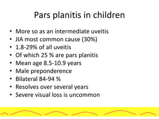
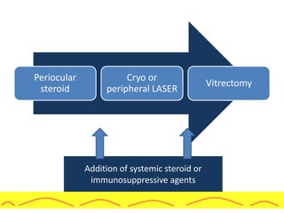
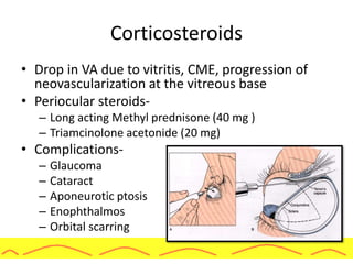
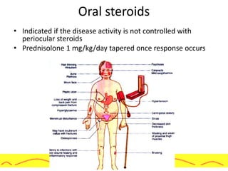
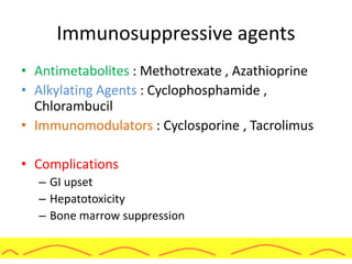
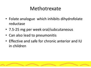
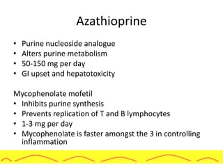
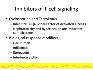
PARS PLANITIS
Shah-Noor Hassan FCPS,FRCS(Glasgow)
Vitreo-Retina Consultant
Bangladesh Eye Hospital & Institute
History
• Cyclitis- Fuchs in 1908, Duke-Elder 1941
• Peripheral uveitis- Schepens-1950
• Peripheral cyclitis- Brockhurst et.al. - 1960
• Pars planitis- Welch et.al. - 1960
• Chronic cyclitis- Hogan & Kimura in 1961
• Vitritis- Gass et.al. - 1968
• Intermediate uveitis- IUSG- 1987
• SUN working group-2004
Nomenclature
• Standardization of Uveitis
Nomenclature working
group classification
• Idiopathic form of
intermediate uveitis
• Includes snowballs and
snowbanking
• If associated with diseases
like Sarcoidosis and Lyme
disease then included in
intermediate uveitis
Epidemiology
• 10-25 % of all the uveitis cases
• Children and young adults
• Can occur at any age
• Both sexes are equally affected
• 80% are bilateral
• Less in Chinese and Japanese population
Etiology
• Idiopathic
• No known hereditary or environmental factors
• Some isolated cases of familial pars planitis
• Associated with various systemic diseases
• Most common- multiple sclerosis, sarcoidosis
Pathogenesis
• Immune mediated response
• But the antigenic stimulus remains speculative
• Davis and colleagues
– Stage 1- immunologically mediated
– Stage 2- Non specific breakdown of intraocular
regulatory mechanisms
(Not necessarily an autoimmune mechanism but
even exogenous viral or bacterial antigens
may be responsible)
Pathogenesis
• Escape from regulatory control of Helper T cells
directed against these antigens
• Defective intraocular T cell regulation of B cells
• Decreased helper to suppressor T cell ratios in aqueous
and peripheral blood
• Other mechanisms
– Anterior chamber associated immune deviation
– Auto retinal antibodies
– Related to Demyelination
– HLA-DR15 and HLA-A28 positivity
– Nucleoporin like protien-nup36
Pathology
• Peripheral retina and ciliary body
demonstrate condensed vitreous ,
fibroblasts, spindle cells, lymphocytes
and blood vessels
• Prominent lymphocyte cuffing of retinal
veins
• Pars plana exudates
– Loose fibrovascular layer containing
scattered mononuclear inflammatory cells
and a few fibrocyte like cells
– Fibroglial tissue consists of vitreous
collagen, mullers cells and probable
fibrous astrocytes
Clinical features
• Floaters and hazy vision
• No pain, photophobia, redness
• First episode is associated with a more
severe and symptomatic iridocyclitis
• Subsequent episodes have a chronic
course…….
• One eye symptomatic other eye may be
asymptomatic and even show signs of
active disease
Presentation
• VISION LOSS
• CME, ERM
• PSC
• Vitreous Opacification
• Membranes
• Retinal Detachment
• Vitreous Hemorrhage
Presentation
• Cells, flare, KPs in AC, synechiae (Spill
over anterior segment inflammation)
• Snow balls (organized vitreous
inflammatory cells )
• Snow banking (exudates at pars plana)
• May be localised to inferior half
Presentation
• Peripheral vasculitis
• CME, Peripapillary retinal edema
• Vitritis, Cyclitis
• Vitreous hemorrhage
• Band shaped keratopathy
Effect on macula
• Macular edema (CME) and maculopathy (12-
82 %)
• Most common cause of visual loss
• Incidence increases with duration and severity
of disease
Vitreous involvement
• Vitritis
• Snowball formation
• Vitreous membranes and
floaters
• Vitreous hemorrhage
Retinal involvement
• Retinal vascular changes
– Tortuosity of arterioles and venules
– Peripheral vascular sheathing
(Periphlebitis-16-36 %)
– Neovascularizations (6.5%)
– Retinal detachment (2.2-51 %)
• Causes of RD
– Vitreous traction due to long standing
inflammation and subsequent hole
formation
– Exudative detachment secondary to
uvietis inflammation
Optic nerve involvement
• Disc edema- 3-38%
• Optic neuritis with or without multiple
sclerosis was seen in 7.4 %
Complications
• Glaucoma
– Acute uveitis- 7.6 %
– Chronic – 6.5% at one year, 11.1 at 5 years
• Causes of glaucoma
– Active inflammation
– Steroid usage
– Increasing age
– Number of years since diagnosis
Cataract
• 15-50% of eyes
• Posterior or anterior subcapsular
• At times posterior cortical even posterior
polar have been reported
• Incidence increases with duration and
severity of disease
• If treated earlier with immunosuppressive
rather than corticosteroids cataract
formation is less severe
Types Of Retinal Detachment
• Exudative RD in 5-17%
• Vitreoretinal traction - in 3-22% TRRD
• Brockhurst and Schepens – 4 types of RRD
Type I:
- Low lying, chronic, associated with demarcation
lines
- Small breaks near ora with exudates
- Benign course
Types Of Retinal Detachment
Type II:
- Large dialysis at the posterior edge of the pars plana exudate
- Slowly progressive
- May resolve spontaneously if VR exudation occludes the break
- Seen in pts with a mild chronic inflammatory course
Type III:
- Rapidly progressive
- Large breaks associated with NVVB and circumferential pars
plana exudates.
- Associated with severe chronic uveitis.
Pars planitis in children
• More so as an intermediate uveitis
• JIA most common cause (30%)
• 1.8-29% of all uveitis
• Of which 25 % are pars planitis
• Mean age 8.5-10.9 years
• Male preponderence
• Bilateral 84-94 %
• Resolves over several years
• Severe visual loss is uncommon
STANDARDIZATION OF UVEITIS
NOMENCLATURE
Natural course
Self limited
10 %
Smoldering
59%
Recurrent
31 %
Diagnosis
CLINICAL FEATURES
OPHTHALMIC
INVESTIGATIIONS
TO RULE OUT SECONDARY
CAUSES
Diagnosis: Clinical
• History
• Clinical findings
• Duration of symptoms, recurrences
• Fever , fatigue or night sweats are typical signs -
Sarcoidosis & TB
• Loss of sensitivity or paresthesias of hands, arms
or legs - Multiple sclerosis
• Dermatitis, Arthritis– Lyme
• Contact with cats – possibility of Bartonella
infection
Ophthalmic investigations
• V/A
• SL biomicroscopy
• IOP and
• Fundus examination with scleral depression
• Amsler grid
• OCT - Macular oedema
• Fluorescein Angiogram-
Vasculitis ,CNP areas ,
New vessels & CME
• B scan (Hazy media)
• UBM
• Diagnostic vitrectomy
Ophthalmic investigations
To rule out secondary causes…
• Complete hemogram
• ELISA for tuberculosis and toxoplasma
• CXR
• Galium Scan and Chest CT
Lab Inv:
- ACE levels- elevated in 60-90% of active sarcoid
patients
- Lysozyme level - Elevated in granulomatous disorders
viz sarcoid, TB, and leprosy
- Elevated antibody titre against Borrelia burgdorferi
• Sarcoidosis
• Tuberculosis.
Differential diagnosis
• Non infectious
– Multiple sclerosis (3-27 %)
– Sarcoidosis (23-26%, IU
developing sarcoidosis- 2-
10%)
– Intraocular lymphoma
(PCNSL- 10-20% have
vitreous inflam)
• Infectious conditions
– Tuberculosis
– Syphilis (10.3%)
– Lyme disease
– Toxoplasma
– Toxocariasis
– HTLV-1, EBV, Cat scratch
disease
– Endogenous
endophthalmitis
– ARN, Eales, VKH, Fuch’s
MANAGEMENT
Four Step Approach (Kaplan et al)
Modified 5 step program: S.Foster et al
Topical +/ Periocular corticosteroids
Oral +/ Topical NSAID
After 3rd injection
Systemic C steroids
Inflammation persists or recurs
Peripheral retinal cryopexy /BIOL
Recur following 6th regional steroid injection
PPV/ Immunosupression
Recalcitrant inflammation
Addition of systemic steroid or
immunosuppressive agents
Periocular
steroid
Cryo or
peripheral LASER
Vitrectomy
Corticosteroids
• Drop in VA due to vitritis, CME, progression of
neovascularization at the vitreous base
• Periocular steroids-
– Long acting Methyl prednisone (40 mg )
– Triamcinolone acetonide (20 mg)
• Complications-
– Glaucoma
– Cataract
– Aponeurotic ptosis
– Enophthalmos
– Orbital scarring
Corticosteroids
• IVTA can be given in cases of severe macular
edema
• Complications
Cataract
Glaucoma
Endophthalmitis
Oral steroids
• Indicated if the disease activity is not controlled with
periocular steroids
• Prednisolone 1 mg/kg/day tapered once response occurs
Immunosuppressive agents
• Antimetabolites : Methotrexate , Azathioprine
• Alkylating Agents : Cyclophosphamide ,
Chlorambucil
• Immunomodulators : Cyclosporine , Tacrolimus
• Complications
– GI upset
– Hepatotoxicity
– Bone marrow suppression
Methotrexate
• Folate analogue which inhibits dihydrofolate
reductase
• 7.5-25 mg per week oral/subcutaneous
• Can also lead to pneumonitis
• Effective and safe for chronic anterior and IU
in children
Azathioprine
• Purine nucleoside analogue
• Alters purine metabolism
• 50-150 mg per day
• GI upset and hepatotoxicity
Mycophenolate mofetil
• Inhibits purine synthesis
• Prevents replication of T and B lymphocytes
• 1-3 mg per day
• Mycophenolate is faster amongst the 3 in controlling
inflammation
Inhibitors of T-cell signaling
• Cyclosporine and Tacrolimus
– Inhibit NF-AT (Nuclear Factor of Activated T-cells )
– Nephrotoxicity and hypertension are important
complications
• Biological response modifiers
– Daclizumab
– Infliximab
– Eternacept
– Interferon alpha
Biological response modifiers
• Daclizumab
– Humanized monoclonal ant-IL-2 receptor alpha
antibody
– Suppresses auto reactive T-cells
– 1 mg/kg IV every 2 weeks for 5 doses
– Increase risk of infection
Biological response modifiers
• Infliximab
– Binds to TNF and prevents its action on
target tissues
• Eternacept
– Dimeric, fully human, soluble TNF receptor
– Binds tightly and specifically to circulating
and cell-bound TNF
• Adalimumab
– Can be self administered as a subcutaneous
injection
– Fully humanized so less chances of
antibody formation
• Disseminated tuberculosis is one of the
fatal complications
Newer steroid implants
• Retisert
– Fluocinolone acetonide implant
– Duration of 30 months
• Ozurdex
– Dexamethasone implant
Ablative procedures
• Failed drug therapy
• At times cryotherapy is preferred before
immunosuppressive Rx
• Aim
– To treat neovascularization associated with the
exudates
– To destroy the peripheral vessels which bring in
the inflammatory mediators
• Double row ,single freeze
• Apply to pars plana and posterior to it
• CONFLUENT BURNS
• Extend 1 clock hr on either side of all areas
affected by inflammation
• EFFECTS
– Decreases vitritis and improves VA
– Decrease in fluorescein in the treated area
– Induce regression of this NVVB and
consequently stabilize inflammation
Cryo ablation
LASER ablation
• LASER photocoagulation works as effective as
cryo
• 3-4 rows of burns are placed at the pars plana
and peripheral retina
• Works on the same mechanism as cryo
Vitrectomy
• Vitrectomy for uveitis began in late 1970s
• Aims
– Get rid of inflammatory mediators and immunologically
competent cells
– Clear the media
• Indications
– Refractory uveitis
– Vision loss due to densely opacified vitreous
– Scar tissue pulling on ciliary body causing hypotony
– CME, ERM
– Dense PCO
– TRD
MANAGEMENT OF CATARACT:
• Eye - quiet for 3 months
– Preoperative – Start steroids 3 days prior
– Postoperative - slow taper.
• Technique –
– As preferred by surgeon
– Minimal trauma
– Preferably heparin coated IOL
What’s new….
• Anti VEGF agents are being evaluated in cases
of uveitis with macular edema
• Lucentis and Avastin have been proved to be
effective in cases of uveitic CME
Nevanac in pars planitis
• Case 1: - Short term benefit in cases of
recurrent intermediate uveitis
Case 2
• Rapid resolution of vitritis in uncomplicated
case of intermediate uveitis
Case 3
• Fresh case of pars planitis with CME
• Nevanac improved the CME
Summary
• Examination of pars plana
• Diagnose macular edema
• Rule out secondary causes
• Plan appropriate treatment modility
• Bold use of steroids and immunosuppressive
agents to prevent vision loss due to macular
involvement
• Look out for complications
• Surgical management in resistant cases and to
clear the media
Thank you…

Pars Planitis

  • 1.
    PARS PLANITIS Shah-Noor HassanFCPS,FRCS(Glasgow) Vitreo-Retina Consultant Bangladesh Eye Hospital & Institute
  • 2.
    History • Cyclitis- Fuchsin 1908, Duke-Elder 1941 • Peripheral uveitis- Schepens-1950 • Peripheral cyclitis- Brockhurst et.al. - 1960 • Pars planitis- Welch et.al. - 1960 • Chronic cyclitis- Hogan & Kimura in 1961 • Vitritis- Gass et.al. - 1968 • Intermediate uveitis- IUSG- 1987 • SUN working group-2004
  • 3.
    Nomenclature • Standardization ofUveitis Nomenclature working group classification • Idiopathic form of intermediate uveitis • Includes snowballs and snowbanking • If associated with diseases like Sarcoidosis and Lyme disease then included in intermediate uveitis
  • 4.
    Epidemiology • 10-25 %of all the uveitis cases • Children and young adults • Can occur at any age • Both sexes are equally affected • 80% are bilateral • Less in Chinese and Japanese population
  • 5.
    Etiology • Idiopathic • Noknown hereditary or environmental factors • Some isolated cases of familial pars planitis • Associated with various systemic diseases • Most common- multiple sclerosis, sarcoidosis
  • 6.
    Pathogenesis • Immune mediatedresponse • But the antigenic stimulus remains speculative • Davis and colleagues – Stage 1- immunologically mediated – Stage 2- Non specific breakdown of intraocular regulatory mechanisms (Not necessarily an autoimmune mechanism but even exogenous viral or bacterial antigens may be responsible)
  • 7.
    Pathogenesis • Escape fromregulatory control of Helper T cells directed against these antigens • Defective intraocular T cell regulation of B cells • Decreased helper to suppressor T cell ratios in aqueous and peripheral blood • Other mechanisms – Anterior chamber associated immune deviation – Auto retinal antibodies – Related to Demyelination – HLA-DR15 and HLA-A28 positivity – Nucleoporin like protien-nup36
  • 8.
    Pathology • Peripheral retinaand ciliary body demonstrate condensed vitreous , fibroblasts, spindle cells, lymphocytes and blood vessels • Prominent lymphocyte cuffing of retinal veins • Pars plana exudates – Loose fibrovascular layer containing scattered mononuclear inflammatory cells and a few fibrocyte like cells – Fibroglial tissue consists of vitreous collagen, mullers cells and probable fibrous astrocytes
  • 9.
    Clinical features • Floatersand hazy vision • No pain, photophobia, redness • First episode is associated with a more severe and symptomatic iridocyclitis • Subsequent episodes have a chronic course……. • One eye symptomatic other eye may be asymptomatic and even show signs of active disease
  • 10.
    Presentation • VISION LOSS •CME, ERM • PSC • Vitreous Opacification • Membranes • Retinal Detachment • Vitreous Hemorrhage
  • 11.
    Presentation • Cells, flare,KPs in AC, synechiae (Spill over anterior segment inflammation) • Snow balls (organized vitreous inflammatory cells ) • Snow banking (exudates at pars plana) • May be localised to inferior half
  • 12.
    Presentation • Peripheral vasculitis •CME, Peripapillary retinal edema • Vitritis, Cyclitis • Vitreous hemorrhage • Band shaped keratopathy
  • 13.
    Effect on macula •Macular edema (CME) and maculopathy (12- 82 %) • Most common cause of visual loss • Incidence increases with duration and severity of disease
  • 14.
    Vitreous involvement • Vitritis •Snowball formation • Vitreous membranes and floaters • Vitreous hemorrhage
  • 15.
    Retinal involvement • Retinalvascular changes – Tortuosity of arterioles and venules – Peripheral vascular sheathing (Periphlebitis-16-36 %) – Neovascularizations (6.5%) – Retinal detachment (2.2-51 %) • Causes of RD – Vitreous traction due to long standing inflammation and subsequent hole formation – Exudative detachment secondary to uvietis inflammation
  • 16.
    Optic nerve involvement •Disc edema- 3-38% • Optic neuritis with or without multiple sclerosis was seen in 7.4 %
  • 17.
    Complications • Glaucoma – Acuteuveitis- 7.6 % – Chronic – 6.5% at one year, 11.1 at 5 years • Causes of glaucoma – Active inflammation – Steroid usage – Increasing age – Number of years since diagnosis
  • 18.
    Cataract • 15-50% ofeyes • Posterior or anterior subcapsular • At times posterior cortical even posterior polar have been reported • Incidence increases with duration and severity of disease • If treated earlier with immunosuppressive rather than corticosteroids cataract formation is less severe
  • 19.
    Types Of RetinalDetachment • Exudative RD in 5-17% • Vitreoretinal traction - in 3-22% TRRD • Brockhurst and Schepens – 4 types of RRD Type I: - Low lying, chronic, associated with demarcation lines - Small breaks near ora with exudates - Benign course
  • 20.
    Types Of RetinalDetachment Type II: - Large dialysis at the posterior edge of the pars plana exudate - Slowly progressive - May resolve spontaneously if VR exudation occludes the break - Seen in pts with a mild chronic inflammatory course Type III: - Rapidly progressive - Large breaks associated with NVVB and circumferential pars plana exudates. - Associated with severe chronic uveitis.
  • 21.
    Pars planitis inchildren • More so as an intermediate uveitis • JIA most common cause (30%) • 1.8-29% of all uveitis • Of which 25 % are pars planitis • Mean age 8.5-10.9 years • Male preponderence • Bilateral 84-94 % • Resolves over several years • Severe visual loss is uncommon
  • 22.
  • 23.
    Natural course Self limited 10% Smoldering 59% Recurrent 31 %
  • 24.
  • 25.
    Diagnosis: Clinical • History •Clinical findings • Duration of symptoms, recurrences • Fever , fatigue or night sweats are typical signs - Sarcoidosis & TB • Loss of sensitivity or paresthesias of hands, arms or legs - Multiple sclerosis • Dermatitis, Arthritis– Lyme • Contact with cats – possibility of Bartonella infection
  • 26.
    Ophthalmic investigations • V/A •SL biomicroscopy • IOP and • Fundus examination with scleral depression • Amsler grid
  • 27.
    • OCT -Macular oedema • Fluorescein Angiogram- Vasculitis ,CNP areas , New vessels & CME • B scan (Hazy media) • UBM • Diagnostic vitrectomy Ophthalmic investigations
  • 28.
    To rule outsecondary causes… • Complete hemogram • ELISA for tuberculosis and toxoplasma • CXR • Galium Scan and Chest CT Lab Inv: - ACE levels- elevated in 60-90% of active sarcoid patients - Lysozyme level - Elevated in granulomatous disorders viz sarcoid, TB, and leprosy - Elevated antibody titre against Borrelia burgdorferi • Sarcoidosis • Tuberculosis.
  • 29.
    Differential diagnosis • Noninfectious – Multiple sclerosis (3-27 %) – Sarcoidosis (23-26%, IU developing sarcoidosis- 2- 10%) – Intraocular lymphoma (PCNSL- 10-20% have vitreous inflam) • Infectious conditions – Tuberculosis – Syphilis (10.3%) – Lyme disease – Toxoplasma – Toxocariasis – HTLV-1, EBV, Cat scratch disease – Endogenous endophthalmitis – ARN, Eales, VKH, Fuch’s
  • 30.
  • 31.
    Four Step Approach(Kaplan et al)
  • 32.
    Modified 5 stepprogram: S.Foster et al Topical +/ Periocular corticosteroids Oral +/ Topical NSAID After 3rd injection Systemic C steroids Inflammation persists or recurs Peripheral retinal cryopexy /BIOL Recur following 6th regional steroid injection PPV/ Immunosupression Recalcitrant inflammation
  • 33.
    Addition of systemicsteroid or immunosuppressive agents Periocular steroid Cryo or peripheral LASER Vitrectomy
  • 34.
    Corticosteroids • Drop inVA due to vitritis, CME, progression of neovascularization at the vitreous base • Periocular steroids- – Long acting Methyl prednisone (40 mg ) – Triamcinolone acetonide (20 mg) • Complications- – Glaucoma – Cataract – Aponeurotic ptosis – Enophthalmos – Orbital scarring
  • 35.
    Corticosteroids • IVTA canbe given in cases of severe macular edema • Complications Cataract Glaucoma Endophthalmitis
  • 36.
    Oral steroids • Indicatedif the disease activity is not controlled with periocular steroids • Prednisolone 1 mg/kg/day tapered once response occurs
  • 37.
    Immunosuppressive agents • Antimetabolites: Methotrexate , Azathioprine • Alkylating Agents : Cyclophosphamide , Chlorambucil • Immunomodulators : Cyclosporine , Tacrolimus • Complications – GI upset – Hepatotoxicity – Bone marrow suppression
  • 38.
    Methotrexate • Folate analoguewhich inhibits dihydrofolate reductase • 7.5-25 mg per week oral/subcutaneous • Can also lead to pneumonitis • Effective and safe for chronic anterior and IU in children
  • 39.
    Azathioprine • Purine nucleosideanalogue • Alters purine metabolism • 50-150 mg per day • GI upset and hepatotoxicity Mycophenolate mofetil • Inhibits purine synthesis • Prevents replication of T and B lymphocytes • 1-3 mg per day • Mycophenolate is faster amongst the 3 in controlling inflammation
  • 40.
    Inhibitors of T-cellsignaling • Cyclosporine and Tacrolimus – Inhibit NF-AT (Nuclear Factor of Activated T-cells ) – Nephrotoxicity and hypertension are important complications • Biological response modifiers – Daclizumab – Infliximab – Eternacept – Interferon alpha
  • 41.
    Biological response modifiers •Daclizumab – Humanized monoclonal ant-IL-2 receptor alpha antibody – Suppresses auto reactive T-cells – 1 mg/kg IV every 2 weeks for 5 doses – Increase risk of infection
  • 42.
    Biological response modifiers •Infliximab – Binds to TNF and prevents its action on target tissues • Eternacept – Dimeric, fully human, soluble TNF receptor – Binds tightly and specifically to circulating and cell-bound TNF • Adalimumab – Can be self administered as a subcutaneous injection – Fully humanized so less chances of antibody formation • Disseminated tuberculosis is one of the fatal complications
  • 43.
    Newer steroid implants •Retisert – Fluocinolone acetonide implant – Duration of 30 months • Ozurdex – Dexamethasone implant
  • 44.
    Ablative procedures • Faileddrug therapy • At times cryotherapy is preferred before immunosuppressive Rx • Aim – To treat neovascularization associated with the exudates – To destroy the peripheral vessels which bring in the inflammatory mediators
  • 45.
    • Double row,single freeze • Apply to pars plana and posterior to it • CONFLUENT BURNS • Extend 1 clock hr on either side of all areas affected by inflammation • EFFECTS – Decreases vitritis and improves VA – Decrease in fluorescein in the treated area – Induce regression of this NVVB and consequently stabilize inflammation Cryo ablation
  • 46.
    LASER ablation • LASERphotocoagulation works as effective as cryo • 3-4 rows of burns are placed at the pars plana and peripheral retina • Works on the same mechanism as cryo
  • 47.
    Vitrectomy • Vitrectomy foruveitis began in late 1970s • Aims – Get rid of inflammatory mediators and immunologically competent cells – Clear the media • Indications – Refractory uveitis – Vision loss due to densely opacified vitreous – Scar tissue pulling on ciliary body causing hypotony – CME, ERM – Dense PCO – TRD
  • 48.
    MANAGEMENT OF CATARACT: •Eye - quiet for 3 months – Preoperative – Start steroids 3 days prior – Postoperative - slow taper. • Technique – – As preferred by surgeon – Minimal trauma – Preferably heparin coated IOL
  • 49.
    What’s new…. • AntiVEGF agents are being evaluated in cases of uveitis with macular edema • Lucentis and Avastin have been proved to be effective in cases of uveitic CME
  • 50.
    Nevanac in parsplanitis • Case 1: - Short term benefit in cases of recurrent intermediate uveitis
  • 51.
    Case 2 • Rapidresolution of vitritis in uncomplicated case of intermediate uveitis
  • 52.
    Case 3 • Freshcase of pars planitis with CME • Nevanac improved the CME
  • 53.
    Summary • Examination ofpars plana • Diagnose macular edema • Rule out secondary causes • Plan appropriate treatment modility • Bold use of steroids and immunosuppressive agents to prevent vision loss due to macular involvement • Look out for complications • Surgical management in resistant cases and to clear the media
  • 54.