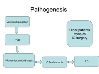
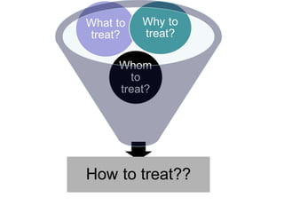
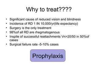
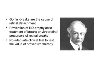
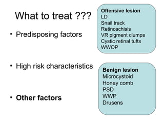
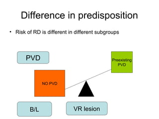
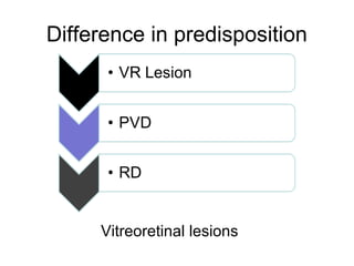
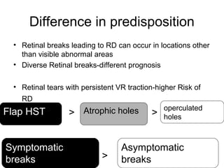
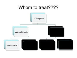
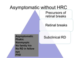
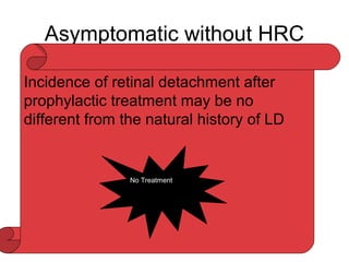
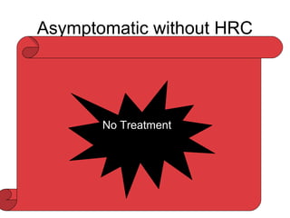
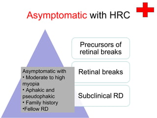
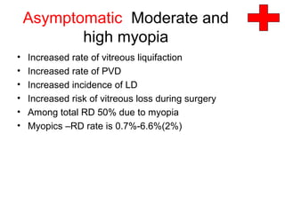
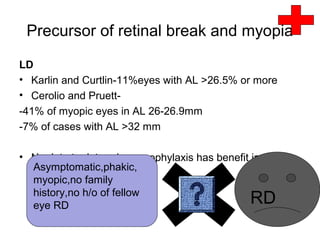
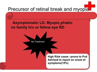
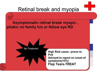
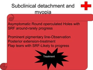
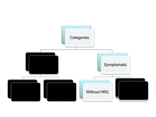
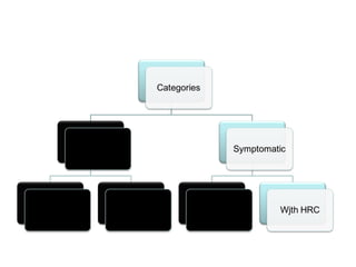
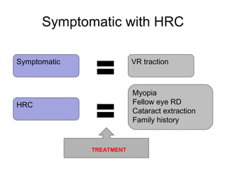
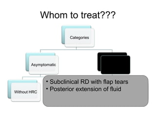
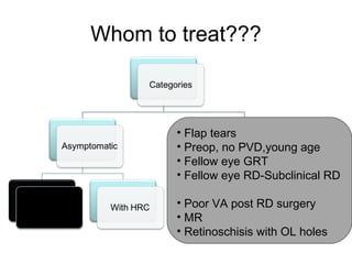
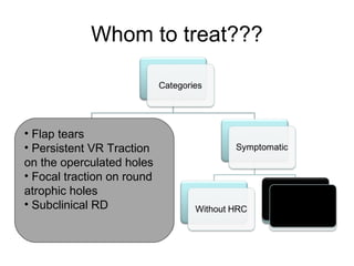
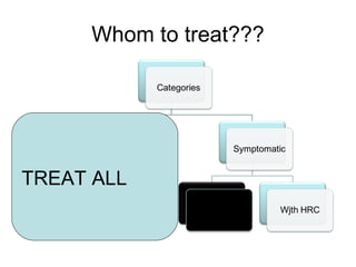
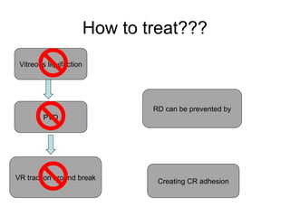
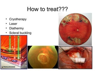
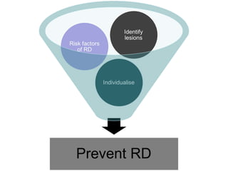
DR.SHAH-NOOR HASSAN
Prophylaxis of RD
Pathogenesis
Vitreous liquifaction
VR traction around break
PVD
IO fluid currents RD
Older patients
Myopics
IO surgery
Why to treat????
• Significant cause of reduced vision and blindness
• Incidence of RD 1 IN 10,000/yr(life expectancy)
• Surgery is the only treatment
• 98%of all RD are rhegmatogenous
• Inspite of successful reattachments Vn>20/50 in 50%of
cases
• Surgical failure rate -5-10% cases
Prophylaxis
• Gonin -breaks are the cause of
retinal detachment
• Prevention of RD-prophylactic
treatment of breaks or vitreoretinal
precursors of retinal breaks
• No adequate clinical trial to test
the value of preventive therapy
Vitreous liquifaction
VR traction around break
PVD
RD can be prevented by
Creating CR adhesion
What to treat ???
• Predisposing factors
• High risk characteristics
• Other factors
Vitreous liquifaction
Absence of PVD
Type of break
Presence of VR traction
What to treat ???
• Predisposing factors
• High risk characteristics
• Other factors
Risk factors
• Vitreoretinal abnormalities
• Symptomatic patient
• Myopia
• Previous history of RD in other eye(12%)
• Family history
• Systemic disease-Sticklers syndrome,Marfans syndrome
• Intraocular procedures -Cataract extraction,YAG
capsulotomy
• Presence or absence of PVD
What to treat ???
• Predisposing factors
• High risk characteristics
• Other factors
Offensive lesion
LD
Snail track
Retinoschisis
VR pigment clumps
Cystic retinal tufts
WWOP
Benign lesion
Microcystoid
Honey comb
PSD
WWP
Drusens
Difference in predisposition
• Risk of RD is different in different subgroups
PVD
NO PVD
Preexisting
PVD
B/L VR lesion
Difference in predisposition
Vitreoretinal lesions
Difference in predisposition
• Retinal breaks leading to RD can occur in locations other
than visible abnormal areas
• Diverse Retinal breaks-different prognosis
• Retinal tears with persistent VR traction-higher Risk of
RD
• > >
• >
Atrophic holesFlap HST operculated
holes
Symptomatic
breaks
Asymptomatic
breaks
Localised RD
• Localised RD-SRF more than 1 DD from the edge of the
break
• Subclinical RD-Anterior equator,no VF loss,<2DD of SRF
Why not treat all the VR lesions
• LD -8% of general population
• Present in 20-30%of retinal detachments
• LD leading to RD-0.3%-0.5%
• If all the LD are treated – overtreatment
• 1 in 10000/yr incidence of RD
• Presence of lesion = prophylaxis
Myopia, aphakia,
h/o RD in fellow eye
With LD
Normal eyes with LD
Whom to treat????
NOT TO TREAT TREAT
In evaluation of risk of RD
or natural history of retinal
breaks,or precursors of
retinal breakscases
should be categorised
with regards to presence
of high risk factors
Whom to treat????
Whom to treat????
Asymptomatic without HRC
Asymptomatic
Phakic
Nonmyopic
No family h/o
No RD in fellow
eye
PVD
Asymptomatic without HRC
LD:
• 8% of general population
• Present 20-30% RD
• LD rare cause of RD without HRC-
• Retinal breaks associated with LD =64% to 83% of
cases
• Tears along the posterior and lateral margin of LD
• Round atrophic holes-within LD-RD-young phakic
myopic eyes without PVD-1 IN 365 cases
• Prophylactic failure is reported-2.2%Tasman and
jaguers ,2.9%Boniuk etal
Incidence of retinal detachment after
prophylactic treatment may be no
different from the natural history of LD
No Treatment
Asymptomatic without HRC
• Cystic Retinal Tufts:
• Retinal breaks at time of PVD
• 10% of clinical RD
• Byer –Chance of RD in eyes with cystic
retinal tuft 1 in 357
No Treatment
Asymptomatic without HRC
Degenerative retinoschisis
• Splitting of retina at OPL or NFL
• RD occurs if there are holes in both the layers
• Previously outerlayer breaks –considered for prophylaxis
• Byer-natural history of retinoschisis-symptomatic retinal
detachment progression is rareNo Treatment
Asymptomatic without HRC
• Retinal breaks
• Autopsy studies
-break due to VR traction -3%
-break without VR traction-4%
• Clinical Studies
-14%-Rutnin and Schepens(102 pt)
-3.3%-Byer(3400 Eyes)
• No RD in 231eyes with asymptomatic breaks-Byer
• Neumann etal-asymptomatic retinal break:RD=218:1
- -
No Treatment
Asymptomatic retinal
breaks and
asymptomatic focal
RD due to round
holes within LD
low risk of
progression
RD
SLOW
Asymptomatic without HRC
Subclinical RD:
• First introduced by Schepens in 1952
• Jesberg,Okun ,Cibis
• Davis-1DD from the edge of the break and
not more than 2 DD posterior to equator
• Rarely progress to symptomatic RD-if
cause is hole within the LD
Round operculated Holes with SRF
around-rarely progress
Prominent pigmentary line-Observation
Flap tears with SRF-Likely to progress
Treatment
Asymptomatic with HRC
Asymptomatic with
• Moderate to high
myopia
• Aphakic and
pseudophakic
• Family history
•Fellow RD
Asymptomatic Moderate and
high myopia
• Increased rate of vitreous liquifaction
• Increased rate of PVD
• Increased incidence of LD
• Increased risk of vitreous loss during surgery
• Among total RD 50% due to myopia
• Myopics –RD rate is 0.7%-6.6%(2%)
Precursor of retinal break and myopia
LD
• Karlin and Curtlin-11%eyes with AL >26.5% or more
• Cerolio and Pruett-
-41% of myopic eyes in AL 26-26.9mm
-7% of cases with AL >32 mm
• No data to determine –prophylaxis has benefit in-
Asymptomatic,phakic,
myopic,no family
history,no h/o of fellow
eye RD RD
Precursor of retinal break and myopia
• Cystic retinal tuft-no relation with refractive
error
• Retinal tears common with cystic retinal
tufts and myopia with PVD
• Retinoschisis not related to myopia
• Waldoff and Hagler-RD with retinoschisis
in myopes<RD with myopes.
Asymptomatic LD. Myopic,phakic
no family h/o or fellow eye RD
No Treatment
High Risk cases –prone to Pvd
Advised to report on onset of
symptoms(15%)
Retinal break and myopia
• Karlin and Curtin
• Neumann etal
AL % OF RETINAL
BREAK
<25 mm 3%
>25 mm 11%
AL HOLE:RD
MYOPIA 1:13.5
NONMYOPIC 1:218
Asymptomatic retinal break myopic ,
phakic no family h/o or fellow eye RD
No Treatment
High Risk cases –prone to
Pvd
Advised to report on onset of
symptoms(15%)
Flap Tears-TREAT
Subclinical detachment and
myopia
Asymptomatic,
phakic,myopic,
no h/o of RD in
fellow eye
RD
Asymptomatic Round operculated Holes with
SRF around-rarely progress
Prominent pigmentary line-Observation
Posterior extension-treatment
Flap tears with SRF-Likely to progress
Treatment
Asymptomatic aphakic and
pseudophakic
• Removal of crystalline lens-increased rates of retinal
tears
• Liquifaction of vitreous gel
• PVD occurs sooner
• 40%of RD occurs after cataract surgery
• Davis etal-183 eyes-asymptomatic retinal breaks in
aphakic eyes -50% RD
• Aphakia with myopia risk of RD is 2X
Aphakic and precursors of
retinal breaks
• Incidence of precursors of retinal breaks and in aphakia
is same as in general population
• Prevalence of LD in aphakic detachment decreases with
advancing age
• RD and retinal breaks less likely to be associated with
LD after cataract extraction in comparison with phakic
RD
Prophylactic treatment –
Not recommended for cystic retinal tufts
Not recommended for retinoschisis
Recommended preoperatiely for LD,no
PVD,young age
Aphakic/p’phakic and retinal
breaks
• Incidence of RD comparable with
asymptomatic phakic RD
• RD occur in sizeable number of cases
despite treatment
• Value of prophylactic therapy uncertain
• NO adequate data in patient –
aphakic,asymptomatic,no family history,or
fellow eye RD
Not recommended Retinal holes with old
age with PVD
Recommended for flap HST,no
PVD,young age(formation of new retinal
breaks at vitreous base not prevented)
Extensive treament-GRT in Fellow eye
Treatment
Aphakic/p’phakic and
subclinical RD
• No adequate data
• Decision of treatment-amount of SRF and type of retinal
break
Treatment
• Subclinical RD due to HST
• SRF approaches the equator
NoTreatment
• Subclinical RD due to atrophic holes within LD
Asymptomatic aphakic and
pseudophakic
RD rates higher if
aphakia associated
myopia or family history
or fellow eye RD
Treatment
Asymptomatic with familial
predisposition
• No adequate data for the prophylaxis
• Sticklers syndrome-hgh risk of RD
• Radial and perivascular lattices
• Poor prognosis post RD surgery
• Widespread treatment is required
• Prophylaxis in Radial and perivascular lattices-different
tractional forces-prophylaxis failure
• Treatment recommended-history of previous RD in
fellow eye
RD in fellow eye
• Pathological vitreous changes bilateral
• Incidence of RD in second eye 25%to 40%
• Symptomatic retinal tears with persistent vitreoretinal
traction-high risk of RD
• Prompt treatment
Benefits less convincing
• Precursors of retinal tears
• Asymptomatic breaks
• Symptomatic breaks without persistent VR traction
Asymptomatic with fellow eye RD
Precursors of retinal breaks
APHAKICPHAKIC
LD
Cystic retinal tufts
Degenerative retinoschisis
Retinal breaks
Subclinical RD
LD
Cystic retinal tufts
Degenerative retinoschisis
Retinal breaks
Subclinical RD
Asymptomatic with fellow eye RD
Precursors of retinal breaks
APHAKICPHAKIC
LD
Cystic retinal tufts
Degenerative retinoschisis
Retinal breaks
Subclinical RD
LD
Cystic retinal tufts
Degenerative retinoschisis
Retinal breaks
Subclinical RD
Asymptomatic with fellow eye
RD
• Precursors of retinal breaks in phakics
1. LD:
• LD in fellow eyes 9-34% cases
• Folk etal-6-15%of ld develop RD
• Folk etal-full treatment of LD had fewer retinal breaks
• Treatment not beneficial in high myope with LD>6 clock
hr
• Treatment
Cases with poor results post RD surgery with LD in
fellow eyes,mentally retarded patients
Asymptomatic with fellow eye
RD
• Precursors of retinal breaks in phakics
2.Cystic retinal tufts:
• cystic retinal tufts in RD=5%
• No data on prophylaxis
• No treatment
3.Degenerative retinoschisis:
No satisfactory evidence
Treatment
Localised areas of RD due to outer layer holes
Asymptomatic with fellow eye
RD
Retinal breaks in phakics
• Neumann etal-
Treatment :HST,Round holes without PVD,Subclinical
RD,Fellow eye of GRT
Round holes with operculum can be observed
AL RD:retinal break
Fellow eye RD 1:1.9
Myopic 1:13.5
NonMyopic 1:218
Asymptomatic with fellow eye RD
Precursors of retinal breaks
APHAKICPHAKIC
LD
Cystic retinal tufts
Degenerative retinoschisis
Retinal breaks
Subclinical RD
LD
Cystic retinal tufts
Degenerative retinoschisis
Retinal breaks
Subclinical RD
Asymptomatic with fellow eye
RD
• Precursors of retinal breaks in aphakics
1. LD:
LD in aphakic RD-25% to 34% of cases
Treatment recommende unless preexisting PVD
2.Cystic retinal tufts:treated the same way as LD
Asymptomatic with fellow eye
RD
Retinal breaks in aphakics
1. Flap tears-Treatment
2. Round holes with no PVD-Treatment
3. Round holes with PVD-Observe
4. Round holes with free operculum-Observe
Subclinical RD-
• Extensive RD as compared to phakics
• Scleral buckling
Prophylaxis recommended in fellow eye GRT
Symptomatic without HRC
• Symptoms
• Acute contraction of vitreous gel
PVD
Traction on the retina
Photopsia,release of pigments-floaters
• 15% of symptomatic PVD –retinal tears
• Risk of RD more in presence of HRC
Precursor of retinal breaks
Retinal breaks
Subclinical detachment
Symptomatic eyes
No HRC
Symptomatic without HRC
Symptomatic Eyes with precursors of retinal
breaks
LD-with symptomatic PVD-flap tears in 66%
cases
Treatment recommended
After PVD with no HRC-Mere presence of LD not
an indication for prophylaxis
Symptomatic eye with retinal breaks
• Flap tears
• Retinal breaks with free operculum
• Atrophic holes
Symptomatic eye with retinal breaks
• Flap tears
• Retinal breaks with free operculum
• Atrophic holes
Persistent vitreous traction
Progress to RD in 33%to55%
Treatment reduces the risk 48% to 4%
Symptomatic eye with retinal breaks
• Flap tears
• Retinal breaks with free operculum
• Atrophic holes
Operculated break-traction released
Treatment not required
If traction persist –similar to HST
Treatment required
Symptomatic eye with retinal breaks
• Flap tears
• Retinal breaks with free operculum
• Atrophic holes
Within LD
RD seen in young myopic without PVD
Examined for signs of focal traction
Eyes with focal traction are treated
Symptomatic with HRC
Symptomatic
Myopia
Fellow eye RD
Cataract extraction
Family history
VR traction
HRC
TREATMENT
Whom to treat???
• Subclinical RD with flap tears
• Posterior extension of fluid
Whom to treat???
• Flap tears
• Preop, no PVD,young age
• Fellow eye GRT
• Fellow eye RD-Subclinical RD
• Poor VA post RD surgery
• MR
• Retinoschisis with OL holes
Whom to treat???
• Flap tears
• Persistent VR Traction
on the operculated holes
• Focal traction on round
atrophic holes
• Subclinical RD
Whom to treat???
TREAT ALL
How to treat???
Vitreous liquifaction
VR traction around break
PVD
RD can be prevented by
Creating CR adhesion
How to treat???
• Cryotherapy
• Laser
• Diathermy
• Scleral buckling
Technique
• Scatter treatment
• Contiguous treatment
• Around the lesion
• 360 preequatorial scarring
Cryotherapy
• Transconjunctival or transscleral
• Contiguous treatment
• Avoid breaks
• Grade 2 burns
Cryotherapy
Advantages
• Hazy media
• Cost effective
• Anterior lesion
• No damage to sclera ,nerve,vessels
Disadvantage
• CME,
• pigment fallout,
• CD,chemosis,
• painful,LA required
Laser photocoagulation
Advantages:
• Topical anaesthesia
• Less painful
• No pigment fall out
• Posterior lesions
• Less inflammation
• Precise treatment
Laser photocoagulation
Disadvantages
• High cost
• Blonde fundus
• Foveal damage
• Corneal and lens burns
Scleral buckling
• Widespread LD withfellow eye RD with other HRC
• Aphakic subclinical RD
• Radial or encirclage
Failure and complication
• RD inspite of prophylaxis
• Complication-treatment outcome worse than the natural
course of the disease
• ERM -1-2%
• Retinal breaks release pigment and limited PVR
Failure rates
Methods to avoid failure
• Medium intensity burns
• Away from the lesion
• Cover both anterior and posterior margins of the lesion
• Join the treatment area till ora in anterior lesion
Complications
• Haemorrhage-PVD
• Bruchs memrane related-SRNVM
• Inflammation
• Anaesthesia related
After treatment
• Follow up:
2 weeks,2months,6-12 months
Patient education
• Symptoms PVD
• Symptoms RD
• Peripheral visual field examination
Prophylaxis of RD
Prophylaxis of RD

Prophylaxis of RD

  • 1.
  • 2.
    Pathogenesis Vitreous liquifaction VR tractionaround break PVD IO fluid currents RD Older patients Myopics IO surgery
  • 4.
    Why to treat???? •Significant cause of reduced vision and blindness • Incidence of RD 1 IN 10,000/yr(life expectancy) • Surgery is the only treatment • 98%of all RD are rhegmatogenous • Inspite of successful reattachments Vn>20/50 in 50%of cases • Surgical failure rate -5-10% cases Prophylaxis
  • 5.
    • Gonin -breaksare the cause of retinal detachment • Prevention of RD-prophylactic treatment of breaks or vitreoretinal precursors of retinal breaks • No adequate clinical trial to test the value of preventive therapy
  • 6.
    Vitreous liquifaction VR tractionaround break PVD RD can be prevented by Creating CR adhesion
  • 7.
    What to treat??? • Predisposing factors • High risk characteristics • Other factors Vitreous liquifaction Absence of PVD Type of break Presence of VR traction
  • 8.
    What to treat??? • Predisposing factors • High risk characteristics • Other factors
  • 9.
    Risk factors • Vitreoretinalabnormalities • Symptomatic patient • Myopia • Previous history of RD in other eye(12%) • Family history • Systemic disease-Sticklers syndrome,Marfans syndrome • Intraocular procedures -Cataract extraction,YAG capsulotomy • Presence or absence of PVD
  • 10.
    What to treat??? • Predisposing factors • High risk characteristics • Other factors Offensive lesion LD Snail track Retinoschisis VR pigment clumps Cystic retinal tufts WWOP Benign lesion Microcystoid Honey comb PSD WWP Drusens
  • 11.
    Difference in predisposition •Risk of RD is different in different subgroups PVD NO PVD Preexisting PVD B/L VR lesion
  • 12.
  • 13.
    Difference in predisposition •Retinal breaks leading to RD can occur in locations other than visible abnormal areas • Diverse Retinal breaks-different prognosis • Retinal tears with persistent VR traction-higher Risk of RD • > > • > Atrophic holesFlap HST operculated holes Symptomatic breaks Asymptomatic breaks
  • 14.
    Localised RD • LocalisedRD-SRF more than 1 DD from the edge of the break • Subclinical RD-Anterior equator,no VF loss,<2DD of SRF
  • 15.
    Why not treatall the VR lesions • LD -8% of general population • Present in 20-30%of retinal detachments • LD leading to RD-0.3%-0.5% • If all the LD are treated – overtreatment • 1 in 10000/yr incidence of RD • Presence of lesion = prophylaxis Myopia, aphakia, h/o RD in fellow eye With LD Normal eyes with LD
  • 16.
    Whom to treat???? NOTTO TREAT TREAT In evaluation of risk of RD or natural history of retinal breaks,or precursors of retinal breakscases should be categorised with regards to presence of high risk factors
  • 17.
  • 18.
  • 19.
  • 20.
    Asymptomatic without HRC LD: •8% of general population • Present 20-30% RD • LD rare cause of RD without HRC- • Retinal breaks associated with LD =64% to 83% of cases • Tears along the posterior and lateral margin of LD • Round atrophic holes-within LD-RD-young phakic myopic eyes without PVD-1 IN 365 cases • Prophylactic failure is reported-2.2%Tasman and jaguers ,2.9%Boniuk etal Incidence of retinal detachment after prophylactic treatment may be no different from the natural history of LD No Treatment
  • 21.
    Asymptomatic without HRC •Cystic Retinal Tufts: • Retinal breaks at time of PVD • 10% of clinical RD • Byer –Chance of RD in eyes with cystic retinal tuft 1 in 357 No Treatment
  • 22.
    Asymptomatic without HRC Degenerativeretinoschisis • Splitting of retina at OPL or NFL • RD occurs if there are holes in both the layers • Previously outerlayer breaks –considered for prophylaxis • Byer-natural history of retinoschisis-symptomatic retinal detachment progression is rareNo Treatment
  • 23.
    Asymptomatic without HRC •Retinal breaks • Autopsy studies -break due to VR traction -3% -break without VR traction-4% • Clinical Studies -14%-Rutnin and Schepens(102 pt) -3.3%-Byer(3400 Eyes) • No RD in 231eyes with asymptomatic breaks-Byer • Neumann etal-asymptomatic retinal break:RD=218:1 - - No Treatment Asymptomatic retinal breaks and asymptomatic focal RD due to round holes within LD low risk of progression RD SLOW
  • 24.
    Asymptomatic without HRC SubclinicalRD: • First introduced by Schepens in 1952 • Jesberg,Okun ,Cibis • Davis-1DD from the edge of the break and not more than 2 DD posterior to equator • Rarely progress to symptomatic RD-if cause is hole within the LD Round operculated Holes with SRF around-rarely progress Prominent pigmentary line-Observation Flap tears with SRF-Likely to progress Treatment
  • 26.
    Asymptomatic with HRC Asymptomaticwith • Moderate to high myopia • Aphakic and pseudophakic • Family history •Fellow RD
  • 27.
    Asymptomatic Moderate and highmyopia • Increased rate of vitreous liquifaction • Increased rate of PVD • Increased incidence of LD • Increased risk of vitreous loss during surgery • Among total RD 50% due to myopia • Myopics –RD rate is 0.7%-6.6%(2%)
  • 28.
    Precursor of retinalbreak and myopia LD • Karlin and Curtlin-11%eyes with AL >26.5% or more • Cerolio and Pruett- -41% of myopic eyes in AL 26-26.9mm -7% of cases with AL >32 mm • No data to determine –prophylaxis has benefit in- Asymptomatic,phakic, myopic,no family history,no h/o of fellow eye RD RD
  • 29.
    Precursor of retinalbreak and myopia • Cystic retinal tuft-no relation with refractive error • Retinal tears common with cystic retinal tufts and myopia with PVD • Retinoschisis not related to myopia • Waldoff and Hagler-RD with retinoschisis in myopes<RD with myopes. Asymptomatic LD. Myopic,phakic no family h/o or fellow eye RD No Treatment High Risk cases –prone to Pvd Advised to report on onset of symptoms(15%)
  • 30.
    Retinal break andmyopia • Karlin and Curtin • Neumann etal AL % OF RETINAL BREAK <25 mm 3% >25 mm 11% AL HOLE:RD MYOPIA 1:13.5 NONMYOPIC 1:218 Asymptomatic retinal break myopic , phakic no family h/o or fellow eye RD No Treatment High Risk cases –prone to Pvd Advised to report on onset of symptoms(15%) Flap Tears-TREAT
  • 31.
    Subclinical detachment and myopia Asymptomatic, phakic,myopic, noh/o of RD in fellow eye RD Asymptomatic Round operculated Holes with SRF around-rarely progress Prominent pigmentary line-Observation Posterior extension-treatment Flap tears with SRF-Likely to progress Treatment
  • 32.
    Asymptomatic aphakic and pseudophakic •Removal of crystalline lens-increased rates of retinal tears • Liquifaction of vitreous gel • PVD occurs sooner • 40%of RD occurs after cataract surgery • Davis etal-183 eyes-asymptomatic retinal breaks in aphakic eyes -50% RD • Aphakia with myopia risk of RD is 2X
  • 33.
    Aphakic and precursorsof retinal breaks • Incidence of precursors of retinal breaks and in aphakia is same as in general population • Prevalence of LD in aphakic detachment decreases with advancing age • RD and retinal breaks less likely to be associated with LD after cataract extraction in comparison with phakic RD Prophylactic treatment – Not recommended for cystic retinal tufts Not recommended for retinoschisis Recommended preoperatiely for LD,no PVD,young age
  • 34.
    Aphakic/p’phakic and retinal breaks •Incidence of RD comparable with asymptomatic phakic RD • RD occur in sizeable number of cases despite treatment • Value of prophylactic therapy uncertain • NO adequate data in patient – aphakic,asymptomatic,no family history,or fellow eye RD Not recommended Retinal holes with old age with PVD Recommended for flap HST,no PVD,young age(formation of new retinal breaks at vitreous base not prevented) Extensive treament-GRT in Fellow eye Treatment
  • 35.
    Aphakic/p’phakic and subclinical RD •No adequate data • Decision of treatment-amount of SRF and type of retinal break Treatment • Subclinical RD due to HST • SRF approaches the equator NoTreatment • Subclinical RD due to atrophic holes within LD
  • 36.
    Asymptomatic aphakic and pseudophakic RDrates higher if aphakia associated myopia or family history or fellow eye RD Treatment
  • 37.
    Asymptomatic with familial predisposition •No adequate data for the prophylaxis • Sticklers syndrome-hgh risk of RD • Radial and perivascular lattices • Poor prognosis post RD surgery • Widespread treatment is required • Prophylaxis in Radial and perivascular lattices-different tractional forces-prophylaxis failure • Treatment recommended-history of previous RD in fellow eye
  • 38.
    RD in felloweye • Pathological vitreous changes bilateral • Incidence of RD in second eye 25%to 40% • Symptomatic retinal tears with persistent vitreoretinal traction-high risk of RD • Prompt treatment Benefits less convincing • Precursors of retinal tears • Asymptomatic breaks • Symptomatic breaks without persistent VR traction
  • 39.
    Asymptomatic with felloweye RD Precursors of retinal breaks APHAKICPHAKIC LD Cystic retinal tufts Degenerative retinoschisis Retinal breaks Subclinical RD LD Cystic retinal tufts Degenerative retinoschisis Retinal breaks Subclinical RD
  • 40.
    Asymptomatic with felloweye RD Precursors of retinal breaks APHAKICPHAKIC LD Cystic retinal tufts Degenerative retinoschisis Retinal breaks Subclinical RD LD Cystic retinal tufts Degenerative retinoschisis Retinal breaks Subclinical RD
  • 41.
    Asymptomatic with felloweye RD • Precursors of retinal breaks in phakics 1. LD: • LD in fellow eyes 9-34% cases • Folk etal-6-15%of ld develop RD • Folk etal-full treatment of LD had fewer retinal breaks • Treatment not beneficial in high myope with LD>6 clock hr • Treatment Cases with poor results post RD surgery with LD in fellow eyes,mentally retarded patients
  • 42.
    Asymptomatic with felloweye RD • Precursors of retinal breaks in phakics 2.Cystic retinal tufts: • cystic retinal tufts in RD=5% • No data on prophylaxis • No treatment 3.Degenerative retinoschisis: No satisfactory evidence Treatment Localised areas of RD due to outer layer holes
  • 43.
    Asymptomatic with felloweye RD Retinal breaks in phakics • Neumann etal- Treatment :HST,Round holes without PVD,Subclinical RD,Fellow eye of GRT Round holes with operculum can be observed AL RD:retinal break Fellow eye RD 1:1.9 Myopic 1:13.5 NonMyopic 1:218
  • 44.
    Asymptomatic with felloweye RD Precursors of retinal breaks APHAKICPHAKIC LD Cystic retinal tufts Degenerative retinoschisis Retinal breaks Subclinical RD LD Cystic retinal tufts Degenerative retinoschisis Retinal breaks Subclinical RD
  • 45.
    Asymptomatic with felloweye RD • Precursors of retinal breaks in aphakics 1. LD: LD in aphakic RD-25% to 34% of cases Treatment recommende unless preexisting PVD 2.Cystic retinal tufts:treated the same way as LD
  • 46.
    Asymptomatic with felloweye RD Retinal breaks in aphakics 1. Flap tears-Treatment 2. Round holes with no PVD-Treatment 3. Round holes with PVD-Observe 4. Round holes with free operculum-Observe Subclinical RD- • Extensive RD as compared to phakics • Scleral buckling Prophylaxis recommended in fellow eye GRT
  • 48.
    Symptomatic without HRC •Symptoms • Acute contraction of vitreous gel PVD Traction on the retina Photopsia,release of pigments-floaters • 15% of symptomatic PVD –retinal tears • Risk of RD more in presence of HRC
  • 49.
    Precursor of retinalbreaks Retinal breaks Subclinical detachment Symptomatic eyes No HRC Symptomatic without HRC
  • 50.
    Symptomatic Eyes withprecursors of retinal breaks LD-with symptomatic PVD-flap tears in 66% cases Treatment recommended After PVD with no HRC-Mere presence of LD not an indication for prophylaxis
  • 51.
    Symptomatic eye withretinal breaks • Flap tears • Retinal breaks with free operculum • Atrophic holes
  • 52.
    Symptomatic eye withretinal breaks • Flap tears • Retinal breaks with free operculum • Atrophic holes Persistent vitreous traction Progress to RD in 33%to55% Treatment reduces the risk 48% to 4%
  • 53.
    Symptomatic eye withretinal breaks • Flap tears • Retinal breaks with free operculum • Atrophic holes Operculated break-traction released Treatment not required If traction persist –similar to HST Treatment required
  • 54.
    Symptomatic eye withretinal breaks • Flap tears • Retinal breaks with free operculum • Atrophic holes Within LD RD seen in young myopic without PVD Examined for signs of focal traction Eyes with focal traction are treated
  • 56.
    Symptomatic with HRC Symptomatic Myopia Felloweye RD Cataract extraction Family history VR traction HRC TREATMENT
  • 57.
    Whom to treat??? •Subclinical RD with flap tears • Posterior extension of fluid
  • 58.
    Whom to treat??? •Flap tears • Preop, no PVD,young age • Fellow eye GRT • Fellow eye RD-Subclinical RD • Poor VA post RD surgery • MR • Retinoschisis with OL holes
  • 59.
    Whom to treat??? •Flap tears • Persistent VR Traction on the operculated holes • Focal traction on round atrophic holes • Subclinical RD
  • 60.
  • 61.
    How to treat??? Vitreousliquifaction VR traction around break PVD RD can be prevented by Creating CR adhesion
  • 62.
    How to treat??? •Cryotherapy • Laser • Diathermy • Scleral buckling
  • 63.
    Technique • Scatter treatment •Contiguous treatment • Around the lesion • 360 preequatorial scarring
  • 64.
    Cryotherapy • Transconjunctival ortransscleral • Contiguous treatment • Avoid breaks • Grade 2 burns
  • 65.
    Cryotherapy Advantages • Hazy media •Cost effective • Anterior lesion • No damage to sclera ,nerve,vessels Disadvantage • CME, • pigment fallout, • CD,chemosis, • painful,LA required
  • 66.
    Laser photocoagulation Advantages: • Topicalanaesthesia • Less painful • No pigment fall out • Posterior lesions • Less inflammation • Precise treatment
  • 67.
    Laser photocoagulation Disadvantages • Highcost • Blonde fundus • Foveal damage • Corneal and lens burns
  • 68.
    Scleral buckling • WidespreadLD withfellow eye RD with other HRC • Aphakic subclinical RD • Radial or encirclage
  • 69.
    Failure and complication •RD inspite of prophylaxis • Complication-treatment outcome worse than the natural course of the disease • ERM -1-2% • Retinal breaks release pigment and limited PVR
  • 70.
  • 71.
    Methods to avoidfailure • Medium intensity burns • Away from the lesion • Cover both anterior and posterior margins of the lesion • Join the treatment area till ora in anterior lesion
  • 72.
    Complications • Haemorrhage-PVD • Bruchsmemrane related-SRNVM • Inflammation • Anaesthesia related
  • 73.
    After treatment • Followup: 2 weeks,2months,6-12 months Patient education • Symptoms PVD • Symptoms RD • Peripheral visual field examination

Editor's Notes

  • #7 Vr traction can be relieved with vitrectomy and scleral buckling ,prophylactic vitrectomy is ha
  • #24 Asymptomatic retinal breaks and asymptomatic focal RD due to round holes within LD low risk of progression phakic nonmyopic eyes with no h/o RD in fellow eye
  • #25 Flap tears with SRF-likely to progress-Prohylaxis
  • #35 Not recommended Retinal holes with old age with PVD Recommended for flap HST,no PVD,young age
  • #62 Vr traction can be relieved with vitrectomy and scleral buckling ,prophylactic vitrectomy is ha