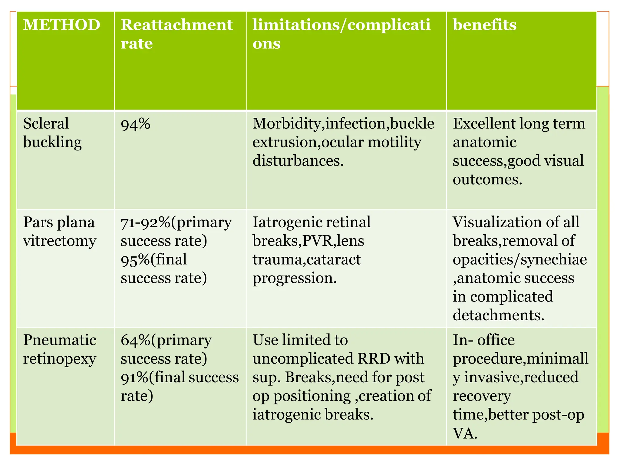
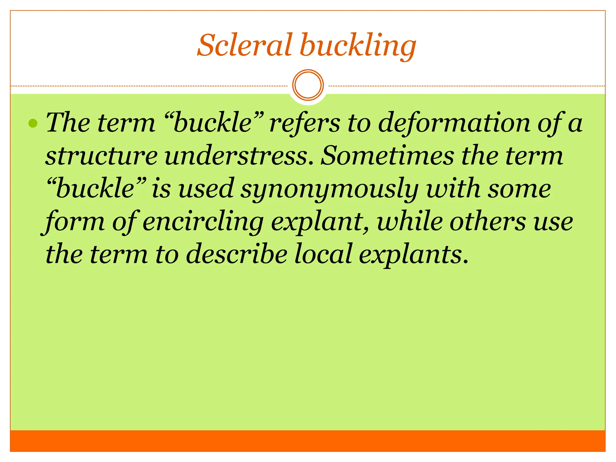
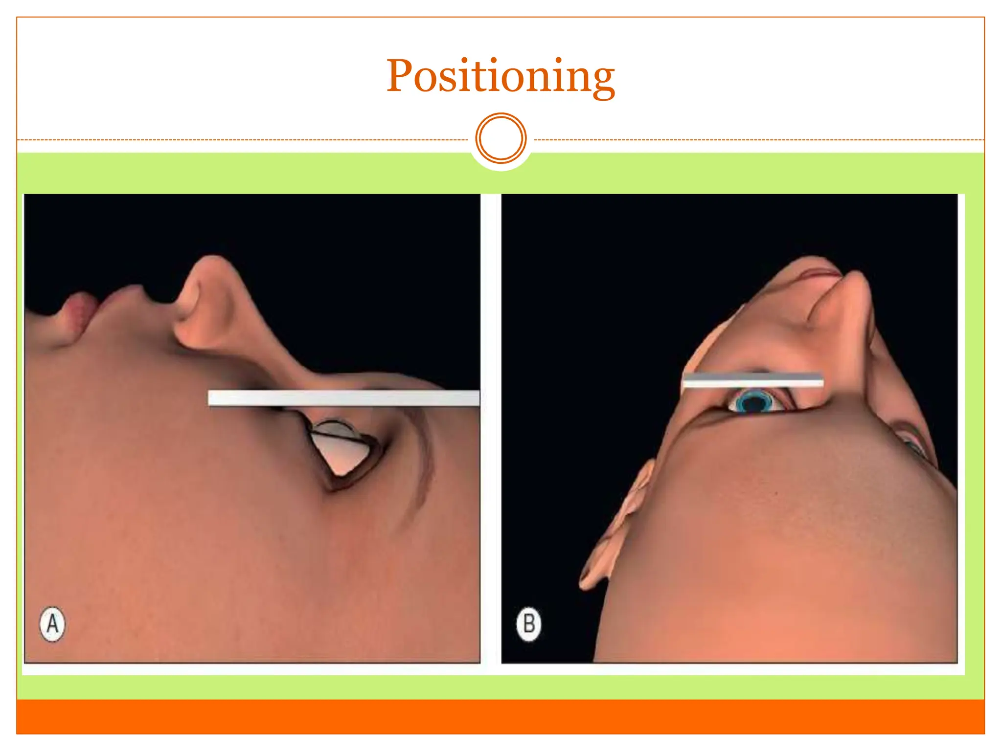
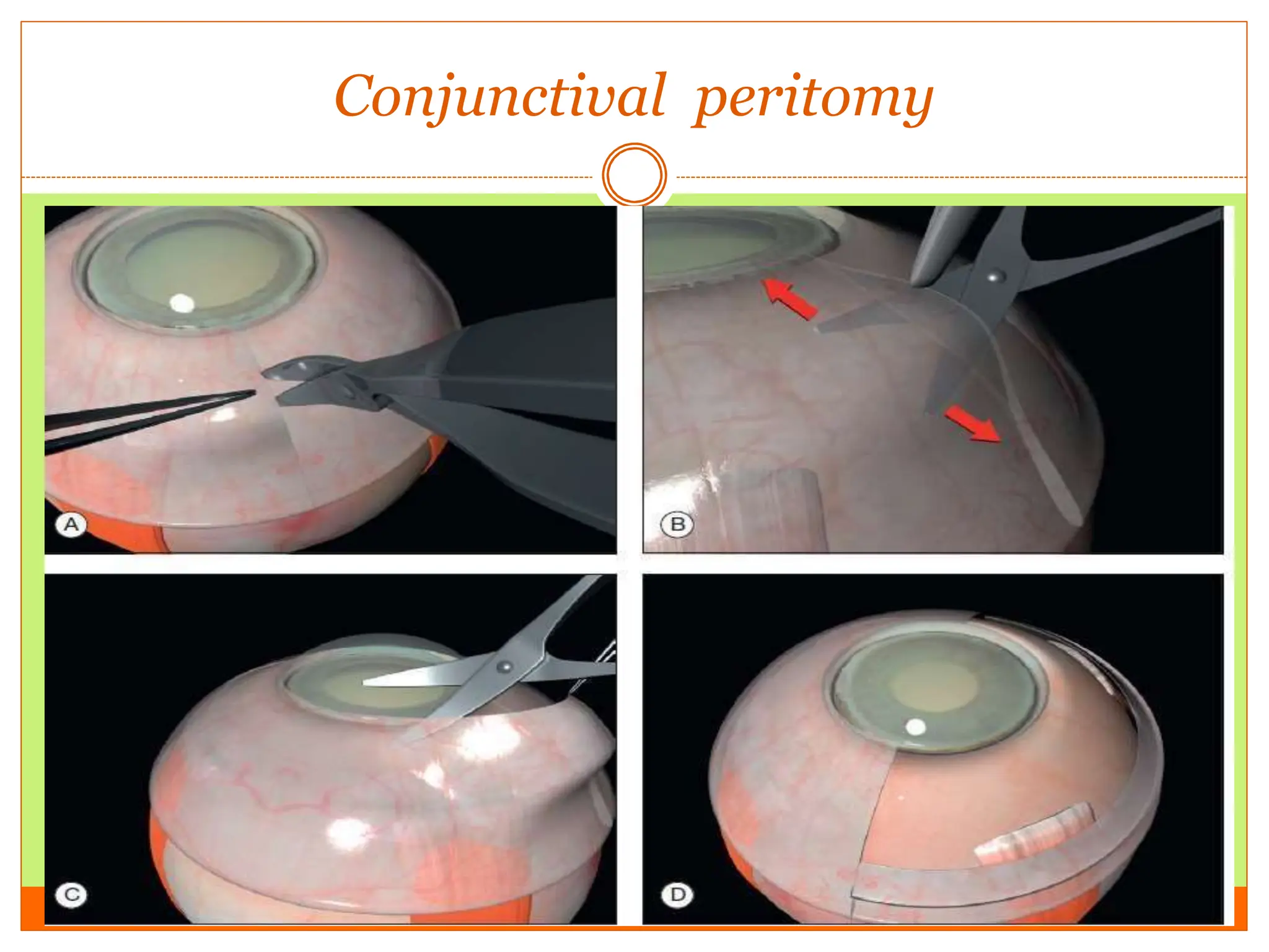
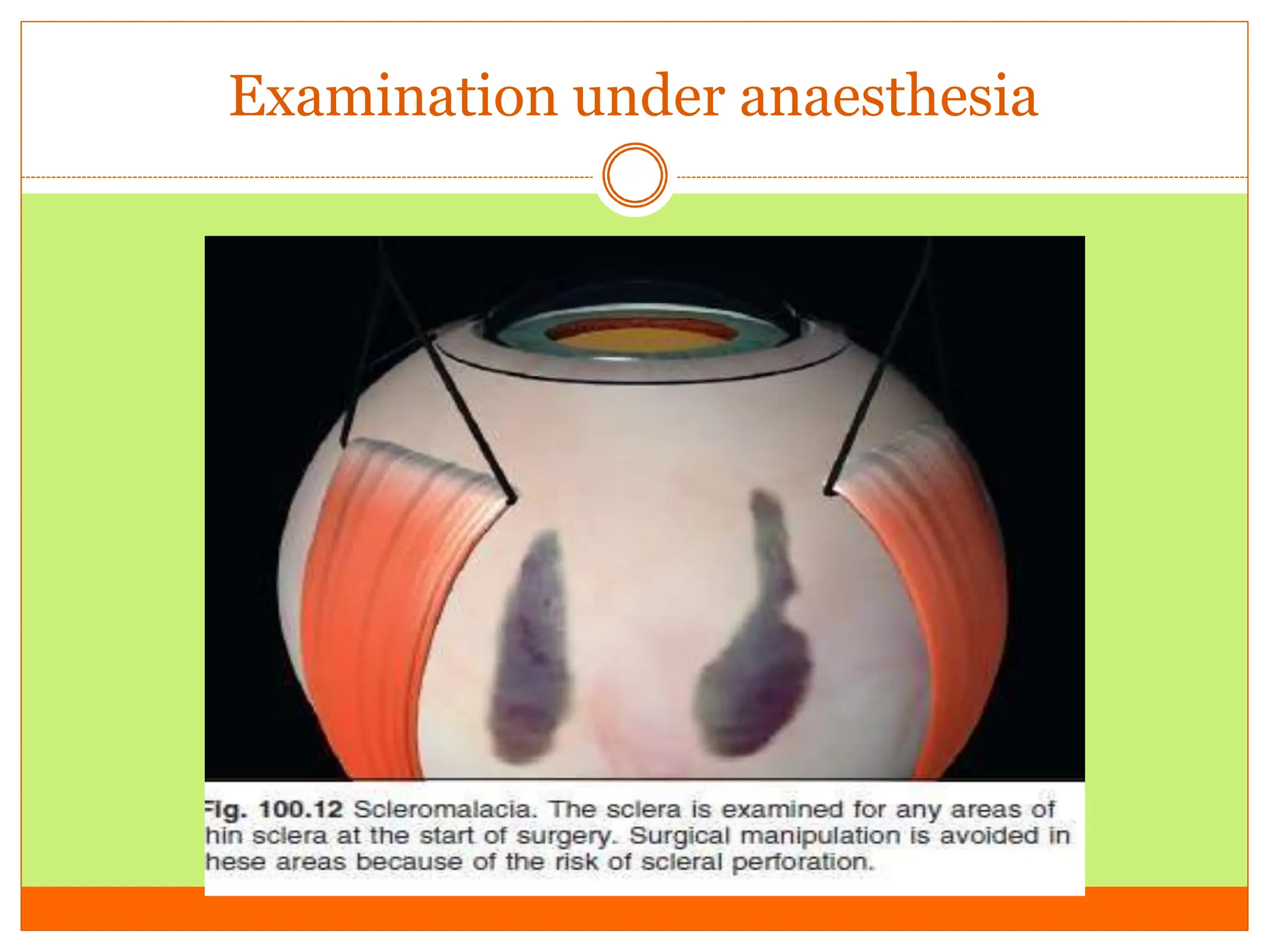
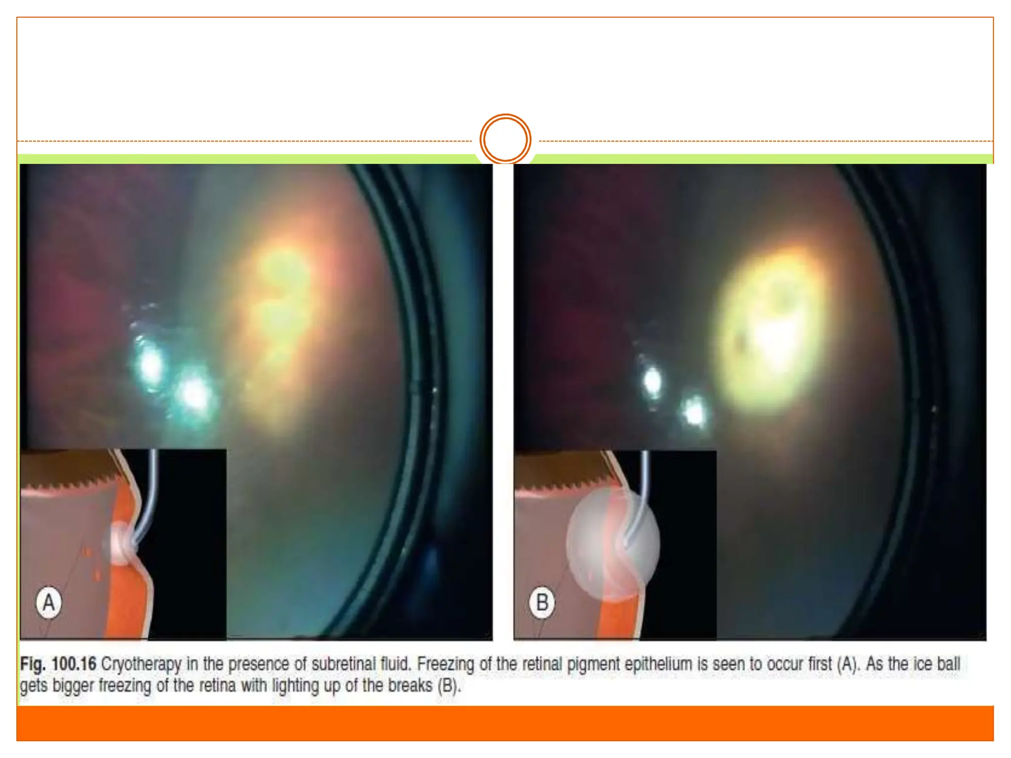
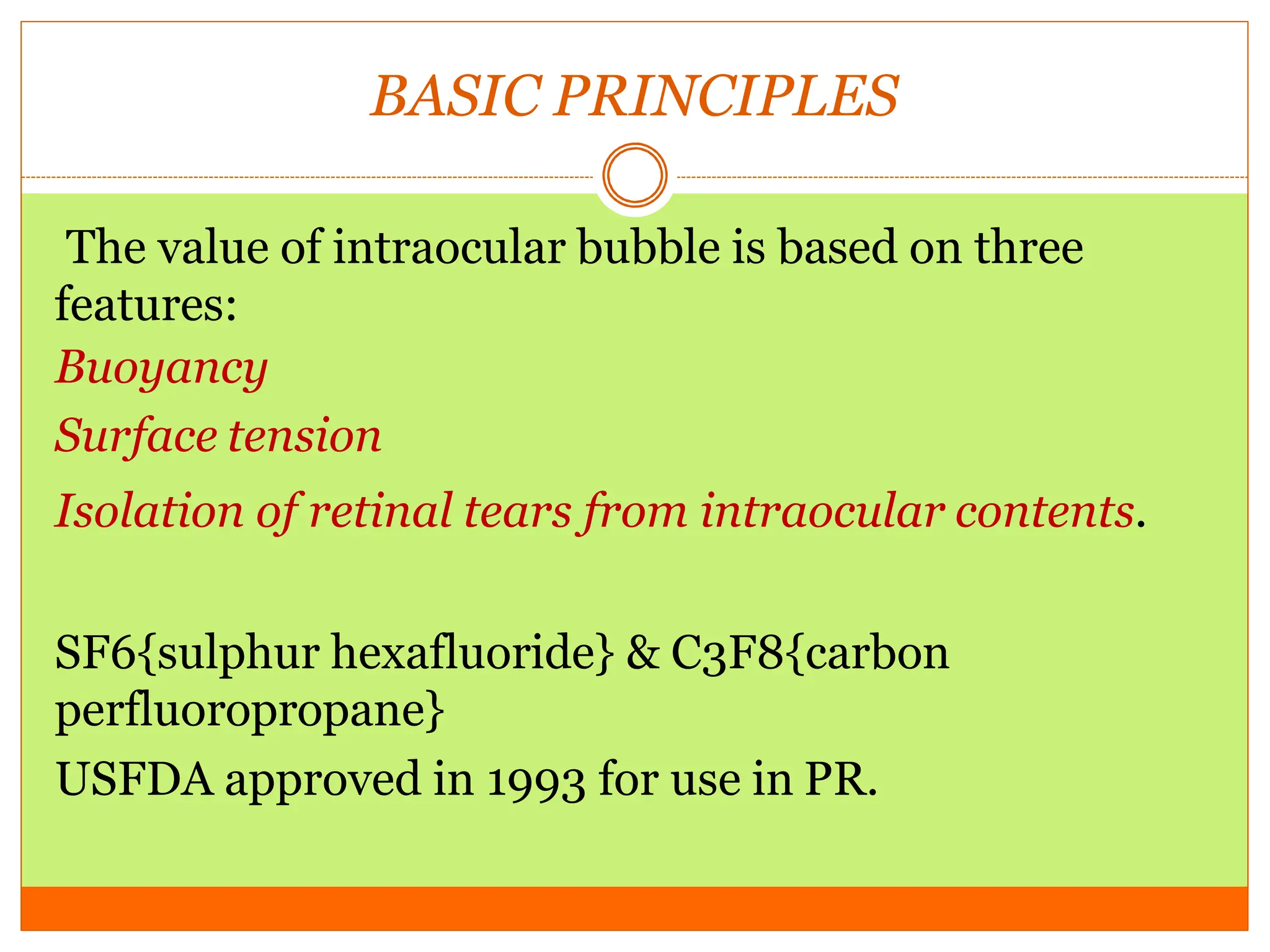
SlideShare a Scribd company logo
DR. SALMAN AHMAD
PGR RYR UNIT 2
SHL LHR
MANAGEMENT OF RETINAL
DETACHMENT
GLOSSARY:-
 Approach of treatment
 Decision
 Instrumentation
 Surgical procedures
Treatment guidelines for retinal breaks
Aphakia /
pseudophaki
a
treat
Treat some
treat
observe
Type of Phakic High myopia Fellow eye
break
HST treat treat treat
symptomatic
HST observe Treat some treat
asymptomatic
Operculated Treat some treat treat
symptomatic
Operculated observe Treat few observe
asymptomatic
Round hole observe observe Treat some
asymptomatic
observe
Lattice observe observe
without holes
Treat some
unless lattice
more than 6
clock hours
observe
Lattice with
round holes
observe observe observe
Selection of the surgical procedure to re-attach
the retina with least morbidity.
 Scleral buckling.
 Vitrectomy( classical/sutureless/using gas/silicone
oil & if needed , an encircling silicone band)
 Pneumatic retinopexy.
METHOD Reattachment
rate
limitations/complicati
ons
benefits
Scleral
buckling
94% Morbidity,infection,buckle
extrusion,ocular motility
disturbances.
Excellent long term
anatomic
success,good visual
outcomes.
Pars plana
vitrectomy
71-92%(primary
success rate)
95%(final
success rate)
Iatrogenic retinal
breaks,PVR,lens
trauma,cataract
progression.
Visualization of all
breaks,removal of
opacities/synechiae
,anatomic success
in complicated
detachments.
Pneumatic
retinopexy
64%(primary
success rate)
91%(final success
rate)
Use limited to
uncomplicated RRD with
sup. Breaks,need for post
op positioning ,creation of
iatrogenic breaks.
In- office
procedure,minimall
y invasive,reduced
recovery
time,better post-op
VA.
Scleral buckling
 The term “buckle” refers to deformation of a
structure understress. Sometimes the term
“buckle” is used synonymously with some
form of encircling explant, while others use
the term to describe local explants.
mechanics
 Biomechanics :alters the shape of the eye
depending on the type of buckling
material.
The location
The tension of the scleral sutures.
The circumferential tightening of an encircling buckle.
Changes after buckling:- AL change
astigmatism
volume of the eye
patient compliance
scleral buckle placement
Surgical anatomy
Extraocular muscles
Pre operative assesment
Macular involvement
 Features suggesting that the retinal detachment is
nonrhegmatogenous .
 The presence of vitreous detachment
 Significant ocular co-pathology, which may affect
management (e.g., glaucomatous optic neuropathy,
aphakia with vitreous in the anterior chamber, a
history of strabismus surgery)
 The number and position of the retinal breaks
Preparation for surgery
 Anaesthesia: peribulbar
 Positioning of the head
 Preparation & draping
Positioning
Surgical steps
 Conjunctival peritomy
 Slinging rectus muscle
Conjunctival peritomy
Slinging rectus muscle
Examination under anaesthesia
RETINOPEXY
 The indent from the explants helps in closing the
retinal breaks .
 This is done in order to increase the bond between
retina & retinal pigment epithelium .
 Even if the indent disappears.
CRYOTHERAPY
AIM: to produce freezing of
healthy retina surrounding all
the retinal breaks.
OBSERVATIONS:- whitening
of the retina after few seconds.
Break is seen as the darker area
over the white freezed area
which confirms the success of
cryo.
Explants
Scleral sutures
SRF DRAINAGE
 TIMING
 LOCATION OF DRAINAGE SITE
 TECHNIQUES
TECHNIQUES
 NON INVASIVE: ACETAZOLAMIDE
DIAMOX
 INVASIVE: PARACENTESIS
CUT DOWN TECHNIQUE
CUT DOWN TECHNIQUE
 A scleral incision 3 mm in length is made in the
sclera, repeatedly spreading the edges, then incising
the base of the resulting groove the choroid becomes
increasingly visible in the base of the incision. Finally
a small knuckle of bare choroid protrudes slightly
s
Post operative complications:
 Recurrent retinal detachment
 Glaucoma
 Presence of epiretinal membrane
 Extrusion /infection
Pneumato retinopexy
 History:-
Ohm performed the first intravitreal air injection for
retinal detachment in 1911.
In 1938 rosengren
In 1973 norton reported use of sulfur hexafloride SF6
At 1985 meeting of american association of
ophthalmology Hilton & Grizard introduced the term
“PNEUMATIC RETINOPEXY”
BASIC PRINCIPLES
The value of intraocular bubble is based on three
features:
Buoyancy
Surface tension
Isolation of retinal tears from intraocular contents.
SF6{sulphur hexafluoride} & C3F8{carbon
perfluoropropane}
USFDA approved in 1993 for use in PR.
Gases triad in viteroretinal surgery
NON EXPANSILE EXPANSILE
Air SF6
Nitrogen C4F10
Helium CF4
Oxygen C2F6
Argon C3F8
Xenon C4F10
Krypton C5F12
FEATURES
 SF6 & C3F8 are
chemically inert
colorless
odorless
GAS Avg. duration Largest size of
the
bubble(duratio
n)
Average
expansion
Non expansile
concentration
Air 3 days Immediate No expansion --
Sf6 12 days 36 hrs 2 times 18%
C3F8 38 days 72 hrs 4 times 14%
Surgical technique
 Anaesthesia :
Topical anaesthesia usually with subconjunctival
anesthesia or application of lidocaine soaked pledgets
may be adequate
NO should not be used in case of general anaesthesia
if intraocular gas is to be usesd.
STERILIZATION
 Sterile lid speculum
 Several drops of povidine & iodine .
 The injection site is dried with a cotton tipped
applicator & is ready for paracentesis & injection.
Preparation of the gas
A 30 guage ,1/2 inch {12 mm} needle is then placed tightly &
excess gas is expelled out, gas should not be kept for more than
few minutes as it can get diluted with the room air.
Pure gas is stored in a
cylinder with a regulatory
valve. Two sterile filters
should be connected
between the cylinder and the
syringe in use.
As physiological dead
space exists within the
system, accuracy maybe
affected by the air
contained within these
spaces. Pure gas should
then be drawn from
the cylinder and the
syringe flushed a few
times to ensure
complete
evacuation of air from
the dead space.
Appropriate amount of
pure
gas is then drawn into
the syringe.
The syringe with one
filter is
then disconnected. The
three-way tap is then
turned to the other
unused filter, and air is
drawn in to achieve the
appropriate
concentration of air–gas
mixture.
PARACENTESIS
 Performed before injection
 Requires:- half inch, 30 guage needle mounted on 1
ml syringe without a plunger.
 Site: paracentesis should be performed through the
limbus in phakic patients.
Otherwise it should be done from the pars plana .
INJECTION OF THE GAS
Fish eggs
Gas entrapment at injection site
Silicone oil
Introduction
 Silicone oil (SO) was first introduced as an internal
tamponade agent in the early 1960s.
 Clinical usage of SO in treating retinal detachment
was first introduced by Paul Cibis in the 1960s,
before the introduction of pars plana vitrectomy.
 Indications of SO
giant retinal tears, viral retinitis, traumatic retinal
detachments , (PDR), complicated pediatric retinal
detachments, macular hole surgeries, and
endophthalmitis.
Physical property of SO
 specific gravity: same as that of aqueous(1.00).
 BUOYANCY : it is small which makes the SO to take
aspherical shape.
 Surface & interfacial tension: interfacial tension
refers to the force that tends to keep a bubble as a
whole It has been found that an oil bubble remains
intact as long as the interfacial tension is above 6
mN/m (milli-Newton/meter).
viscosity
Complication
 Oil in anterior chamber
 Glaucoma
 Chronic hypotony
 Cataract formation
 Emulsification
 Keratopathy
 Recurrent retinal detachment
MANAGEMENT OF
RD
PART 2
vitrectomy
Viterosurgical anatomy
MECHANICS
 Peeling:Force along the axis of a collagen fiber
bundle causes non-elastic collagen fibers to slightly
stretch and ultimately to fail. Membrane peeling
requires force perpendicular and tangential to the
retina which causes failure of the attachment at the
vitreoretinal interface by elongation.
Fig. 101.2 Forceps designed to place one
blade under the epiretinal
membrane (ERM) damage the retinal
surface. Similarly, pics and
membrane scrapers damage the retinal
surface.
SHEAR
 Shear cutting occurs when force is applied along two
opposing parallel edges moving past each other.
Vitreous cutters and scissors use shearing to cut
tissue.
Fig. 101.3 Scissors create a push-out
force; if they are inserted open
and then closed they tear the retina at the
epiretinal membrane
attachment points
FATIGUE FAILURE
Fatigue failure occurs when repetitive motion,
elongation, and compression weaken tissue structure
and cause failure. Ultrasonic cavitation
(fragmentation, phacoemulsification) is an
example of this mode of cutting.
An
excessively steep
entry angle creates
a long scleral
tunnel, but
increases the risk
of infusing into the
suprachoroidal or
subretinal space
Infusion into the suprachoroidal space
causes expansion of
the peripheral choroid.
If the infusion cannula does not extend
into the vitreous
cavity, a 25G MVR blade can be used to
incise the tissue covering
the tip.
Infusion through a 25G needle will
compress the choroid
and cause egress of the suprachoroidal
fluid around the cannula.
MICROSCOPE
Vitreous cutters
All current vitreous cutters utilize suction and
inclusive shearingi deal tissue cutting is defined as that
producing zero displacement of the tissue to be
removed and no vitreoretinal
traction.
SCLEROTOMY
Sclerotomies should be located to avoid
conjunctival
scars, filtering blebs, regions of abnormal
pars plana, and allow the
greatest degree of intraocular access.
The 25G vitrectomy systems utilize
transconjunctival
trocars, which eliminate suturing, reduce
surgery times, and increase
patient comfort. Conjunctival displacement
ensures that the
conjunctival wound does not overlay the
scleral wound.
PATIENT SELECTION
 Patients with wide and bullous RD.
 Older patients with presence of RD.
 The presence of RD with marked traction with
different anterior posterior depth of breaks.
 The presence of breaks in multiple quadrants, or
the
 Absence of an apparent retinal break in a
pseudophakic patient,a liquefied vitreous.
PRINCIPLES:
 Removal of the vitreous gel and preretinal tractional
membrane.
 Intraoperative flattening of the detached retina.
 Application of retinopexy.
 Placement of a tamponade in the vitreous cavity.
SURGICAL TECHNIQUES
 Primary vitrectomy is commonly performed using a
wide-angle viewing system attached to an operating
microscope.
CREATES THREE PORTS THROUGH PARS PLANA:
Firmly insert the infusion cannula. Irrigation pressure is set
around 20–35 mmHg, depending on the choice of operating
system gauge. Confirm that the infusion cannula is in the vitreous
cavity by examining its position using an exterior light pipe.
CORE VITRECTOMY:
 The central vitreous is removed.
Detached retina with posterior vitreous
detachment is
shown. Core vitrectomy is performed
PERIPHERAL VITRECTOMY
A bubble of perfluorocarbon
liquid (PFCL) has been
injected to displace posterior
subretinal fluid. While holding
down the
detached posterior retina,
peripheral vitreous base is
safely shaved
and the flap of the retinal break
is cut to release the
vitreoretinal
traction.
FLUID AIR EXCHANGE
Air–PFCL exchange is
performed and air is
replaced with
SF6, C2F6 gas.
PHOTOCOAGULATION/CRYOPEXY
Endolaser retinopexy is
applied around the
retinal break
under the PFCL bubble.
 The posterior vitreous membrane can be removed
using a diamond-dusted scraper; this prevents
secondary macular pucker.
ERA OF SUTURELESS
MICROINSICION VITRECTOMY
SURGERY
MIVS
Core
vitrectomy has been
performed and a
small bubble of
PFCL was injected
to hold down the
posterior retina.
After
peripheral vitreous
base dissection,
more PFCL has
been injected to the
level of peripheral
tear. Subretinal fluid
was first displaced
anteriorly by PFCL,
and then aspirated
through the
peripheral
retinal break.
Endolaser
retinopexy was
applied around the
retinal breaks under
the PFCL bubble
Endolaser
retinopexy was
applied around the
retinal breaks under
the PFCL bubble.
Air–
PFCL exchange
was performed
while draining
subretinal fluid
through the
peripheral
retinal break.
COMPLICATIONS
 Nuclear sclerosis cataract
 Secondary glaucoma
 endophthalmitis

More Related Content

Similar to RETINAL DETACHMENT SURGERY 2.pptx

Cataract extraction (manual) and cataract surgery
Cataract extraction (manual) and cataract surgeryCataract extraction (manual) and cataract surgery
Cataract extraction (manual) and cataract surgery
Bipin Bista
 
mgmtofpcr-171026135845iooojjjjjikjjjjjkkkk
mgmtofpcr-171026135845iooojjjjjikjjjjjkkkkmgmtofpcr-171026135845iooojjjjjikjjjjjkkkk
mgmtofpcr-171026135845iooojjjjjikjjjjjkkkk
preetiagarwal53
 
Mgmt of pcr
Mgmt of pcrMgmt of pcr
Mgmt of pcr
Poonam Shrestha
 
Apeceoctomy traditional and new concepts
Apeceoctomy traditional and new conceptsApeceoctomy traditional and new concepts
Apeceoctomy traditional and new concepts
Ahmed Alrashedi
 
Pneumatic retinopexy
Pneumatic retinopexyPneumatic retinopexy
Pneumatic retinopexy
Nikhil Rp
 
Manual small incision cataract surgery
Manual small incision cataract surgeryManual small incision cataract surgery
Manual small incision cataract surgery
medusae1
 
LASIK.pptx
LASIK.pptxLASIK.pptx
Femtosecond laser assisted cataract surgery
Femtosecond laser assisted cataract surgeryFemtosecond laser assisted cataract surgery
Femtosecond laser assisted cataract surgery
VIMSAROPHTHALMOLOGYD
 
Lacrimal sac surgery
Lacrimal sac surgeryLacrimal sac surgery
Lacrimal sac surgery
SSSIHMS-PG
 
Intracapsular Cataract extraction
Intracapsular Cataract extraction Intracapsular Cataract extraction
Intracapsular Cataract extraction
JESLIN JOSE
 
Keratoprosthesis
KeratoprosthesisKeratoprosthesis
Keratoprosthesis
peterroy90
 
Complex cases in Cataract surgery and its management.pptx
 Complex cases in Cataract surgery and its management.pptx Complex cases in Cataract surgery and its management.pptx
Complex cases in Cataract surgery and its management.pptx
MadhumitaBooks
 
Dalk
DalkDalk
Corneal surgery
Corneal surgeryCorneal surgery
Corneal surgery
shree sagar
 
Corneal surgery
Corneal surgeryCorneal surgery
Corneal surgery
shree sagar
 
RETINAL%20DETACHMENT.pptx
RETINAL%20DETACHMENT.pptxRETINAL%20DETACHMENT.pptx
RETINAL%20DETACHMENT.pptx
NehaPandey199
 
Gingivectomy
GingivectomyGingivectomy
Gingivectomy
Ahmed Elgamal
 
Dacryocystorhinostomy
DacryocystorhinostomyDacryocystorhinostomy
Dacryocystorhinostomy
Fateh Bal Eye Hospital
 
Tissue Adhesive In Ophthalmology
 Tissue Adhesive In Ophthalmology Tissue Adhesive In Ophthalmology
Tissue Adhesive In Ophthalmology
DiyarAlzubaidy
 
corneal-surgery-PRANAV KOHLI.pptx
corneal-surgery-PRANAV KOHLI.pptxcorneal-surgery-PRANAV KOHLI.pptx
corneal-surgery-PRANAV KOHLI.pptx
PranavKohli7
 

Similar to RETINAL DETACHMENT SURGERY 2.pptx (20)

Cataract extraction (manual) and cataract surgery
Cataract extraction (manual) and cataract surgeryCataract extraction (manual) and cataract surgery
Cataract extraction (manual) and cataract surgery
 
mgmtofpcr-171026135845iooojjjjjikjjjjjkkkk
mgmtofpcr-171026135845iooojjjjjikjjjjjkkkkmgmtofpcr-171026135845iooojjjjjikjjjjjkkkk
mgmtofpcr-171026135845iooojjjjjikjjjjjkkkk
 
Mgmt of pcr
Mgmt of pcrMgmt of pcr
Mgmt of pcr
 
Apeceoctomy traditional and new concepts
Apeceoctomy traditional and new conceptsApeceoctomy traditional and new concepts
Apeceoctomy traditional and new concepts
 
Pneumatic retinopexy
Pneumatic retinopexyPneumatic retinopexy
Pneumatic retinopexy
 
Manual small incision cataract surgery
Manual small incision cataract surgeryManual small incision cataract surgery
Manual small incision cataract surgery
 
LASIK.pptx
LASIK.pptxLASIK.pptx
LASIK.pptx
 
Femtosecond laser assisted cataract surgery
Femtosecond laser assisted cataract surgeryFemtosecond laser assisted cataract surgery
Femtosecond laser assisted cataract surgery
 
Lacrimal sac surgery
Lacrimal sac surgeryLacrimal sac surgery
Lacrimal sac surgery
 
Intracapsular Cataract extraction
Intracapsular Cataract extraction Intracapsular Cataract extraction
Intracapsular Cataract extraction
 
Keratoprosthesis
KeratoprosthesisKeratoprosthesis
Keratoprosthesis
 
Complex cases in Cataract surgery and its management.pptx
 Complex cases in Cataract surgery and its management.pptx Complex cases in Cataract surgery and its management.pptx
Complex cases in Cataract surgery and its management.pptx
 
Dalk
DalkDalk
Dalk
 
Corneal surgery
Corneal surgeryCorneal surgery
Corneal surgery
 
Corneal surgery
Corneal surgeryCorneal surgery
Corneal surgery
 
RETINAL%20DETACHMENT.pptx
RETINAL%20DETACHMENT.pptxRETINAL%20DETACHMENT.pptx
RETINAL%20DETACHMENT.pptx
 
Gingivectomy
GingivectomyGingivectomy
Gingivectomy
 
Dacryocystorhinostomy
DacryocystorhinostomyDacryocystorhinostomy
Dacryocystorhinostomy
 
Tissue Adhesive In Ophthalmology
 Tissue Adhesive In Ophthalmology Tissue Adhesive In Ophthalmology
Tissue Adhesive In Ophthalmology
 
corneal-surgery-PRANAV KOHLI.pptx
corneal-surgery-PRANAV KOHLI.pptxcorneal-surgery-PRANAV KOHLI.pptx
corneal-surgery-PRANAV KOHLI.pptx
 

More from Salman Khan

PRIMARY CONGENITAL GLAUCOMA.ppt
PRIMARY CONGENITAL GLAUCOMA.pptPRIMARY CONGENITAL GLAUCOMA.ppt
PRIMARY CONGENITAL GLAUCOMA.ppt
Salman Khan
 
ROP salman.pptx
ROP salman.pptxROP salman.pptx
ROP salman.pptx
Salman Khan
 
stereopsis final.pptx
stereopsis final.pptxstereopsis final.pptx
stereopsis final.pptx
Salman Khan
 
CHOROIDAL MELANOMA.ppt
CHOROIDAL MELANOMA.pptCHOROIDAL MELANOMA.ppt
CHOROIDAL MELANOMA.ppt
Salman Khan
 
Ectropion and Entropion.pptx
Ectropion and Entropion.pptxEctropion and Entropion.pptx
Ectropion and Entropion.pptx
Salman Khan
 
CRVO final.ppt
CRVO final.pptCRVO final.ppt
CRVO final.ppt
Salman Khan
 
CHEMICAL INJURIES.pptx
CHEMICAL INJURIES.pptxCHEMICAL INJURIES.pptx
CHEMICAL INJURIES.pptx
Salman Khan
 
endopthalmitis-161126082828.pptx
endopthalmitis-161126082828.pptxendopthalmitis-161126082828.pptx
endopthalmitis-161126082828.pptx
Salman Khan
 
final VHL presentation.ppt
final VHL presentation.pptfinal VHL presentation.ppt
final VHL presentation.ppt
Salman Khan
 
NeoVascular Glaucoma final.pptx
NeoVascular Glaucoma final.pptxNeoVascular Glaucoma final.pptx
NeoVascular Glaucoma final.pptx
Salman Khan
 

More from Salman Khan (10)

PRIMARY CONGENITAL GLAUCOMA.ppt
PRIMARY CONGENITAL GLAUCOMA.pptPRIMARY CONGENITAL GLAUCOMA.ppt
PRIMARY CONGENITAL GLAUCOMA.ppt
 
ROP salman.pptx
ROP salman.pptxROP salman.pptx
ROP salman.pptx
 
stereopsis final.pptx
stereopsis final.pptxstereopsis final.pptx
stereopsis final.pptx
 
CHOROIDAL MELANOMA.ppt
CHOROIDAL MELANOMA.pptCHOROIDAL MELANOMA.ppt
CHOROIDAL MELANOMA.ppt
 
Ectropion and Entropion.pptx
Ectropion and Entropion.pptxEctropion and Entropion.pptx
Ectropion and Entropion.pptx
 
CRVO final.ppt
CRVO final.pptCRVO final.ppt
CRVO final.ppt
 
CHEMICAL INJURIES.pptx
CHEMICAL INJURIES.pptxCHEMICAL INJURIES.pptx
CHEMICAL INJURIES.pptx
 
endopthalmitis-161126082828.pptx
endopthalmitis-161126082828.pptxendopthalmitis-161126082828.pptx
endopthalmitis-161126082828.pptx
 
final VHL presentation.ppt
final VHL presentation.pptfinal VHL presentation.ppt
final VHL presentation.ppt
 
NeoVascular Glaucoma final.pptx
NeoVascular Glaucoma final.pptxNeoVascular Glaucoma final.pptx
NeoVascular Glaucoma final.pptx
 

Recently uploaded

Orion Air Quality Monitoring Systems - CWS
Orion Air Quality Monitoring Systems - CWSOrion Air Quality Monitoring Systems - CWS
Orion Air Quality Monitoring Systems - CWS
Columbia Weather Systems
 
Salas, V. (2024) "John of St. Thomas (Poinsot) on the Science of Sacred Theol...
Salas, V. (2024) "John of St. Thomas (Poinsot) on the Science of Sacred Theol...Salas, V. (2024) "John of St. Thomas (Poinsot) on the Science of Sacred Theol...
Salas, V. (2024) "John of St. Thomas (Poinsot) on the Science of Sacred Theol...
Studia Poinsotiana
 
Body fluids_tonicity_dehydration_hypovolemia_hypervolemia.pptx
Body fluids_tonicity_dehydration_hypovolemia_hypervolemia.pptxBody fluids_tonicity_dehydration_hypovolemia_hypervolemia.pptx
Body fluids_tonicity_dehydration_hypovolemia_hypervolemia.pptx
muralinath2
 
PRESENTATION ABOUT PRINCIPLE OF COSMATIC EVALUATION
PRESENTATION ABOUT PRINCIPLE OF COSMATIC EVALUATIONPRESENTATION ABOUT PRINCIPLE OF COSMATIC EVALUATION
PRESENTATION ABOUT PRINCIPLE OF COSMATIC EVALUATION
ChetanK57
 
platelets_clotting_biogenesis.clot retractionpptx
platelets_clotting_biogenesis.clot retractionpptxplatelets_clotting_biogenesis.clot retractionpptx
platelets_clotting_biogenesis.clot retractionpptx
muralinath2
 
Nutraceutical market, scope and growth: Herbal drug technology
Nutraceutical market, scope and growth: Herbal drug technologyNutraceutical market, scope and growth: Herbal drug technology
Nutraceutical market, scope and growth: Herbal drug technology
Lokesh Patil
 
Earliest Galaxies in the JADES Origins Field: Luminosity Function and Cosmic ...
Earliest Galaxies in the JADES Origins Field: Luminosity Function and Cosmic ...Earliest Galaxies in the JADES Origins Field: Luminosity Function and Cosmic ...
Earliest Galaxies in the JADES Origins Field: Luminosity Function and Cosmic ...
Sérgio Sacani
 
原版制作(carleton毕业证书)卡尔顿大学毕业证硕士文凭原版一模一样
原版制作(carleton毕业证书)卡尔顿大学毕业证硕士文凭原版一模一样原版制作(carleton毕业证书)卡尔顿大学毕业证硕士文凭原版一模一样
原版制作(carleton毕业证书)卡尔顿大学毕业证硕士文凭原版一模一样
yqqaatn0
 
Deep Software Variability and Frictionless Reproducibility
Deep Software Variability and Frictionless ReproducibilityDeep Software Variability and Frictionless Reproducibility
Deep Software Variability and Frictionless Reproducibility
University of Rennes, INSA Rennes, Inria/IRISA, CNRS
 
in vitro propagation of plants lecture note.pptx
in vitro propagation of plants lecture note.pptxin vitro propagation of plants lecture note.pptx
in vitro propagation of plants lecture note.pptx
yusufzako14
 
role of pramana in research.pptx in science
role of pramana in research.pptx in sciencerole of pramana in research.pptx in science
role of pramana in research.pptx in science
sonaliswain16
 
DERIVATION OF MODIFIED BERNOULLI EQUATION WITH VISCOUS EFFECTS AND TERMINAL V...
DERIVATION OF MODIFIED BERNOULLI EQUATION WITH VISCOUS EFFECTS AND TERMINAL V...DERIVATION OF MODIFIED BERNOULLI EQUATION WITH VISCOUS EFFECTS AND TERMINAL V...
DERIVATION OF MODIFIED BERNOULLI EQUATION WITH VISCOUS EFFECTS AND TERMINAL V...
Wasswaderrick3
 
Nucleic Acid-its structural and functional complexity.
Nucleic Acid-its structural and functional complexity.Nucleic Acid-its structural and functional complexity.
Nucleic Acid-its structural and functional complexity.
Nistarini College, Purulia (W.B) India
 
Seminar of U.V. Spectroscopy by SAMIR PANDA
 Seminar of U.V. Spectroscopy by SAMIR PANDA Seminar of U.V. Spectroscopy by SAMIR PANDA
Seminar of U.V. Spectroscopy by SAMIR PANDA
SAMIR PANDA
 
Richard's aventures in two entangled wonderlands
Richard's aventures in two entangled wonderlandsRichard's aventures in two entangled wonderlands
Richard's aventures in two entangled wonderlands
Richard Gill
 
Hemostasis_importance& clinical significance.pptx
Hemostasis_importance& clinical significance.pptxHemostasis_importance& clinical significance.pptx
Hemostasis_importance& clinical significance.pptx
muralinath2
 
Deep Behavioral Phenotyping in Systems Neuroscience for Functional Atlasing a...
Deep Behavioral Phenotyping in Systems Neuroscience for Functional Atlasing a...Deep Behavioral Phenotyping in Systems Neuroscience for Functional Atlasing a...
Deep Behavioral Phenotyping in Systems Neuroscience for Functional Atlasing a...
Ana Luísa Pinho
 
GBSN- Microbiology (Lab 3) Gram Staining
GBSN- Microbiology (Lab 3) Gram StainingGBSN- Microbiology (Lab 3) Gram Staining
GBSN- Microbiology (Lab 3) Gram Staining
Areesha Ahmad
 
Leaf Initiation, Growth and Differentiation.pdf
Leaf Initiation, Growth and Differentiation.pdfLeaf Initiation, Growth and Differentiation.pdf
Leaf Initiation, Growth and Differentiation.pdf
RenuJangid3
 
GBSN - Biochemistry (Unit 5) Chemistry of Lipids
GBSN - Biochemistry (Unit 5) Chemistry of LipidsGBSN - Biochemistry (Unit 5) Chemistry of Lipids
GBSN - Biochemistry (Unit 5) Chemistry of Lipids
Areesha Ahmad
 

Recently uploaded (20)

Orion Air Quality Monitoring Systems - CWS
Orion Air Quality Monitoring Systems - CWSOrion Air Quality Monitoring Systems - CWS
Orion Air Quality Monitoring Systems - CWS
 
Salas, V. (2024) "John of St. Thomas (Poinsot) on the Science of Sacred Theol...
Salas, V. (2024) "John of St. Thomas (Poinsot) on the Science of Sacred Theol...Salas, V. (2024) "John of St. Thomas (Poinsot) on the Science of Sacred Theol...
Salas, V. (2024) "John of St. Thomas (Poinsot) on the Science of Sacred Theol...
 
Body fluids_tonicity_dehydration_hypovolemia_hypervolemia.pptx
Body fluids_tonicity_dehydration_hypovolemia_hypervolemia.pptxBody fluids_tonicity_dehydration_hypovolemia_hypervolemia.pptx
Body fluids_tonicity_dehydration_hypovolemia_hypervolemia.pptx
 
PRESENTATION ABOUT PRINCIPLE OF COSMATIC EVALUATION
PRESENTATION ABOUT PRINCIPLE OF COSMATIC EVALUATIONPRESENTATION ABOUT PRINCIPLE OF COSMATIC EVALUATION
PRESENTATION ABOUT PRINCIPLE OF COSMATIC EVALUATION
 
platelets_clotting_biogenesis.clot retractionpptx
platelets_clotting_biogenesis.clot retractionpptxplatelets_clotting_biogenesis.clot retractionpptx
platelets_clotting_biogenesis.clot retractionpptx
 
Nutraceutical market, scope and growth: Herbal drug technology
Nutraceutical market, scope and growth: Herbal drug technologyNutraceutical market, scope and growth: Herbal drug technology
Nutraceutical market, scope and growth: Herbal drug technology
 
Earliest Galaxies in the JADES Origins Field: Luminosity Function and Cosmic ...
Earliest Galaxies in the JADES Origins Field: Luminosity Function and Cosmic ...Earliest Galaxies in the JADES Origins Field: Luminosity Function and Cosmic ...
Earliest Galaxies in the JADES Origins Field: Luminosity Function and Cosmic ...
 
原版制作(carleton毕业证书)卡尔顿大学毕业证硕士文凭原版一模一样
原版制作(carleton毕业证书)卡尔顿大学毕业证硕士文凭原版一模一样原版制作(carleton毕业证书)卡尔顿大学毕业证硕士文凭原版一模一样
原版制作(carleton毕业证书)卡尔顿大学毕业证硕士文凭原版一模一样
 
Deep Software Variability and Frictionless Reproducibility
Deep Software Variability and Frictionless ReproducibilityDeep Software Variability and Frictionless Reproducibility
Deep Software Variability and Frictionless Reproducibility
 
in vitro propagation of plants lecture note.pptx
in vitro propagation of plants lecture note.pptxin vitro propagation of plants lecture note.pptx
in vitro propagation of plants lecture note.pptx
 
role of pramana in research.pptx in science
role of pramana in research.pptx in sciencerole of pramana in research.pptx in science
role of pramana in research.pptx in science
 
DERIVATION OF MODIFIED BERNOULLI EQUATION WITH VISCOUS EFFECTS AND TERMINAL V...
DERIVATION OF MODIFIED BERNOULLI EQUATION WITH VISCOUS EFFECTS AND TERMINAL V...DERIVATION OF MODIFIED BERNOULLI EQUATION WITH VISCOUS EFFECTS AND TERMINAL V...
DERIVATION OF MODIFIED BERNOULLI EQUATION WITH VISCOUS EFFECTS AND TERMINAL V...
 
Nucleic Acid-its structural and functional complexity.
Nucleic Acid-its structural and functional complexity.Nucleic Acid-its structural and functional complexity.
Nucleic Acid-its structural and functional complexity.
 
Seminar of U.V. Spectroscopy by SAMIR PANDA
 Seminar of U.V. Spectroscopy by SAMIR PANDA Seminar of U.V. Spectroscopy by SAMIR PANDA
Seminar of U.V. Spectroscopy by SAMIR PANDA
 
Richard's aventures in two entangled wonderlands
Richard's aventures in two entangled wonderlandsRichard's aventures in two entangled wonderlands
Richard's aventures in two entangled wonderlands
 
Hemostasis_importance& clinical significance.pptx
Hemostasis_importance& clinical significance.pptxHemostasis_importance& clinical significance.pptx
Hemostasis_importance& clinical significance.pptx
 
Deep Behavioral Phenotyping in Systems Neuroscience for Functional Atlasing a...
Deep Behavioral Phenotyping in Systems Neuroscience for Functional Atlasing a...Deep Behavioral Phenotyping in Systems Neuroscience for Functional Atlasing a...
Deep Behavioral Phenotyping in Systems Neuroscience for Functional Atlasing a...
 
GBSN- Microbiology (Lab 3) Gram Staining
GBSN- Microbiology (Lab 3) Gram StainingGBSN- Microbiology (Lab 3) Gram Staining
GBSN- Microbiology (Lab 3) Gram Staining
 
Leaf Initiation, Growth and Differentiation.pdf
Leaf Initiation, Growth and Differentiation.pdfLeaf Initiation, Growth and Differentiation.pdf
Leaf Initiation, Growth and Differentiation.pdf
 
GBSN - Biochemistry (Unit 5) Chemistry of Lipids
GBSN - Biochemistry (Unit 5) Chemistry of LipidsGBSN - Biochemistry (Unit 5) Chemistry of Lipids
GBSN - Biochemistry (Unit 5) Chemistry of Lipids
 

RETINAL DETACHMENT SURGERY 2.pptx

  • 1. DR. SALMAN AHMAD PGR RYR UNIT 2 SHL LHR MANAGEMENT OF RETINAL DETACHMENT
  • 2. GLOSSARY:-  Approach of treatment  Decision  Instrumentation  Surgical procedures
  • 3. Treatment guidelines for retinal breaks Aphakia / pseudophaki a treat Treat some treat observe Type of Phakic High myopia Fellow eye break HST treat treat treat symptomatic HST observe Treat some treat asymptomatic Operculated Treat some treat treat symptomatic Operculated observe Treat few observe asymptomatic Round hole observe observe Treat some asymptomatic observe Lattice observe observe without holes Treat some unless lattice more than 6 clock hours observe Lattice with round holes observe observe observe
  • 4. Selection of the surgical procedure to re-attach the retina with least morbidity.  Scleral buckling.  Vitrectomy( classical/sutureless/using gas/silicone oil & if needed , an encircling silicone band)  Pneumatic retinopexy.
  • 5. METHOD Reattachment rate limitations/complicati ons benefits Scleral buckling 94% Morbidity,infection,buckle extrusion,ocular motility disturbances. Excellent long term anatomic success,good visual outcomes. Pars plana vitrectomy 71-92%(primary success rate) 95%(final success rate) Iatrogenic retinal breaks,PVR,lens trauma,cataract progression. Visualization of all breaks,removal of opacities/synechiae ,anatomic success in complicated detachments. Pneumatic retinopexy 64%(primary success rate) 91%(final success rate) Use limited to uncomplicated RRD with sup. Breaks,need for post op positioning ,creation of iatrogenic breaks. In- office procedure,minimall y invasive,reduced recovery time,better post-op VA.
  • 6. Scleral buckling  The term “buckle” refers to deformation of a structure understress. Sometimes the term “buckle” is used synonymously with some form of encircling explant, while others use the term to describe local explants.
  • 7. mechanics  Biomechanics :alters the shape of the eye depending on the type of buckling material. The location The tension of the scleral sutures. The circumferential tightening of an encircling buckle. Changes after buckling:- AL change astigmatism volume of the eye patient compliance scleral buckle placement
  • 8.
  • 11.
  • 12. Pre operative assesment Macular involvement  Features suggesting that the retinal detachment is nonrhegmatogenous .  The presence of vitreous detachment  Significant ocular co-pathology, which may affect management (e.g., glaucomatous optic neuropathy, aphakia with vitreous in the anterior chamber, a history of strabismus surgery)  The number and position of the retinal breaks
  • 13.
  • 15.  Anaesthesia: peribulbar  Positioning of the head  Preparation & draping
  • 17. Surgical steps  Conjunctival peritomy  Slinging rectus muscle
  • 19.
  • 20.
  • 23. RETINOPEXY  The indent from the explants helps in closing the retinal breaks .  This is done in order to increase the bond between retina & retinal pigment epithelium .  Even if the indent disappears.
  • 24. CRYOTHERAPY AIM: to produce freezing of healthy retina surrounding all the retinal breaks. OBSERVATIONS:- whitening of the retina after few seconds. Break is seen as the darker area over the white freezed area which confirms the success of cryo.
  • 25.
  • 27.
  • 28.
  • 29.
  • 30.
  • 32.
  • 33.
  • 34. SRF DRAINAGE  TIMING  LOCATION OF DRAINAGE SITE  TECHNIQUES
  • 35. TECHNIQUES  NON INVASIVE: ACETAZOLAMIDE DIAMOX  INVASIVE: PARACENTESIS CUT DOWN TECHNIQUE
  • 36. CUT DOWN TECHNIQUE  A scleral incision 3 mm in length is made in the sclera, repeatedly spreading the edges, then incising the base of the resulting groove the choroid becomes increasingly visible in the base of the incision. Finally a small knuckle of bare choroid protrudes slightly
  • 37. s
  • 38.
  • 39.
  • 40. Post operative complications:  Recurrent retinal detachment  Glaucoma  Presence of epiretinal membrane  Extrusion /infection
  • 41.
  • 42. Pneumato retinopexy  History:- Ohm performed the first intravitreal air injection for retinal detachment in 1911. In 1938 rosengren In 1973 norton reported use of sulfur hexafloride SF6 At 1985 meeting of american association of ophthalmology Hilton & Grizard introduced the term “PNEUMATIC RETINOPEXY”
  • 43. BASIC PRINCIPLES The value of intraocular bubble is based on three features: Buoyancy Surface tension Isolation of retinal tears from intraocular contents. SF6{sulphur hexafluoride} & C3F8{carbon perfluoropropane} USFDA approved in 1993 for use in PR.
  • 44.
  • 45. Gases triad in viteroretinal surgery NON EXPANSILE EXPANSILE Air SF6 Nitrogen C4F10 Helium CF4 Oxygen C2F6 Argon C3F8 Xenon C4F10 Krypton C5F12
  • 46. FEATURES  SF6 & C3F8 are chemically inert colorless odorless
  • 47. GAS Avg. duration Largest size of the bubble(duratio n) Average expansion Non expansile concentration Air 3 days Immediate No expansion -- Sf6 12 days 36 hrs 2 times 18% C3F8 38 days 72 hrs 4 times 14%
  • 48. Surgical technique  Anaesthesia : Topical anaesthesia usually with subconjunctival anesthesia or application of lidocaine soaked pledgets may be adequate NO should not be used in case of general anaesthesia if intraocular gas is to be usesd.
  • 49. STERILIZATION  Sterile lid speculum  Several drops of povidine & iodine .  The injection site is dried with a cotton tipped applicator & is ready for paracentesis & injection.
  • 50. Preparation of the gas A 30 guage ,1/2 inch {12 mm} needle is then placed tightly & excess gas is expelled out, gas should not be kept for more than few minutes as it can get diluted with the room air.
  • 51. Pure gas is stored in a cylinder with a regulatory valve. Two sterile filters should be connected between the cylinder and the syringe in use.
  • 52. As physiological dead space exists within the system, accuracy maybe affected by the air contained within these spaces. Pure gas should then be drawn from the cylinder and the syringe flushed a few times to ensure complete evacuation of air from the dead space. Appropriate amount of pure gas is then drawn into the syringe.
  • 53. The syringe with one filter is then disconnected. The three-way tap is then turned to the other unused filter, and air is drawn in to achieve the appropriate concentration of air–gas mixture.
  • 54. PARACENTESIS  Performed before injection  Requires:- half inch, 30 guage needle mounted on 1 ml syringe without a plunger.  Site: paracentesis should be performed through the limbus in phakic patients. Otherwise it should be done from the pars plana .
  • 55.
  • 58. Gas entrapment at injection site
  • 60. Introduction  Silicone oil (SO) was first introduced as an internal tamponade agent in the early 1960s.  Clinical usage of SO in treating retinal detachment was first introduced by Paul Cibis in the 1960s, before the introduction of pars plana vitrectomy.  Indications of SO giant retinal tears, viral retinitis, traumatic retinal detachments , (PDR), complicated pediatric retinal detachments, macular hole surgeries, and endophthalmitis.
  • 61. Physical property of SO  specific gravity: same as that of aqueous(1.00).  BUOYANCY : it is small which makes the SO to take aspherical shape.  Surface & interfacial tension: interfacial tension refers to the force that tends to keep a bubble as a whole It has been found that an oil bubble remains intact as long as the interfacial tension is above 6 mN/m (milli-Newton/meter).
  • 62.
  • 64. Complication  Oil in anterior chamber  Glaucoma  Chronic hypotony  Cataract formation  Emulsification  Keratopathy  Recurrent retinal detachment
  • 68. MECHANICS  Peeling:Force along the axis of a collagen fiber bundle causes non-elastic collagen fibers to slightly stretch and ultimately to fail. Membrane peeling requires force perpendicular and tangential to the retina which causes failure of the attachment at the vitreoretinal interface by elongation.
  • 69. Fig. 101.2 Forceps designed to place one blade under the epiretinal membrane (ERM) damage the retinal surface. Similarly, pics and membrane scrapers damage the retinal surface.
  • 70. SHEAR  Shear cutting occurs when force is applied along two opposing parallel edges moving past each other. Vitreous cutters and scissors use shearing to cut tissue.
  • 71.
  • 72. Fig. 101.3 Scissors create a push-out force; if they are inserted open and then closed they tear the retina at the epiretinal membrane attachment points
  • 73.
  • 74. FATIGUE FAILURE Fatigue failure occurs when repetitive motion, elongation, and compression weaken tissue structure and cause failure. Ultrasonic cavitation (fragmentation, phacoemulsification) is an example of this mode of cutting.
  • 75. An excessively steep entry angle creates a long scleral tunnel, but increases the risk of infusing into the suprachoroidal or subretinal space
  • 76. Infusion into the suprachoroidal space causes expansion of the peripheral choroid.
  • 77. If the infusion cannula does not extend into the vitreous cavity, a 25G MVR blade can be used to incise the tissue covering the tip.
  • 78. Infusion through a 25G needle will compress the choroid and cause egress of the suprachoroidal fluid around the cannula.
  • 80.
  • 81. Vitreous cutters All current vitreous cutters utilize suction and inclusive shearingi deal tissue cutting is defined as that producing zero displacement of the tissue to be removed and no vitreoretinal traction.
  • 82.
  • 83.
  • 84. SCLEROTOMY Sclerotomies should be located to avoid conjunctival scars, filtering blebs, regions of abnormal pars plana, and allow the greatest degree of intraocular access.
  • 85. The 25G vitrectomy systems utilize transconjunctival trocars, which eliminate suturing, reduce surgery times, and increase patient comfort. Conjunctival displacement ensures that the conjunctival wound does not overlay the scleral wound.
  • 86.
  • 87. PATIENT SELECTION  Patients with wide and bullous RD.  Older patients with presence of RD.  The presence of RD with marked traction with different anterior posterior depth of breaks.  The presence of breaks in multiple quadrants, or the  Absence of an apparent retinal break in a pseudophakic patient,a liquefied vitreous.
  • 88. PRINCIPLES:  Removal of the vitreous gel and preretinal tractional membrane.  Intraoperative flattening of the detached retina.  Application of retinopexy.  Placement of a tamponade in the vitreous cavity.
  • 89. SURGICAL TECHNIQUES  Primary vitrectomy is commonly performed using a wide-angle viewing system attached to an operating microscope. CREATES THREE PORTS THROUGH PARS PLANA: Firmly insert the infusion cannula. Irrigation pressure is set around 20–35 mmHg, depending on the choice of operating system gauge. Confirm that the infusion cannula is in the vitreous cavity by examining its position using an exterior light pipe.
  • 90. CORE VITRECTOMY:  The central vitreous is removed. Detached retina with posterior vitreous detachment is shown. Core vitrectomy is performed
  • 91. PERIPHERAL VITRECTOMY A bubble of perfluorocarbon liquid (PFCL) has been injected to displace posterior subretinal fluid. While holding down the detached posterior retina, peripheral vitreous base is safely shaved and the flap of the retinal break is cut to release the vitreoretinal traction.
  • 92. FLUID AIR EXCHANGE Air–PFCL exchange is performed and air is replaced with SF6, C2F6 gas.
  • 93. PHOTOCOAGULATION/CRYOPEXY Endolaser retinopexy is applied around the retinal break under the PFCL bubble.
  • 94.  The posterior vitreous membrane can be removed using a diamond-dusted scraper; this prevents secondary macular pucker.
  • 95. ERA OF SUTURELESS MICROINSICION VITRECTOMY SURGERY MIVS
  • 96. Core vitrectomy has been performed and a small bubble of PFCL was injected to hold down the posterior retina.
  • 97. After peripheral vitreous base dissection, more PFCL has been injected to the level of peripheral tear. Subretinal fluid was first displaced anteriorly by PFCL, and then aspirated through the peripheral retinal break.
  • 98. Endolaser retinopexy was applied around the retinal breaks under the PFCL bubble
  • 99. Endolaser retinopexy was applied around the retinal breaks under the PFCL bubble.
  • 100. Air– PFCL exchange was performed while draining subretinal fluid through the peripheral retinal break.
  • 101. COMPLICATIONS  Nuclear sclerosis cataract  Secondary glaucoma  endophthalmitis