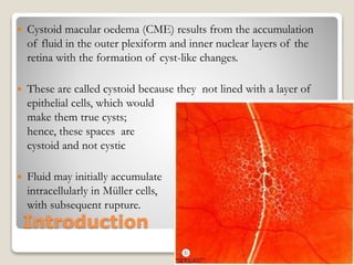
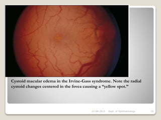
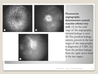
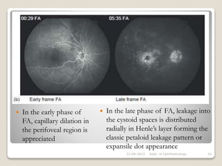
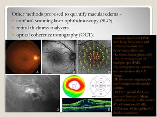
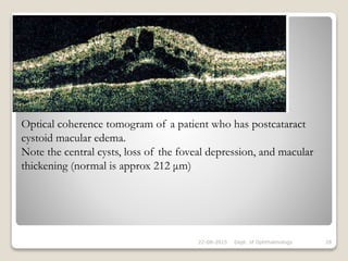
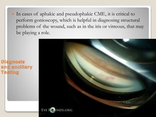
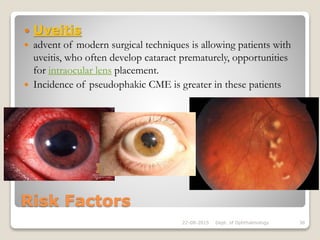
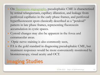
This document discusses cystoid macular edema (CME), including its pathogenesis, etiology, associated ocular conditions, manifestations, diagnosis and testing. Specifically, it focuses on pseudophakic or Irvine-Gass syndrome CME, which can occur after cataract surgery. The summary discusses how CME results from fluid accumulation in the retina, its appearance on fluorescein angiography, risk factors for pseudophakic CME like vitreous loss during surgery, and how it is diagnosed using techniques like optical coherence tomography.
Branched Retinal Vein Occlusion (BRVO) for undergraduate MBBS Students.
Covers the basics of Aetiology, pathophysiology, clinical features, types, associated conditions and management of BRVO.
Also encompasses salient points for PGMEE
Central Retinal Artery Occlusion (CRAO) for undergraduate MBBS Students.
Covers the basics of Aetiology, pathophysiology, clinical features, types, associated conditions and management of CRAO.
Also encompasses salient points for PGMEE
This ppt describe about the incidence, diagnosis and management of maculopathy in caaes of pathological myopia.
Data collected and created by Vivek Chaudhary
For queries : vivek977optom@gmail.com
This lecture is part of the yearly Basic Course Lectures in Ophthalmology given by the Dept of Ophthalmology and Visual Sciences at the Philippine General Hospital.
Originally given by Dr Pearl Tamesis-Villalon, it is a 1:30:00 hour lecture on the pathologic lesions seen in the vitreous, retina and choroid. It is meant for the general physician and the beginning ophthalmology resident who is interested in the basics of retinal pathology.
It includes pathologic changes seen in hypertension, diabetes, vaso occlusive disease, vitreous, membranes, choroid, retinal pigment epithelium, retinal detachments, etc. Lesions such as hemorrhages, cotton wool spots, hard exudates and their location in the retinal layers are explained. Fluorescein angiogram and OCT images are also incorporated.
Some images were grabbed from the internet, apologies for not making the necessary acknowledgements.
Congenital pit is an atypical coloboma usually located on the temporal edge of the disc, associated with irregular defects in the juxtapapillary choroid and pigment epithelium. Macular fibers passing through this area often are affected and corresponding changes in the retinal ganglion cell layer and in the visual field occur.
Background: Idiopathic Macular Telangiectasia type 2 (IMT2) is a relatively uncommon clinical condition with an estimated prevalence of 1% within the general population. This condition can be challenging to precisely identify in early stages but advancements in clinical imaging to include fl uorescein angiography and Optical Coherence Tomography Angiography (OCTA) can allow for timely diagnosis and prompt intervention that may leads to improved long-term clinical outcomes. Emerging literature has recognized the role of Macular
Pigment (MP) in IMT2 in terms of Henle fi ber layer deposition mechanisms and potential mitigation of infl ammatory and oxidative stress. Primary care optometrists are in a unique position to facilitate early detection and manage through close evaluation and individualized lutein supplementation
Leukocoria ( or white pupillary reflex) is an abnormal white reflection from the eye.
Leukocoria is a medical sign for a number of several conditions.
- this presentation at annual conference of the Ophthalmic department, faculty of medicine - Al-Azhar University in association with DOS & EOS Cairo, Egypt January 2017
Explore natural remedies for syphilis treatment in Singapore. Discover alternative therapies, herbal remedies, and lifestyle changes that may complement conventional treatments. Learn about holistic approaches to managing syphilis symptoms and supporting overall health.
ARTIFICIAL INTELLIGENCE IN HEALTHCARE.pdfAnujkumaranit
Artificial intelligence (AI) refers to the simulation of human intelligence processes by machines, especially computer systems. It encompasses tasks such as learning, reasoning, problem-solving, perception, and language understanding. AI technologies are revolutionizing various fields, from healthcare to finance, by enabling machines to perform tasks that typically require human intelligence.
Title: Sense of Taste
Presenter: Dr. Faiza, Assistant Professor of Physiology
Qualifications:
MBBS (Best Graduate, AIMC Lahore)
FCPS Physiology
ICMT, CHPE, DHPE (STMU)
MPH (GC University, Faisalabad)
MBA (Virtual University of Pakistan)
Learning Objectives:
Describe the structure and function of taste buds.
Describe the relationship between the taste threshold and taste index of common substances.
Explain the chemical basis and signal transduction of taste perception for each type of primary taste sensation.
Recognize different abnormalities of taste perception and their causes.
Key Topics:
Significance of Taste Sensation:
Differentiation between pleasant and harmful food
Influence on behavior
Selection of food based on metabolic needs
Receptors of Taste:
Taste buds on the tongue
Influence of sense of smell, texture of food, and pain stimulation (e.g., by pepper)
Primary and Secondary Taste Sensations:
Primary taste sensations: Sweet, Sour, Salty, Bitter, Umami
Chemical basis and signal transduction mechanisms for each taste
Taste Threshold and Index:
Taste threshold values for Sweet (sucrose), Salty (NaCl), Sour (HCl), and Bitter (Quinine)
Taste index relationship: Inversely proportional to taste threshold
Taste Blindness:
Inability to taste certain substances, particularly thiourea compounds
Example: Phenylthiocarbamide
Structure and Function of Taste Buds:
Composition: Epithelial cells, Sustentacular/Supporting cells, Taste cells, Basal cells
Features: Taste pores, Taste hairs/microvilli, and Taste nerve fibers
Location of Taste Buds:
Found in papillae of the tongue (Fungiform, Circumvallate, Foliate)
Also present on the palate, tonsillar pillars, epiglottis, and proximal esophagus
Mechanism of Taste Stimulation:
Interaction of taste substances with receptors on microvilli
Signal transduction pathways for Umami, Sweet, Bitter, Sour, and Salty tastes
Taste Sensitivity and Adaptation:
Decrease in sensitivity with age
Rapid adaptation of taste sensation
Role of Saliva in Taste:
Dissolution of tastants to reach receptors
Washing away the stimulus
Taste Preferences and Aversions:
Mechanisms behind taste preference and aversion
Influence of receptors and neural pathways
Impact of Sensory Nerve Damage:
Degeneration of taste buds if the sensory nerve fiber is cut
Abnormalities of Taste Detection:
Conditions: Ageusia, Hypogeusia, Dysgeusia (parageusia)
Causes: Nerve damage, neurological disorders, infections, poor oral hygiene, adverse drug effects, deficiencies, aging, tobacco use, altered neurotransmitter levels
Neurotransmitters and Taste Threshold:
Effects of serotonin (5-HT) and norepinephrine (NE) on taste sensitivity
Supertasters:
25% of the population with heightened sensitivity to taste, especially bitterness
Increased number of fungiform papillae
Anti ulcer drugs and their Advance pharmacology ||
Anti-ulcer drugs are medications used to prevent and treat ulcers in the stomach and upper part of the small intestine (duodenal ulcers). These ulcers are often caused by an imbalance between stomach acid and the mucosal lining, which protects the stomach lining.
||Scope: Overview of various classes of anti-ulcer drugs, their mechanisms of action, indications, side effects, and clinical considerations.
- Video recording of this lecture in English language: https://youtu.be/lK81BzxMqdo
- Video recording of this lecture in Arabic language: https://youtu.be/Ve4P0COk9OI
- Link to download the book free: https://nephrotube.blogspot.com/p/nephrotube-nephrology-books.html
- Link to NephroTube website: www.NephroTube.com
- Link to NephroTube social media accounts: https://nephrotube.blogspot.com/p/join-nephrotube-on-social-media.html
MANAGEMENT OF ATRIOVENTRICULAR CONDUCTION BLOCK.pdfJim Jacob Roy
Cardiac conduction defects can occur due to various causes.
Atrioventricular conduction blocks ( AV blocks ) are classified into 3 types.
This document describes the acute management of AV block.
Report Back from SGO 2024: What’s the Latest in Cervical Cancer?bkling
Are you curious about what’s new in cervical cancer research or unsure what the findings mean? Join Dr. Emily Ko, a gynecologic oncologist at Penn Medicine, to learn about the latest updates from the Society of Gynecologic Oncology (SGO) 2024 Annual Meeting on Women’s Cancer. Dr. Ko will discuss what the research presented at the conference means for you and answer your questions about the new developments.
The prostate is an exocrine gland of the male mammalian reproductive system
It is a walnut-sized gland that forms part of the male reproductive system and is located in front of the rectum and just below the urinary bladder
Function is to store and secrete a clear, slightly alkaline fluid that constitutes 10-30% of the volume of the seminal fluid that along with the spermatozoa, constitutes semen
A healthy human prostate measures (4cm-vertical, by 3cm-horizontal, 2cm ant-post ).
It surrounds the urethra just below the urinary bladder. It has anterior, median, posterior and two lateral lobes
It’s work is regulated by androgens which are responsible for male sex characteristics
Generalised disease of the prostate due to hormonal derangement which leads to non malignant enlargement of the gland (increase in the number of epithelial cells and stromal tissue)to cause compression of the urethra leading to symptoms (LUTS
Factory Supply Best Quality Pmk Oil CAS 28578–16–7 PMK Powder in Stockrebeccabio
Factory Supply Best Quality Pmk Oil CAS 28578–16–7 PMK Powder in Stock
Telegram: bmksupplier
signal: +85264872720
threema: TUD4A6YC
You can contact me on Telegram or Threema
Communicate promptly and reply
Free of customs clearance, Double Clearance 100% pass delivery to USA, Canada, Spain, Germany, Netherland, Poland, Italy, Sweden, UK, Czech Republic, Australia, Mexico, Russia, Ukraine, Kazakhstan.Door to door service
Hot Selling Organic intermediates
Lung Cancer: Artificial Intelligence, Synergetics, Complex System Analysis, S...Oleg Kshivets
RESULTS: Overall life span (LS) was 2252.1±1742.5 days and cumulative 5-year survival (5YS) reached 73.2%, 10 years – 64.8%, 20 years – 42.5%. 513 LCP lived more than 5 years (LS=3124.6±1525.6 days), 148 LCP – more than 10 years (LS=5054.4±1504.1 days).199 LCP died because of LC (LS=562.7±374.5 days). 5YS of LCP after bi/lobectomies was significantly superior in comparison with LCP after pneumonectomies (78.1% vs.63.7%, P=0.00001 by log-rank test). AT significantly improved 5YS (66.3% vs. 34.8%) (P=0.00000 by log-rank test) only for LCP with N1-2. Cox modeling displayed that 5YS of LCP significantly depended on: phase transition (PT) early-invasive LC in terms of synergetics, PT N0—N12, cell ratio factors (ratio between cancer cells- CC and blood cells subpopulations), G1-3, histology, glucose, AT, blood cell circuit, prothrombin index, heparin tolerance, recalcification time (P=0.000-0.038). Neural networks, genetic algorithm selection and bootstrap simulation revealed relationships between 5YS and PT early-invasive LC (rank=1), PT N0—N12 (rank=2), thrombocytes/CC (3), erythrocytes/CC (4), eosinophils/CC (5), healthy cells/CC (6), lymphocytes/CC (7), segmented neutrophils/CC (8), stick neutrophils/CC (9), monocytes/CC (10); leucocytes/CC (11). Correct prediction of 5YS was 100% by neural networks computing (area under ROC curve=1.0; error=0.0).
CONCLUSIONS: 5YS of LCP after radical procedures significantly depended on: 1) PT early-invasive cancer; 2) PT N0--N12; 3) cell ratio factors; 4) blood cell circuit; 5) biochemical factors; 6) hemostasis system; 7) AT; 8) LC characteristics; 9) LC cell dynamics; 10) surgery type: lobectomy/pneumonectomy; 11) anthropometric data. Optimal diagnosis and treatment strategies for LC are: 1) screening and early detection of LC; 2) availability of experienced thoracic surgeons because of complexity of radical procedures; 3) aggressive en block surgery and adequate lymph node dissection for completeness; 4) precise prediction; 5) adjuvant chemoimmunoradiotherapy for LCP with unfavorable prognosis.
1. Cystoid Macular
Oedema
Moderator: Dr. Rekha B.K. MS, DOMS, PhD
Professor & Head of Department
Presenter: Dr. Arushi Prakash
3rd year Resident
Department of Ophthalmology
J.N. Medical college, Belgaum
22-08-2015 Dept. of Ophthalmology 1
4. Macula
• anatomic specialization of the neural retina unique to humans
and to higher primates
• defined
• histologically by the presence of multiple layers of retinal
ganglion cells
(extramacular retina- only a single layer of ganglion cells)
• susceptible to such a large number of disorders,
For eg.- the macular degenerations,
or to edema
in a large variety of diseases.
22-08-2015 Dept. of Ophthalmology 4
6. Introduction
Cystoid macular oedema (CME) results from the accumulation
of fluid in the outer plexiform and inner nuclear layers of the
retina with the formation of cyst-like changes.
These are called cystoid because they not lined with a layer of
epithelial cells, which would
make them true cysts;
hence, these spaces are
cystoid and not cystic
Fluid may initially accumulate
intracellularly in Müller cells,
with subsequent rupture.
22-08-2015 Dept. of Ophthalmology 6
7. 22-08-2015 Dept. of Ophthalmology 7
(A) Histology shows cystic spaces in
the outer plexiform and inner nuclear
layer
(B) progression to
lamellar hole formation
In long-standing cases, smaller microcystic spaces coalesce into larger
cavities and may progress to lamellar hole formation at the fovea with
irreversible impairment of central vision
8. Pathogenesis & Etiology
Intraretinal fluid may accumulate into CME by:
Breakdown of normal anatomical barriers:
a) inner blood retinal barrier
b) outer blood retinal barrier
inflammatory mediators such as
prostaglandins, leukotrienes, protein kinase C, nitric oxide, vascular endothelial
growth factor, various other cytokines
may cause incompetence of this barrier
22-08-2015 Dept. of Ophthalmology 8
9. Pathogenesis & Etiology
Another mechanism results from failure of
normal physiological process-
In normal functional retina, RPE cells constantly act to
eliminate fluid from retina.
If this function is compromised, CME may occur
22-08-2015 Dept. of Ophthalmology 9
16. OCULAR MANIFESTATIONS
The major symptom is decreased central visual acuity.
Accompanying symptoms may include metamorphopsia,
micropsia, scotomata, ocular irritation, photophobia, and
conjunctival injection.
Presenting visual acuities usually range from 20/25 (6/8) to
20/80 (6/26) but may be as poor as 20/400 (6/133).
Clinically, CME is seen best using the slit lamp and either a
contact lens (e.g., Goldmann lens) or a handheld, noncontact
lens (e.g.,78 D, 60 D).
22-08-2015 Dept. of Ophthalmology 16
17. OCULAR MANIFESTATIONS
The edema results in light scattering due to the multiple
interfaces created by the separated retinal cells.
This light scattering decreases the neural retina’s translucency so
that the normal retinal pigment epithelial and choroidal
background patterns are blurred.
Individual pockets of fluid in the outer plexiform layer are seen,
with the largest pockets centrally and progressively smaller cysts
peripherally.
22-08-2015 Dept. of Ophthalmology 17
18. OCULAR MANIFESTATIONS
Retroillumination can help to delineate the polycystic spaces.
As these changes can be subtle and media opacity can affect the
view, clinical confirmation of CME may be difficult in certain
eyes.
A yellow spot, believed to be due to diffusion of the luteal
pigment, may be evident in the central macula.
Small intraretinal and intracystic hemorrhages, microaneurysms,
and telangiectasias may be seen as well.
22-08-2015 Dept. of Ophthalmology 18
19. 22-08-2015 Dept. of Ophthalmology 19
Cystoid macular edema in the Irvine-Gass syndrome. Note the radial
cystoid changes centered in the fovea causing a “yellow spot.”
20. OCULAR MANIFESTATIONS
The underlying cause of CME does not alter its appearance, but
the associated ocular findings vary widely depending on etiology.
After cataract surgery, aqueous and/or vitreous cell and flare,
optic nerve head swelling, and a ruptured anterior hyaloid face
may be evident.
Other patients show vitreous traction to anterior chamber
structures.
Epiretinal membranes are concurrently present in about 10% of
eyes.
22-08-2015 Dept. of Ophthalmology 20
21. OCULAR MANIFESTATIONS
Inflammatory, vascular, tractional, inherited, and tumor-related
causes result in the corresponding associated ocular findings.
As a result of CME, a rupture of the inner retinal cyst can occur
to give a lamellar macular hole.
Prolonged CME may induce atrophy of the macular
photoreceptors – visual acuity is poor, but clinical examination
and fluorescein angiography may be grossly normal with the
exception of a blunting of the foveal reflex.
22-08-2015 Dept. of Ophthalmology 21
22. Diagnosis and ancillary Testing
In eyes with clear media, clinical examination usually yields the
diagnosis.
Angiography reveals a typical petalloid pattern in the central
macula secondary to dye leakage from the perifoveal capillaries.
The dye accumulates in cyst-like spaces within the outer
plexiform layer (Henle’s layer).
Late fluorescein angiographic pictures should be taken to allow
time for the dye to accumulate within the anatomical fovea. Also
seen on fluorescein angiography is leakage from the disc and
retinal vessels.
22-08-2015 Dept. of Ophthalmology 22
23. Fluorescein
angiography
demonstrates cystoid
macular edema very
well. (A) In the early
stage of the angiography,
minimal leakage is seen.
(B) The petalloid leakage
pattern, present in the late
stages of the angiography,
is diagnostic of CME. (C)
Note the profuse leakage
from the optic nerve head
in the late stages.
22-08-2015 Dept. of Ophthalmology 23
24. In the early phase of
FA, capillary dilation in
the perifoveal region is
appreciated
22-08-2015 Dept. of Ophthalmology 24
In the late phase of FA, leakage into
the cystoid spaces is distributed
radially in Henle’s layer forming the
classic petaloid leakage pattern or
expansile dot appearance
25. DEPRIVENS is a common mnemonic for risk
factors that cause leakage on FA
Diabetes
Epinephrine
Pars Planitis/Uveitis
Retinitis Pigmentosa (RP)
Irvine-Gass
Vein Occlusion
E2-prostaglandin
Nicotinic acid and Niacin
Surgery
22-08-2015 Dept. of Ophthalmology 25
26. Risk factors that do not demonstrate leakage on
FA include:
Juvenile retinoschisis
Goldmann-Favre disease
certain types of RP
Nicotinic Acid maculopathy
Phototoxicity
Antimicrotubule agents
22-08-2015 Dept. of Ophthalmology 26
27. Other methods proposed to quantify macular edema -
confocal scanning laser ophthalmoscopy (SLO)
retinal thickness analyzers
optical coherence tomography (OCT).
22-08-2015
Clinically significant DME
with large foveal cysts and
subfoveal neuroretinal
detachment (right eye):
(A) Color fundus photo; (B)
FAF showing pattern of
multiple-spot iFAF;
(C) microperimetry sensitivity
map overlaid on the FAF
image;
(D) fluorescein angiography
confirming cystoid macular
edema;
(E) OCT (retinal thickness
map and line scan). Mean
retinal sensitivity in the central
4° (∼1 mm) was 5.5 dB;
BCVA was +0.16 logMar (0.7
Snellen equivalent).
28. 22-08-2015 Dept. of Ophthalmology 28
Optical coherence tomogram of a patient who has postcataract
cystoid macular edema.
Note the central cysts, loss of the foveal depression, and macular
thickening (normal is approx 212 μm)
29. Diagnosis
and ancillary
Testing
In cases of aphakic and pseudophakic CME, it is critical to
perform gonioscopy, which is helpful in diagnosing structural
problems of the wound, such as in the iris or vitreous, that may
be playing a role.
22-08-2015 Dept. of Ophthalmology 29
31. In 1953, Irvine described a cystoid macular edema
(CME) that specifically arised after cataract surgery.
Gass and Norton subsequently studied the
characteristics of the new disease entity with
fluorescein angiography
22-08-2015 Dept. of Ophthalmology 31
32. Definitions
Angiographic pseudophakic CME: seen on fluorescein
angiography (FA)
Clinical pseudophakic CME: associated with decreased visual
acuity
Acute pseudophakic CME: within 6 months
Chronic pseudophakic CME: over 6 months
22-08-2015 Dept. of Ophthalmology 32
33. Incidence
Angiographic CME after intracapsular cataract
extraction: As high as 60%*
Angiographic CME after extracapsular cataract
extraction: 15% to 30%
Clinical CME after small incision phacoemulsification:
0.1% to 2.35%
OCT evidence of CME after small incision
phacoemulsification: 4% to 11%, but also reported to be
as high as 41%
Most patients with CME found via angiography or OCT will not
have visual changes.
Furthermore, most patients with clinical CME will experience
spontaneous improvement by 3 to 12 months
22-08-2015 Dept. of Ophthalmology 33
34. Pathophysiology
inflammatory mediators that are upregulated in
the aqueous and vitreous humors after surgical manipulation
Inflammation breaks down the blood-aqueous and blood-
retinal-barriers, leads to increased vascular permeability
Eosinophilic transudate accumulates in the outer plexiform and
inner nuclear layers of the retina to create cystic spaces that
coalesce to form larger pockets of fluid
In chronic CME, lamellar macular holes and subretinal fluid may
also form
22-08-2015 Dept. of Ophthalmology 34
35. Risk Factors
Surgical
• Vitreous loss
• Vitreous traction at incision sites
• Vitrectomy for retained lens fragments
• Iris trauma
• Posterior capsule rupture
• Intraocular lens dislocation
• Early postoperative capsulotomy
• Iris-fixated intraocular lenses
• Anterior chamber intraocular lenses
22-08-2015 Dept. of Ophthalmology 35
36. Risk Factors
Diabetic Macular Edema
Cataract surgery was historically thought to accelerate
progression of diabetic retinopathy (DR),
However, postoperative macular edema usually develops in
those with a prior history of diabetic macular edema (DME).
DME and severe DR should be thoroughly treated before
undergoing cataract extraction.
22-08-2015 Dept. of Ophthalmology 36
37. Risk Factors
Diabetic Macular Edema
The source of the fluid has been presumed to be due to
excessive retinal capillary leakage, but there is some evidence
that the retinal pigment epithelium may also have dysfunctional
fluid homeostatic
function in diabetes and
contribute to this
appearance.
22-08-2015 Dept. of Ophthalmology 37
38. Risk Factors
Uveitis
advent of modern surgical techniques is allowing patients with
uveitis, who often develop cataract prematurely, opportunities
for intraocular lens placement.
Incidence of pseudophakic CME is greater in these patients
22-08-2015 Dept. of Ophthalmology 38
39. Risk Factors
Glaucoma Medications
Preoperative and postoperative topical glaucoma medications,
specifically latanoprost and timolol, may increase the incidence
of pseudophakic CME.
It appears that a commonly used preservative, benzalkonium
chloride, is cytotoxic and stimulates inflammatory responses.
The effects may be prevented by concomitant use of NSAID
drops.
22-08-2015 Dept. of Ophthalmology 39
40. Other Risk Factors
Previous histories of
retinal vein occlusion (RVO),
epiretinal membrane (ERM),
22-08-2015 Dept. of Ophthalmology 40
41. Diagnosis
History and Clinical Presentation
occurrence peaks at approximately 4-6 weeks postoperatively.
most common presentation is blurry vision.
Less common presentations include central scotomas,
metamorphopsia, and mild photophobia.
On biomicroscopy, retinal thickening and loss of the foveal
depression is usually appreciated.
findings are best observed with fundus contact lens, red-free
light may aide in demonstrating cystic changes.
In severe or chronic cases, optic disc swelling and/or a lamellar
hole may also be seen.
Splinter hemorrhages may also be present.
Biomicroscopy may not show any abnormalities in 5-10% eyes
22-08-2015 Dept. of Ophthalmology 41
42. Imaging Studies
On fluorescein angiography, pseudophakic CME is characterized
by retinal telangiectasis, capillary dilatation, and leakage from
perifoveal capillaries in the early phase frames, and perifoveal
hyperfluorescent spots classically described as a “petaloid”
pattern in late phase frames, representing fluorescein
accumulation in cystic spaces.
Cystoid changes may also be apparent in the fovea and
extramacular areas.
Optic nerve staining is also commonly seen,
FA is the gold standard in diagnosing pseudophakic CME, but
treatment responses would be more conveniently monitored by
biomicroscopy, visual acuity and OCT.
22-08-2015 Dept. of Ophthalmology 42
43. OCT is a relatively new, widely adopted technology that
allows high-resolution cross-sectional imaging of
the macula .
Pseudophakic CME on OCT is characterized by loss
of the foveal depression, retinal thickening, and cystic
hyporeflective areas within the macula.
However, OCT has not replaced FA as the gold
standard in diagnosing pseudophakic CME, because
FA can also rule out other causes of CME such
as diabetic macular edema and retinal vein occlusion .
22-08-2015 Dept. of Ophthalmology 43
44. Differential Diagnosis
Diabetic macular edema
Retinal vein occlusion
Radiation retinopathy
Hypertensive retinopathy
Uveitis
Topical prostaglandins
Retinal dystrophies
Choroidal tumors
Leukemia
Chronic renal failure
Of special note, it is important not to misdiagnose pseudophakic CME
with choroidal neovascularization seen in age-related macular degeneration
(AMD). Many patients undergoing cataract surgery are also within the age
range for developing AMD.
22-08-2015 Dept. of Ophthalmology 44
45. Prophylaxis and Treatment
no standardized treatment or prophylactic protocol for
pseudophakic CME, due a lack of strong randomized
clinical trials and comparative effectiveness studies.
lack of large studies due to fact that most cases of
acute pseudophakic CME spontaneously resolve.
treatment of chronic pseudophakic CME remains a
challenge.
22-08-2015 Dept. of Ophthalmology 45
46. The management of pseudophakic CME is based on its
pathogenesis.
In the inflammation cascade, cell membrane lipids are
converted to arachidonic acid by phospholipase A2,
and prostaglandins are then formed by cyclooxygenases
(COX).
Corticosteroids reduce inflammation by inhibiting
phospholipase A2, and
nonsteroidal anti-inflammatory drugs (NSAIDs) inhibit
COX.
The 2 critical COX isoforms are COX-1 andCOX-2, and
the later is the major isoform expressed in the retina
22-08-2015 Dept. of Ophthalmology 46
47. Cyclo-Oxygenase Inhibitors
Cyclo-oxygenase is inhibited by COI drugs, which prevents the
conversion of arachidonic acid into endoperoxides and, hence,
inhibits PG synthesis. Clinically, COIs decrease capillary leakage.
Topical COIs (Ketorolac tromethamine 0.5%, indomethacin
1%, Nepafenac 0.1%, Bromofenac 0.09%)
Complications associated with the use of topical anti-
inflammatory agents include ocular irritation and discomfort
following application, conjunctival injection, mild punctate
keratopathy, and mydriasis. Allergic and hypersensitivity
reactions have been reported.
22-08-2015 Dept. of Ophthalmology 47
48. Cyclo-Oxygenase
Inhibitors
Systemic COI
associated with multiple side effects.
Gastrointestinal- nausea, vomiting, diarrhea, anorexia,
abdominal discomfort, ulceration, gastrointestinal bleeding.
Can interfere with platelet function and clotting.
Bone marrow suppression, hepatotoxicity, impaired renal
function, and central nervous system symptomatology, including
headache, dizziness, somnolence, depression, fatigue, insomnia,
and confusion, have been reported.
Hypersensitivity and induction of asthmatic attacks can occur.
22-08-2015 Dept. of Ophthalmology 48
49. Cyclo-Oxygenase Inhibitors
Systemic COI
Indomethacin can be deposited in the cornea in a pattern.
Association with optic nerve dysfunction has been reported.
dermatologic reactions include rash, dermatitis, and Stevens-
Johnson syndrome can occur.
The availability of topical formulations that provide good ocular
penetration of the drug make it unnecessary to recommend
systemic therapy for ophthalmic indications, particularly in view
of the many serious side effects of systemic administration
22-08-2015 Dept. of Ophthalmology 49
50. Corticosteroids
Topical
Topical corticosteroids are commonly used in the treatment of
pseudophakic CME.
lipophilic acetate suspensions of prednisolone penetrate the intact
corneal epithelium and reach the anterior chamber in higher
concentration than water-soluble forms, such as dexamethasone
Few studies, however, have examined their efficacy.
Furthermore, its effects are often confounded by the
concomitantly used and more efficacious topical NSAIDs.
Significant potential complications of topical corticosteroid
therapy include glaucoma, posterior subcapsular cataracts,
exacerbations of infections, and corneal problems
22-08-2015 Dept. of Ophthalmology 50
51. Corticosteroids
Topical
Of note, anecdotal evidence indicates that the visual acuity of
“corticosteroid responders” with postcataract CME improves
more than that of patients whose IOP remains normal. This
may be explained by a hydrostatic effect.
22-08-2015 Dept. of Ophthalmology 51
52. Corticosteroids
Periocular
Posterior sub-Tenon's injections may have advantages over
topical application.
exert a maximal, long-lasting response at site of injection,
Water soluble drugs have excellent penetration through the
sclera as opposed to topical application.
Carry carry the risk of inadvertent penetration of the globe.
Contraindications for posterior sub-Tenon's injections include
steroid-induced glaucoma, hypersensitivity to components of
the injected steroid preparation, active
necrotizing scleritis, and active
ocular toxoplasmosis
22-08-2015 Dept. of Ophthalmology 52
53. Corticosteroids
Intravitreal
A single intravitreal injection of triamcinolone induced clinical
and angiographic resolution of CME.
Side effects of treatment were similar to those of periocular
injection including IOP rise and increased rate of cataract
formation.
major limitations of intravitreal corticosteroids is the transient
effect that requires repeated injections.
22-08-2015 Dept. of Ophthalmology 53
54. 22-08-2015 Dept. of Ophthalmology 54
Spectral domain optical coherence tomography (SD-OCT) of a patient with
chronic refractory pseudophakic CME. There is retinal thickening, cystic
intraretinal lesions, and subretinal fluid.
SD-OCT of the same patient 1 month after a sub-Tenon's
injection of triamcinolone. The retinal thickening, cystic
lesions, and subretinal fluid have improved
55. Corticosteroids
Systemic corticosteroids
administered orally or intravenously.
Numerous potential side effects
Ocular complications - elevated IOP, posterior subcapsular
cataract, increased incidence of viral ocular infections, ptosis,
mydriasis, scleral melt, and lid skin atrophy.
Potential short-term systemic effects- peptic ulcer disease, aseptic
necrosis of the femoral head, and mental changes (euphoria,
insomnia, & psychosis).
Long-term systemic side effects- osteoporosis, a cushingoid state,
electrolyte imbalance, reactivation of latent infections such as
tuberculosis, myopathy, suppression of the pituitary-adrenal axis,
and increased severity of preexisting diabetes and hypertension.
22-08-2015 Dept. of Ophthalmology 55
56. Corticosteroids
Drug Delivery Systems
Sustained drug delivery systems (DDS) have been developed to
address this limitation of intravitreal corticosteroid injections.
Ozurdex (Allergan, Irvine, CA) is an injectable, biodegradable
intravitreal DDS that provides sustained release of preservative-
free dexamethasone, a potent corticosteroid.
22-08-2015 Dept. of Ophthalmology 56
57. Anti-VEGF Treatments
VEGF is well known to be a key mediator of angiogenesis, but
it also plays an important role in the inflammation and capillary
permeability that causes CME.
Bevacizumab (Avastin, Genentech, South San Francisco, CA) is a humanized
monoclonal antibody that inhibits VEGF-A
“Triple therapy” with intravitreal triamcinolone, intravitreal
bevacizumab and topical NSAIDs has been shown to be
effective as well, although the effects of the intravitreal
medications were transient.
22-08-2015 Dept. of Ophthalmology 57
58. Carbonic Anhydrase Inhibitors
Oral CAIs may be considered in refractory pseudophakic CME.
CAIs improve the pumping action of the retinal pigment
epithelium, to decrease intraretinal fluid.
initial dose of 250mg daily, increased to 500mg daily if no effect
is apparent.
A trial of several weeks is warranted, with successful outcome
judged by improved visual acuity on careful repeated
measurements or by decreased CME on fluorescein
angiography.
If decreased CME is observed by fluorescein angiography after
several weeks of use, the continuation of acetazolamide may be
considered even if visual acuity has not improved, provided the
patient is able to tolerate the drug
22-08-2015 Dept. of Ophthalmology 58
60. Immunomodulatory Therapy
Recent small pilot studies have started to examine interferon
alpha (IFN-a, Imgenex, San Diego, CA) and intravitreal infliximab (Remicade,
Centocor Ortho Biotech, Horsham, PA) with mixed results.
22-08-2015 Dept. of Ophthalmology 60
61. HYPERBARIC
OXYGEN
Improvement in aphakic CME with hyperbaric oxygen therapy
was reported by Ploff and Thom*.
The dosage given was 2.2atm oxygen for 1.5h twice a day for 7
days, followed by 2h daily for 14 days. The mechanism was
hypothesized to be macular capillary contraction.
22-08-2015 Dept. of Ophthalmology 61
* Williams GA, Haller JA, Kuppermann BD, et al. Dexamethasone posterior-segment drug delivery system in the
treatment of macular edema resulting from uveitis or Irvine-Gass syndrome. Am J Ophthalmol 2009;147(6):1048-1054,
1054 e1041-1042.
62. Surgical
Treatment
Laser Vitreolysis
Neodymium:YAG laser anterior vitreolysis can release vitreous
incarceration in the cataract incision wounds that complicate
pseudophakic CME.
22-08-2015 Dept. of Ophthalmology 62
63. Pars Plana Vitrectomy
may be considered when
pseudophakic CME is
complicated by
vitreoretinal traction,
and/or if the CME is
unresponsive to other
treatments.
Vitrectomy may also
theoretically reduce the
concentration of
inflammatory mediators
and growth factors.
22-08-2015 Dept. of Ophthalmology 63
Surgical Treatment
64. Situation Recommendations/Options
Intracameral lens (within the pupil;
iris suspended)
Remove, consider exchange for
flexible AC IOL, or suture-fixated
PC IOL
Anterior chamber IOL with
distorted pupil and vitreous strands
in AC
Vitrectomy to restore normal pupil.
Leave IOL if flexible, remove if not
Would a sulcus-fixated PC IOL be
safe? Omitting IOL is another
option, as is a suture-fixated PC IOL
ACIOL without distorted pupil or
vitreous in AC
Remove, especially if rigid IOL;
replace with flexible IOL or would
sulcus-fixated PC IOL be safe?
Elevated, isolated vitreous strand
distorting pupil with AC or PC IOL
Consider YAG vitreolysis
22-08-2015 Dept. of Ophthalmology 64
65. Situation Recommendations/Options
PCIOL with pupillary capture Free capture
PC IOL with moderate pupillary
distortion from vitreous strands
Anterior vitrectomy for restoring
pupil to normal and consider leaving
lens
PCIOL, sulcus fixation, normal pupil Consider removing IOL; possibly
exchange for flexible AC IOL, or
leave IOLs out, or use a suture-
fixated PC IOL
PC IOL; “in-the-bag,” normal pupil Pars plana vitrectomy if evidence of
traction on macula (rare). Rule out
other causes of CME
PCIOL with pupillary capture Free capture
PCIOL with moderate pupillary
distortion from vitreous strands
Anterior vitrectomy for restoring
pupil to normal and consider leaving
lens
22-08-2015 Dept. of Ophthalmology 65
66. Retinal Vein Occlusions
Both branch and central vein occlusions can result
in severe macular edema.
Due to hypoxic capillary endothelial damage
secondary to increased intravascular hydrostatic
pressure.
The Branch Vein Occlusion Study demonstrated
visual benefit following application of a grid
pattern of photocoagulation to the area of chronic
macular edema.
The Central Retinal Vein Occlusion Study showed
no visual benefit in the use of grid macular
photocoagulation for the treatment of associated
macular edema.
22-08-2015 Dept. of Ophthalmology 66
However, a possible benefit may be seen in younger patients who undergo treatment. It
is not unusual for CME to overlie a choroidal neovascular membrane or a serous retinal
detachment. Often it goes unnoticed in light of the accompanying severe subretinal
and/or intraretinal abnormalities.
68. Coat’s Disease
Other, more unusual retinal vascular
conditions produce CME. Retinal
telangiectasis (also known as Leber's miliary
aneurysms or Coats' disease) is a unilateral
condition that occurs more commonly in
males. The retinal vasculature is anomalous
and produces leakage with resultant cystoid
and diffuse macular edema.
The vascular anomalies may be local or
widespread.
Cryotherapy or laser treatment is applied if
these lesions threaten macular function.
22-08-2015 Dept. of Ophthalmology 68
69. Idiopathic juxtafoveal telangiectasis
Idiopathic juxtafoveal
telangiectasis can occur
in either a unilateral or
bilateral pattern.
Exudation and CME
result in visual loss.
Photocoagulation may
help to improve vision
if there is no
spontaneous
improvement.
22-08-2015 Dept. of Ophthalmology 69
71. Radiation Retinopathy
Patients who receive radiation treatment involving the head and neck may
develop signs of radiation retinopathy 6 months to 3 years following this
treatment.
A bilateral retinopathy occurs in almost one third of patients treated with
external beam irradiation.
Local plaque therapy requires higher doses than external beam therapy to
produce damage.
The clinical features of radiation retinopathy mirror those of diabetic
retinopathy and include CME.
Although no therapy is
proved, grid laser
photocoagulation is
believed to lessen the
macular edema
22-08-2015 Dept. of Ophthalmology 71
72. Arterial Macroaneurysm
Acquired retinal arterial
macroaneurysms are often
multiple and may thrombose
and close spontaneously.
This entity is treated if lipid
exudate threatens the macula
or if CME develops.
Two modalities of laser
treatment to be considered
are direct laser treatment to
close off the lesions and
indirect laser treatment in a
tight grid pattern around the
macroaneurysm.
22-08-2015 Dept. of Ophthalmology 72
73. Choroidal Tumors
CME with choroidal
tumors, such as nevi,
malignant melanomas,
and cavernous
hemangiomas, can
occur.
Intraretinal cystoid
changes occur over
the tumor and at sites
distant from the
tumor as a result of
lack of oxygenation
of retinal tissue.
22-08-2015 Dept. of Ophthalmology 73
74. Ocular Inflammations
idiopathic uveitis
intermediate uveitis
birdshot retinochoroidopathy
sarcoidosis, toxoplasmosis,
posterior scleritis,
Vogt- Koyanagi-Harada's syndrome
Behçet's syndrome.
The common underlying cause is an
inflammatory-mediated breakdown in the
blood-retina barrier
22-08-2015 Dept. of Ophthalmology 74
75. Retinitis Pigmentosa
Patients who have
retinitis pigmentosa
can also have CME.
Although the
precise
pathophysiology is
not clearly defined,
it is felt that the
perifoveal
capillaries
demonstrate
increased
permeability in
such cases
22-08-2015 Dept. of Ophthalmology 75
76. Epiretinal Membrane
&
Vitreomacular Traction Syndrome
The presence of epiretinal
membranes and the
vitreomacular traction
syndrome are associated with
CME, particularly in areas of
greatest traction and distortion
of retinal blood vessels.
Management of this condition
is to remove the membrane
surgically
22-08-2015 Dept. of Ophthalmology
7
6
77. DOMINANT CYSTOID MACULAR
EDEMA
extremely rare autosomal dominant disease
mapped to chromosome 7q.
Interestingly, autosomal dominant retinitis
pigmentosa, known as RP9, also maps to this region.
Unique macular disorder-
the inner nuclear layer of retina is the site primarily affected.
Müller's cells are the specific cellular constituents thought to be
involved, based on histopathological evidence.
22-08-2015 Dept. of Ophthalmology 77
78. Ophthalmoscopy reveals multilobulated cysts
in the macula.
Peripheral pigmentary changes
may be present.
Ancillary testing with fluorescein angiography
shows capillary leakage, with petaloid dye accumulation in the
macula.
Electrophysiology shows normal electroretinographic findings,
but subnormal electro-oculographic light peak–to–dark trough
ratios have been found, and abnormal dark-adaptation studies
have been documented.
22-08-2015 Dept. of Ophthalmology 78
79. PATHOLOGY
On Histopathology-
macular cysts, disorganized and gliotic
inner nuclear layer, focal Müller's cell necrosis,
epiretinal membrane formation, and
abnormal deposition of basement membrane
in the perivascular space.
Degenerative changes also have been seen in the RPE and
photoreceptors of the macula. Histopathological features seen
in dominantly inherited cystoid macular edema, namely the
involvement of Müller's cells, are quite different from those
features seen in cystoid macular edema secondary to other
causes.
22-08-2015 Dept. of Ophthalmology 79
80. TREATMENT, COURSE, AND OUTCOME
Patients first begin to notice decreasing visual
acuity at about 30 years of age, with slowly
progressive worsening to a moderate or
severe level years later.
Advanced cases have maculae of atrophic
appearance, with window defects seen on
fluorescein angiography.
As in all the macular dystrophies, no known effective treatment
22-08-2015 Dept. of Ophthalmology 80
81. 81
# Ophthalmology. Myron Yanoff & Jay S. Duker. 4th Ed. Pg
625-31
# Cataract Surgery And Its Complications. by Norman S. Jaffe,
Mark S. Jaffe, Gary F. Jaffe. 4th Ed. Pg 426-41
#http://eyewiki.aao.org/Pseudophakic_Cystoid_Macular_Edema
_(Irvine-Gass_Syndrome)
#The Retina and its Disorders, Joseph Besharse, Dean Bok. 1st Ed.
Topical or systemic indomethacin inhibits cyclooxygenase enzyme that decreases the production of prostaglandinsare used postoperatively for aphakic or pseudophakic CME