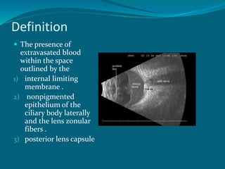
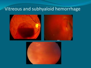
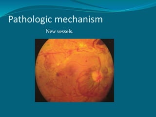
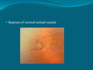
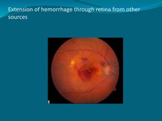
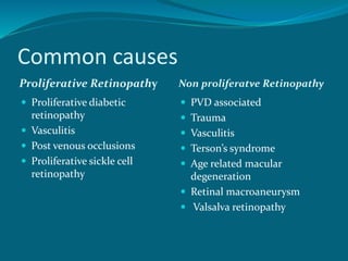
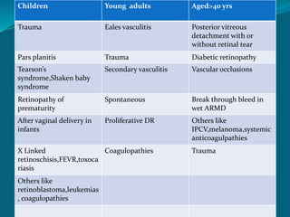
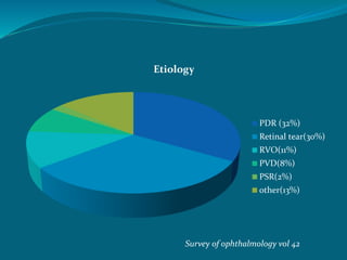
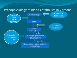
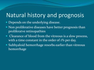
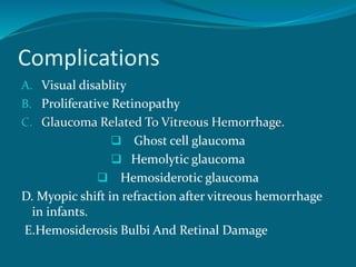
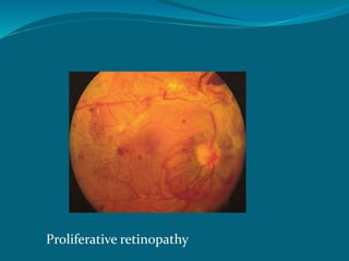
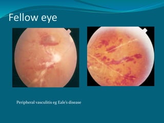
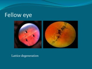
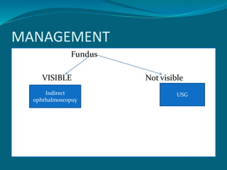
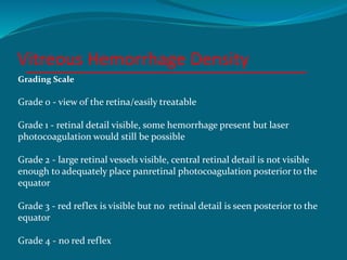
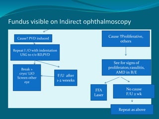
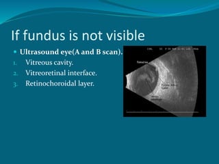
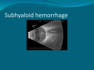
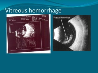
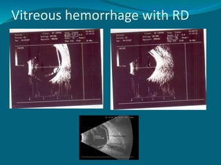
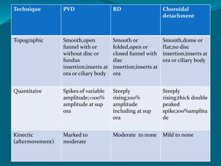
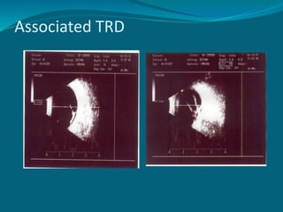
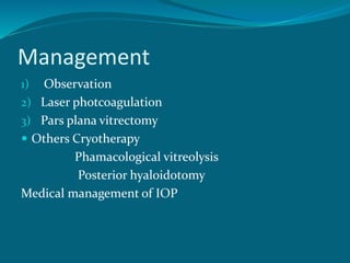
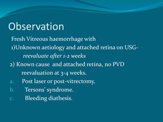
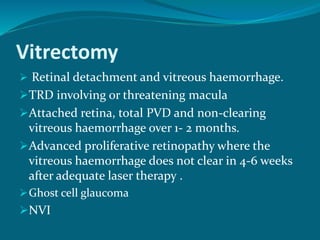
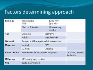
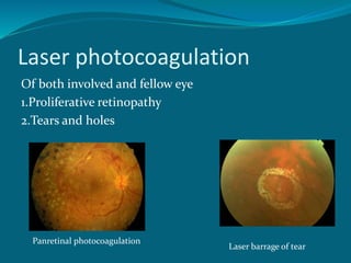
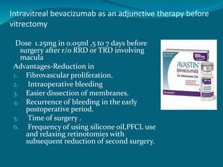
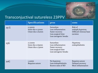
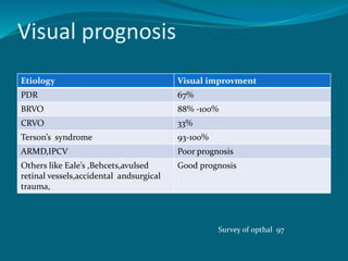
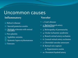
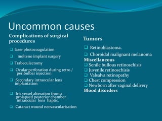
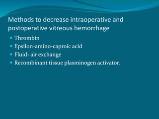
DR.SHAH-NOOR HASSAN
FCPS,FRCS(Glasgow)
Definition
 The presence of
extravasated blood
within the space
outlined by the
1) internal limiting
membrane .
2) nonpigmented
epithelium of the
ciliary body laterally
and the lens zonular
fibers .
3) posterior lens capsule
Vitreous and subhyaloid hemorrhage
Pathologic mechanism
New vessels.
 Rupture of normal retinal vessels.
Extension of hemorrhage through retina from other
sources
Common causes
Proliferative Retinopathy Non proliferatve Retinopathy
 Proliferative diabetic
retinopathy
 Vasculitis
 Post venous occlusions
 Proliferative sickle cell
retinopathy
 PVD associated
 Trauma
 Vasculitis
 Terson’s syndrome
 Age related macular
degeneration
 Retinal macroaneurysm
 Valsalva retinopathy
Age
Children Young adults Aged>40 yrs
Trauma Eales vasculitis Posterior vitreous
detachment with or
without retinal tear
Pars planitis Trauma Diabetic retinopathy
Tearson’s
syndrome,Shaken baby
syndrome
Secondary vasculitis Vascular occlusions
Retinopathy of
prematurity
Spontaneous Break through bleed in
wet ARMD
After vaginal delivery in
infants
Proliferative DR Others like
IPCV,melanoma,systemic
anticoagulpathies
X Linked
retinoschisis,FEVR,toxoca
riasis
Coagulopathies Trauma
Others like
retinoblastoma,leukemias
, coagulopathies
Etiology
PDR (32%)
Retinal tear(30%)
RVO(11%)
PVD(8%)
PSR(2%)
other(13%)
Survey of ophthalmology vol 42
Pathophysiology of Blood Catabolism in Vitreous
Hemorrhage
PMN
Fibrin
dissolution
Macrophages initiate
phagocytosis
Granulation tisssue resorbs
hemorrhage
2 days
2 weeks
Lack of
early
PMN
response
Slow lysis
of fibrin
Extracellul
ar lysis of
RBC
Rapid clot
formation
Natural history and prognosis
 Depends on the underlying disease.
 Non proliferative diseases have better prognosis than
proliferatve retinopathies
 Clearance of blood from the vitreous is a slow process,
with a time constant in the order of 1% per day.
 Subhyaloid hemorrhage resorbs earlier than vitreous
hemorrhage
Complications
A. Visual disablity
B. Proliferative Retinopathy
C. Glaucoma Related To Vitreous Hemorrhage.
 Ghost cell glaucoma
 Hemolytic glaucoma
 Hemosiderotic glaucoma
D. Myopic shift in refraction after vitreous hemorrhage
in infants.
E.Hemosiderosis Bulbi And Retinal Damage
Differential diagnosis
 Endophthalmitis
 Intermediate uveitis
 Asteroid hyalosis
 Dense PCO
 Grade 4 NS
Clinical characteristics
 Symptoms
1. Visual loss sudden ,painless
2.Floaters, cloudiness
Evaluation of a history with
Vitreous Haemorrhage
Present history of
Flashes
Trauma,surgery
Duration
Past history of- Recurrence
Systemic history
Diabetes mellitus
Hypertension
Other systemic illness
Ocular examination1.Recent visual acuity- Reference
baseline for follow-up
examination.
2.Relative afferent pupillary
defect.
? Retinal detachment
3.Intraocular pressure.
Hypotony
Raised IOP RAPD
 Anterior segment
examination
Keratic precipitates, Cells and flare
signs of trauma
 Rubeosis iridis, Angle new
vessels.
 Fundus examination
Involved eye
Fellow eye.
Proliferative retinopathy
Fellow eye
Peripheral vasculitis eg Eale’s disease
Involved eye
Retinal tear with bridging vessels
Fellow eye
Lattice degeneration
MANAGEMENT
Fundus
VISIBLE Not visible
Indirect
ophthalmoscopuy
USG
Vitreous Hemorrhage Density
Grading Scale
Grade 0 - view of the retina/easily treatable
Grade 1 - retinal detail visible, some hemorrhage present but laser
photocoagulation would still be possible
Grade 2 - large retinal vessels visible, central retinal detail is not visible
enough to adequately place panretinal photocoagulation posterior to the
equator
Grade 3 - red reflex is visible but no retinal detail is seen posterior to the
equator
Grade 4 - no red reflex
Fundus visible on Indirect ophthalmoscopy
Cause? PVD induced
Repeat I /O with indentation
USG to r/o RD,PVD
Break +
cryo/ LIO
Screen other
eye
F/U after
1-2 weeeks
Cause ?Proliferative,
others
See for signs of
proliferaton,vasulitis,
AMD in B/E
FFA
Laser
No cause
F/U 2 wk
Repeat as above
If fundus is not visible
 Ultrasound eye(A and B scan).
1. Vitreous cavity.
2. Vitreoretinal interface.
3. Retinochoroidal layer.
Subhyaloid hemorrhage
Vitreous hemorrhage
Vitreous hemorrhage with Total
PVD
Vitreous hemorrhage with RD
Technique PVD RD Choroidal
detachment
Topographic Smooth,open
funnel with or
without disc or
fundus
insertion;inserts at
ora or ciliary body
Smooth or
folded,open or
closed funnel with
disc
insertion;inserts at
ora
Smooth,dome or
flat;no disc
insertion;inserts at
ora or ciliary body
Quantitaive Spikes of variable
amplitude;<100%
amplitude at sup
ora
Steeply
rising;100%
amplitude
including at sup
ora
Steeply
rising;thick double
peaked
spike;100%amplitu
de
Kinectic
(aftermovement)
Marked to
moderate
Moderate to none Mild to none
Associated TRD
Management
1) Observation
2) Laser photcoagulation
3) Pars plana vitrectomy
 Others Cryotherapy
Phamacological vitreolysis
Posterior hyaloidotomy
Medical management of IOP
Observation
Fresh Vitreous haemorrhage with
1)Unknown aetiology and attached retina on USG-
reevaluate after 1-2 weeks
2) Known cause and attached retina, no PVD
reevaluation at 3-4 weeks.
a. Post laser or post-vitrectomy,
b. Tersons’ syndrome.
c. Bleeding diathesis.
Vitrectomy
 Retinal detachment and vitreous haemorrhage.
TRD involving or threatening macula
Attached retina, total PVD and non-clearing
vitreous haemorrhage over 1- 2 months.
Advanced proliferative retinopathy where the
vitreous haemorrhage does not clear in 4-6 weeks
after adequate laser therapy .
Ghost cell glaucoma
NVI
Factors determining approach
Etiology Proliferative
NVI
Early PPV
(4-6 wk)
Non proliferative Observe 2-3
mnths
Age Children Early PPV
Adults Wait for PVD
Tramatic Frequent follow up &early intervention
Duration >4-6wk PPV
<4-6wk Observe
Recent BCVA Poor recent BCVA-poorer prognosis ?CNVM, macula
ischemia
Other eye O/E- early intervention
NVI Early intervention
Laser photocoagulation
Of both involved and fellow eye
1.Proliferative retinopathy
2.Tears and holes
Panretinal photocoagulation
Laser barrage of tear
Cryotherapy
 Post-vitrectomy eyes with recurrent vitreous
haemorrhage from sclerotomy sites or early anterior
hyaloid proliferation.
Intravitreal bevacizumab as an adjunctive therapy before
vitrectomy
Dose 1.25mg in 0.05ml ,5 to 7 days before
surgery after r/o RRD or TRD involving
macula
Advantages-Reduction in
1. Fibrovascular proliferation.
2. Intraoperative bleeding
3. Easier dissection of membranes.
4. Recurrence of bleeding in the early
postoperative period.
5. Time of surgery .
6. Frequency of using silicone oil,PFCL use
and relaxing retinotomies with
subsequent reduction of second surgery.
Disadvantages-Risk of
1. Progression of TRD.
2. Endopthalmitis.
Transconjuctival sutureless 23PPV
Specifications pros cons
25 G 0.55mm
Inner dia-0.57mm
Outer dia-0.62mm
Sutureless
Less inflammation
Faster recovery
Less surgical time
Less iatrogenic breaks
Risk of
endoph,hypotony
Difficult vitreous base
excision
23G 0.72mm
Inner dia-0.65mm
Outer dia-0.75mm
Sutureless
Less inflammation
Faster recovery
Less surgical time
Less iatrogenic breaks
Hypotony
endopthalmitis
20G 1.12mm
Requires suture
No hypotony
Less endopthalmitis
Known rates of RD
Requires suture
Delayed recovery
More inflammation
Because the port on the 23-g cutter is closer to the tip, you can
cut tissue better, and fluidics offer more stability/control
23G PPV Greater instrument stiffness in 23G allows for more
complete anterior vitrectomy and manipulation of the
globe.
 Robust fluidics allow for greater aspiration rates to
generate vitreous detachment and more efficient core
vitrectomy.
 These factors improve both the surgical performance and
safety profile of the vitrectomy system.
 In sum, 23-g vitrectomy systems enjoy the best of both 20-
and 25-g worlds.
Pharmacological vitreolysis
 Microplasmin
 Dispase
 Hyaluronidase
 Chondroitinase
 Plasminogen tissue activator, urokinase,streptokinase,
natokinase, collagenase, plasmin
DRVS
 616 eyes with recent severe diabetic vitreous
hemorrhage reducing visual acuity to 5/200 or less for
at least one month

VA≥5/200 Type 1
( VA≥10/20)
Type 2
( VA≥10/20)
Early PPV 25% 36% 16%
Deferral 15% 12% 18%
DRVS
 The eyes most suitable for early vitrectomy are
1. Fibrous proliferations and at least moderately severe
new vessels are present.
2. Extensive scatter photocoagulation has already been
carried out or is precluded by vitreous hemorrhage
Visual prognosis
Etiology Visual improvment
PDR 67%
BRVO 88% -100%
CRVO 33%
Terson’s syndrome 93-100%
ARMD,IPCV Poor prognosis
Others like Eale’s ,Behcets,avulsed
retinal vessels,accidental andsurgical
trauma,
Good prognosis
Survey of opthal 97
Thank you
Uncommon causes
Inflammatory
 Behcet’s disease
 Sarcoid posterior uveitis
 Multiple sclerosis with retinal
vasculitis
 Pars planitis
 Syphilitic retinitis

 Systemic lupus erythematosus
 Toxocara
Vascular
 1. Coat’s disease
 2. Retinal branch artery
malformation
 3. Retinopathy of prematurity
 4. Ocular ischaemic syndrome
 5. Branch retinal artery occlusion
 6. Central retinal artery occlusion
 7. Choroidal vascular aneurysm
 8. Retinal vein rupture
. 9. Hypertensive uveitis
 10. Persistent hyaloid artery
Uncommon causes
Complications of surgical
procedures Tumors
 laser photocoagulation
 molteno implant surgery
 Trabeculectomy
 Ocular perforation during retro /
peribulbar injection
 Secondary intraocular lens
implantation
 Iris vessel alteration from a
prolapsed posterior chamber
intraocular lens haptic.
 Cataract wound neovascularisation
 Retinoblastoma.
 Choroidal malignant melanoma
Miscellaneous
 Senile bullous retinoschisis
 Juvenile retinoschisis
 Valsalva retinopathy
 Chest compression
 Newborn after vaginal delivery
Blood disorders
Methods to decrease intraoperative and
postoperative vitreous hemorrhage
 Thrombin
 Epsilon-amino-caproic acid
 Fluid- air exchange
 Recombinant tissue plasminogen activator.
Terson’s syndrome
 Sudden intracranial hypertension
Effusion of clear or bloody CSF into the optic nerve sheath
Optic nerve sheath dilates and compresses and obstructs
retinochoroidal anastomoses
Marked reduction in venous drainage of the eye
Subsequent retinal venous hypertension and hemorrhage.
Pathophysiology of Blood
Catabolism in vitreous
Vitreous hemorrhage differs from hemorrhage into other
tissues outside of the eye through the following
characteristics:
1.Rapid clot formation with sharp borders.
Vitreous collagen
 Facilitates platelet aggregation.
 Inhibits passive diffusion of red cells and fibrin.
2.Slow lysis of fibrin.-
 Low level of vitreal tissue fibrinolytic activity (tissue
plasminogen activator).
 Lack of a PMN response.
Fundus not visible on indirect ophthalmoscopy
USG
Indications of PPV PVD status
Abnormal adhesion
TRD/RRD
Mass lesion
IOFB if trauma
Observation for 4
to 6 wks
?
?

Vitreous hemorrhage

  • 1.
  • 2.
    Definition  The presenceof extravasated blood within the space outlined by the 1) internal limiting membrane . 2) nonpigmented epithelium of the ciliary body laterally and the lens zonular fibers . 3) posterior lens capsule
  • 3.
  • 4.
  • 5.
     Rupture ofnormal retinal vessels.
  • 6.
    Extension of hemorrhagethrough retina from other sources
  • 7.
    Common causes Proliferative RetinopathyNon proliferatve Retinopathy  Proliferative diabetic retinopathy  Vasculitis  Post venous occlusions  Proliferative sickle cell retinopathy  PVD associated  Trauma  Vasculitis  Terson’s syndrome  Age related macular degeneration  Retinal macroaneurysm  Valsalva retinopathy
  • 8.
    Age Children Young adultsAged>40 yrs Trauma Eales vasculitis Posterior vitreous detachment with or without retinal tear Pars planitis Trauma Diabetic retinopathy Tearson’s syndrome,Shaken baby syndrome Secondary vasculitis Vascular occlusions Retinopathy of prematurity Spontaneous Break through bleed in wet ARMD After vaginal delivery in infants Proliferative DR Others like IPCV,melanoma,systemic anticoagulpathies X Linked retinoschisis,FEVR,toxoca riasis Coagulopathies Trauma Others like retinoblastoma,leukemias , coagulopathies
  • 9.
  • 10.
    Pathophysiology of BloodCatabolism in Vitreous Hemorrhage PMN Fibrin dissolution Macrophages initiate phagocytosis Granulation tisssue resorbs hemorrhage 2 days 2 weeks Lack of early PMN response Slow lysis of fibrin Extracellul ar lysis of RBC Rapid clot formation
  • 11.
    Natural history andprognosis  Depends on the underlying disease.  Non proliferative diseases have better prognosis than proliferatve retinopathies  Clearance of blood from the vitreous is a slow process, with a time constant in the order of 1% per day.  Subhyaloid hemorrhage resorbs earlier than vitreous hemorrhage
  • 12.
    Complications A. Visual disablity B.Proliferative Retinopathy C. Glaucoma Related To Vitreous Hemorrhage.  Ghost cell glaucoma  Hemolytic glaucoma  Hemosiderotic glaucoma D. Myopic shift in refraction after vitreous hemorrhage in infants. E.Hemosiderosis Bulbi And Retinal Damage
  • 13.
    Differential diagnosis  Endophthalmitis Intermediate uveitis  Asteroid hyalosis  Dense PCO  Grade 4 NS
  • 14.
    Clinical characteristics  Symptoms 1.Visual loss sudden ,painless 2.Floaters, cloudiness
  • 15.
    Evaluation of ahistory with Vitreous Haemorrhage Present history of Flashes Trauma,surgery Duration Past history of- Recurrence Systemic history Diabetes mellitus Hypertension Other systemic illness
  • 16.
    Ocular examination1.Recent visualacuity- Reference baseline for follow-up examination. 2.Relative afferent pupillary defect. ? Retinal detachment 3.Intraocular pressure. Hypotony Raised IOP RAPD
  • 17.
     Anterior segment examination Keraticprecipitates, Cells and flare signs of trauma  Rubeosis iridis, Angle new vessels.  Fundus examination Involved eye Fellow eye.
  • 18.
  • 19.
  • 20.
    Involved eye Retinal tearwith bridging vessels
  • 21.
  • 22.
  • 23.
    Vitreous Hemorrhage Density GradingScale Grade 0 - view of the retina/easily treatable Grade 1 - retinal detail visible, some hemorrhage present but laser photocoagulation would still be possible Grade 2 - large retinal vessels visible, central retinal detail is not visible enough to adequately place panretinal photocoagulation posterior to the equator Grade 3 - red reflex is visible but no retinal detail is seen posterior to the equator Grade 4 - no red reflex
  • 24.
    Fundus visible onIndirect ophthalmoscopy Cause? PVD induced Repeat I /O with indentation USG to r/o RD,PVD Break + cryo/ LIO Screen other eye F/U after 1-2 weeeks Cause ?Proliferative, others See for signs of proliferaton,vasulitis, AMD in B/E FFA Laser No cause F/U 2 wk Repeat as above
  • 25.
    If fundus isnot visible  Ultrasound eye(A and B scan). 1. Vitreous cavity. 2. Vitreoretinal interface. 3. Retinochoroidal layer.
  • 26.
  • 27.
  • 28.
  • 29.
  • 30.
    Technique PVD RDChoroidal detachment Topographic Smooth,open funnel with or without disc or fundus insertion;inserts at ora or ciliary body Smooth or folded,open or closed funnel with disc insertion;inserts at ora Smooth,dome or flat;no disc insertion;inserts at ora or ciliary body Quantitaive Spikes of variable amplitude;<100% amplitude at sup ora Steeply rising;100% amplitude including at sup ora Steeply rising;thick double peaked spike;100%amplitu de Kinectic (aftermovement) Marked to moderate Moderate to none Mild to none
  • 31.
  • 32.
    Management 1) Observation 2) Laserphotcoagulation 3) Pars plana vitrectomy  Others Cryotherapy Phamacological vitreolysis Posterior hyaloidotomy Medical management of IOP
  • 33.
    Observation Fresh Vitreous haemorrhagewith 1)Unknown aetiology and attached retina on USG- reevaluate after 1-2 weeks 2) Known cause and attached retina, no PVD reevaluation at 3-4 weeks. a. Post laser or post-vitrectomy, b. Tersons’ syndrome. c. Bleeding diathesis.
  • 34.
    Vitrectomy  Retinal detachmentand vitreous haemorrhage. TRD involving or threatening macula Attached retina, total PVD and non-clearing vitreous haemorrhage over 1- 2 months. Advanced proliferative retinopathy where the vitreous haemorrhage does not clear in 4-6 weeks after adequate laser therapy . Ghost cell glaucoma NVI
  • 35.
    Factors determining approach EtiologyProliferative NVI Early PPV (4-6 wk) Non proliferative Observe 2-3 mnths Age Children Early PPV Adults Wait for PVD Tramatic Frequent follow up &early intervention Duration >4-6wk PPV <4-6wk Observe Recent BCVA Poor recent BCVA-poorer prognosis ?CNVM, macula ischemia Other eye O/E- early intervention NVI Early intervention
  • 36.
    Laser photocoagulation Of bothinvolved and fellow eye 1.Proliferative retinopathy 2.Tears and holes Panretinal photocoagulation Laser barrage of tear
  • 37.
    Cryotherapy  Post-vitrectomy eyeswith recurrent vitreous haemorrhage from sclerotomy sites or early anterior hyaloid proliferation.
  • 38.
    Intravitreal bevacizumab asan adjunctive therapy before vitrectomy Dose 1.25mg in 0.05ml ,5 to 7 days before surgery after r/o RRD or TRD involving macula Advantages-Reduction in 1. Fibrovascular proliferation. 2. Intraoperative bleeding 3. Easier dissection of membranes. 4. Recurrence of bleeding in the early postoperative period. 5. Time of surgery . 6. Frequency of using silicone oil,PFCL use and relaxing retinotomies with subsequent reduction of second surgery.
  • 39.
    Disadvantages-Risk of 1. Progressionof TRD. 2. Endopthalmitis.
  • 40.
    Transconjuctival sutureless 23PPV Specificationspros cons 25 G 0.55mm Inner dia-0.57mm Outer dia-0.62mm Sutureless Less inflammation Faster recovery Less surgical time Less iatrogenic breaks Risk of endoph,hypotony Difficult vitreous base excision 23G 0.72mm Inner dia-0.65mm Outer dia-0.75mm Sutureless Less inflammation Faster recovery Less surgical time Less iatrogenic breaks Hypotony endopthalmitis 20G 1.12mm Requires suture No hypotony Less endopthalmitis Known rates of RD Requires suture Delayed recovery More inflammation
  • 41.
    Because the porton the 23-g cutter is closer to the tip, you can cut tissue better, and fluidics offer more stability/control
  • 42.
    23G PPV Greaterinstrument stiffness in 23G allows for more complete anterior vitrectomy and manipulation of the globe.  Robust fluidics allow for greater aspiration rates to generate vitreous detachment and more efficient core vitrectomy.  These factors improve both the surgical performance and safety profile of the vitrectomy system.  In sum, 23-g vitrectomy systems enjoy the best of both 20- and 25-g worlds.
  • 43.
    Pharmacological vitreolysis  Microplasmin Dispase  Hyaluronidase  Chondroitinase  Plasminogen tissue activator, urokinase,streptokinase, natokinase, collagenase, plasmin
  • 44.
    DRVS  616 eyeswith recent severe diabetic vitreous hemorrhage reducing visual acuity to 5/200 or less for at least one month  VA≥5/200 Type 1 ( VA≥10/20) Type 2 ( VA≥10/20) Early PPV 25% 36% 16% Deferral 15% 12% 18%
  • 45.
    DRVS  The eyesmost suitable for early vitrectomy are 1. Fibrous proliferations and at least moderately severe new vessels are present. 2. Extensive scatter photocoagulation has already been carried out or is precluded by vitreous hemorrhage
  • 46.
    Visual prognosis Etiology Visualimprovment PDR 67% BRVO 88% -100% CRVO 33% Terson’s syndrome 93-100% ARMD,IPCV Poor prognosis Others like Eale’s ,Behcets,avulsed retinal vessels,accidental andsurgical trauma, Good prognosis Survey of opthal 97
  • 47.
  • 50.
    Uncommon causes Inflammatory  Behcet’sdisease  Sarcoid posterior uveitis  Multiple sclerosis with retinal vasculitis  Pars planitis  Syphilitic retinitis   Systemic lupus erythematosus  Toxocara Vascular  1. Coat’s disease  2. Retinal branch artery malformation  3. Retinopathy of prematurity  4. Ocular ischaemic syndrome  5. Branch retinal artery occlusion  6. Central retinal artery occlusion  7. Choroidal vascular aneurysm  8. Retinal vein rupture . 9. Hypertensive uveitis  10. Persistent hyaloid artery
  • 51.
    Uncommon causes Complications ofsurgical procedures Tumors  laser photocoagulation  molteno implant surgery  Trabeculectomy  Ocular perforation during retro / peribulbar injection  Secondary intraocular lens implantation  Iris vessel alteration from a prolapsed posterior chamber intraocular lens haptic.  Cataract wound neovascularisation  Retinoblastoma.  Choroidal malignant melanoma Miscellaneous  Senile bullous retinoschisis  Juvenile retinoschisis  Valsalva retinopathy  Chest compression  Newborn after vaginal delivery Blood disorders
  • 52.
    Methods to decreaseintraoperative and postoperative vitreous hemorrhage  Thrombin  Epsilon-amino-caproic acid  Fluid- air exchange  Recombinant tissue plasminogen activator.
  • 53.
    Terson’s syndrome  Suddenintracranial hypertension Effusion of clear or bloody CSF into the optic nerve sheath Optic nerve sheath dilates and compresses and obstructs retinochoroidal anastomoses Marked reduction in venous drainage of the eye Subsequent retinal venous hypertension and hemorrhage.
  • 54.
    Pathophysiology of Blood Catabolismin vitreous Vitreous hemorrhage differs from hemorrhage into other tissues outside of the eye through the following characteristics: 1.Rapid clot formation with sharp borders. Vitreous collagen  Facilitates platelet aggregation.  Inhibits passive diffusion of red cells and fibrin.
  • 55.
    2.Slow lysis offibrin.-  Low level of vitreal tissue fibrinolytic activity (tissue plasminogen activator).  Lack of a PMN response.
  • 57.
    Fundus not visibleon indirect ophthalmoscopy USG Indications of PPV PVD status Abnormal adhesion TRD/RRD Mass lesion IOFB if trauma Observation for 4 to 6 wks ? ?