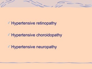
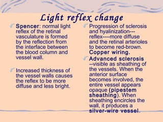
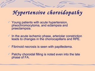
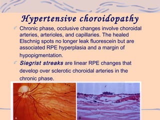
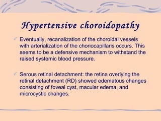
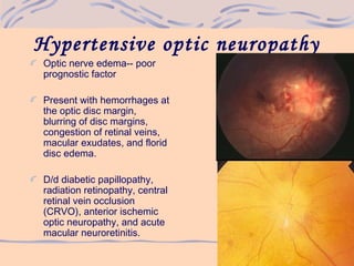
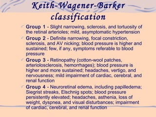
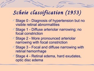
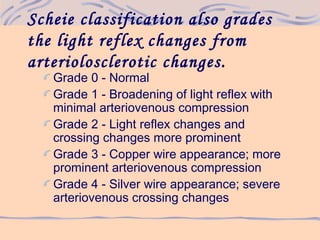
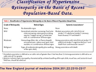
1) Hypertensive retinopathy involves vasoconstrictive and vasospastic responses in the retinal arterioles that can be classified into increasing stages of severity based on observed retinal changes.
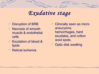
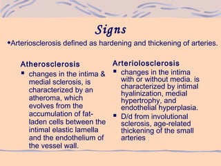
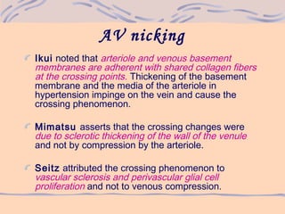
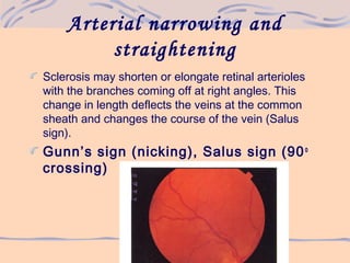
2) As hypertension progresses, it can cause narrowing and sclerosis of retinal arterioles, hemorrhages, exudates, microaneurysms, and cotton wool spots in more severe cases.
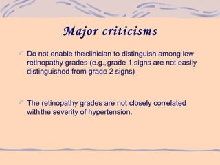
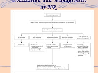
3) Evaluation of hypertensive retinopathy provides insight into the severity of hypertension and risk for end organ damage, with more advanced changes indicating poorly controlled blood pressure that threatens cardiac, cerebral and renal function.
4. Vasoconstructive stage
Vasospasm & increase
in retinal arteriolar tone
Clinically seen as
generalized narrowing
of the retinal arterioles.
Intimal thickening,
hyperplasia of the
medial wall & hyaline
degeneration in
sclerotic stage
Clinically more severe
generalized & focal
areas of arteriolar
narrowing
Changes in A/V junction
Alteration in arteriolar
light reflex
5. Exudative stage
Disruption of BRB
Necrosis of smooth
muscle & endothelial
cells
Exudation of blood &
lipids
Retinal ischemia
Clinically seen as micro
aneurysms,
hemorrhages, hard
exudates, and cotton
wool spots.
Optic disk swelling
8. Signs
Atherosclerosis
changes in the intima &
medial sclerosis, is
characterized by an
atheroma, which
evolves from the
accumulation of fat-
laden cells between the
intimal elastic lamella
and the endothelium of
the vessel wall.
Arteriolosclerosis
changes in the intima
with or without media. is
characterized by intimal
hyalinization, medial
hypertrophy, and
endothelial hyperplasia.
D/d from involutional
sclerosis, age-related
thickening of the small
arteries
•Arteriosclerosis defined as hardening and thickening of arteries.
9. Light reflex change
Spencer: normal light
reflex of the retinal
vasculature is formed
by the reflection from
the interface between
the blood column and
vessel wall.
Increased thickness of
the vessel walls causes
the reflex to be more
diffuse and less bright.
Progression of sclerosis
and hyalinization---
reflex----more diffuse
and the retinal arterioles
to become red-brown.
Copper wiring.
Advanced sclerosis
--visible as sheathing of
the vessels. When the
anterior surface
becomes involved, the
entire vessel appears
opaque (pipestem
sheathing). When
sheathing encircles the
wall, it produces a
silver-wire vessel.
10.
11.
12. AV nicking
Ikui noted that arteriole and venous basement
membranes are adherent with shared collagen fibers
at the crossing points. Thickening of the basement
membrane and the media of the arteriole in
hypertension impinge on the vein and cause the
crossing phenomenon.
Mimatsu asserts that the crossing changes were
due to sclerotic thickening of the wall of the venule
and not by compression by the arteriole.
Seitz attributed the crossing phenomenon to
vascular sclerosis and perivascular glial cell
proliferation and not to venous compression.
13. Arterial narrowing and
straightening
Sclerosis may shorten or elongate retinal arterioles
with the branches coming off at right angles. This
change in length deflects the veins at the common
sheath and changes the course of the vein (Salus
sign).
Gunn’s sign (nicking), Salus sign (90o
crossing)
14. Extra vascular retinal lesions
Microaneurysms
Retinal hemorrhages--
Streak hemorrhages located
in the nerve fiber layer
predominate over the blot
hemorrhages located deeper
in the outer plexiform layer.
Cotton wool spots
Retinal and macular
edema
Retinal lipid deposits
macular star is the most
predominant appearance,
and this appearance is due
to the radially oriented nerve
fiber layer of Henle.
15. Focal intraretinal
periarteriolar transudate
(FIPT)
Coined by Hayreh
The earliest retinal lesions seen in accelerated
malignant hypertension.
These appear as round or oval lesions located in the
deeper retinal layers next to major retinal arterioles
and their major branches.
FA appearance may precede ophthalmoscopic
recognition.
On resolution, no residual lesions are noted.
The proposed mechanism is focal accumulation of
plasmatic molecules in retinal tissue due to
autoregulation failure.
16. Malignant hypertension
Associated with an
idiopathic acute blood
pressure elevation
H/O HTexists,
especially of a
nephrogenic etiology
Most common in
younger adults
Toxemia of pregnancy,
renal disease, and
collagen vascular
disease
17. Malignant hypertension
Necrosis and fibrinoid
deposition in the vessel
wall occurs, more
commonly involving the
choroidal arteries than
retinal arteries.
Retinal edema occurs
from breakdown of the
BOB at the level of the
RPE.
The edema fluid may
have a fibrinous
appearance.
18. Hypertensive choroidopathy
Young patients with acute hypertension,
pheochromocytoma, and eclampsia and
preeclampsia.
In the acute ischemic phase, arteriolar constriction
leads to changes in the choriocapillaris and RPE.
Fibrinoid necrosis is seen with papilledema.
Patchy choroidal filling is noted even into the late
phase of FA.
19. Hypertensive choroidopathy
Acute focal RPE lesions overlying the involved area
are seen as pale pinpoint lesions distributed in
groups. These are called Elschnig spots and leak
fluorescein profusely.
These lesions are distinguished from Hayreh's FIPTs,
since they stain less intensely and for shorter periods
of time, in addition to their subretinal location.
20. Hypertensive choroidopathy
Chronic phase, occlusive changes involve choroidal
arteries, arterioles, and capillaries. The healed
Elschnig spots no longer leak fluorescein but are
associated RPE hyperplasia and a margin of
hypopigmentation.
Siegrist streaks are linear RPE changes that
develop over sclerotic choroidal arteries in the
chronic phase.
21. Hypertensive choroidopathy
Eventually, recanalization of the choroidal vessels
with arterialization of the choriocapillaris occurs. This
seems to be a defensive mechanism to withstand the
raised systemic blood pressure.
Serous retinal detachment: the retina overlying the
retinal detachment (RD) showed edematous changes
consisting of foveal cyst, macular edema, and
microcystic changes.
22. Hypertensive optic neuropathy
Optic nerve edema-- poor
prognostic factor
Present with hemorrhages at
the optic disc margin,
blurring of disc margins,
congestion of retinal veins,
macular exudates, and florid
disc edema.
D/d diabetic papillopathy,
radiation retinopathy, central
retinal vein occlusion
(CRVO), anterior ischemic
optic neuropathy, and acute
macular neuroretinitis.
23. Keith-Wagener-Barker
classification
Group 1 - Slight narrowing, sclerosis, and tortuosity of
the retinal arterioles; mild, asymptomatic hypertension
Group 2 - Definite narrowing, focal constriction,
sclerosis, and AV nicking; blood pressure is higher and
sustained; few, if any, symptoms referable to blood
pressure
Group 3 - Retinopathy (cotton-wool patches,
arteriolosclerosis, hemorrhages); blood pressure is
higher and more sustained; headaches, vertigo, and
nervousness; mild impairment of cardiac, cerebral, and
renal function
Group 4 - Neuroretinal edema, including papilledema;
Siegrist streaks, Elschnig spots; blood pressure
persistently elevated; headaches, asthenia, loss of
weight, dyspnea, and visual disturbances; impairment
of cardiac, cerebral, and renal function
24. Scheie classification (1953)
Stage 0 - Diagnosis of hypertension but no
visible retinal abnormalities
Stage 1 - Diffuse arteriolar narrowing; no
focal constriction
Stage 2 - More pronounced arteriolar
narrowing with focal constriction
Stage 3 - Focal and diffuse narrowing with
retinal hemorrhage
Stage 4 - Retinal edema, hard exudates,
optic disc edema
25. Scheie classification also grades
the light reflex changes from
arteriolosclerotic changes.
Grade 0 - Normal
Grade 1 - Broadening of light reflex with
minimal arteriovenous compression
Grade 2 - Light reflex changes and
crossing changes more prominent
Grade 3 - Copper wire appearance; more
prominent arteriovenous compression
Grade 4 - Silver wire appearance; severe
arteriovenous crossing changes
27. Major criticisms
Do not enable theclinician to distinguish among low
retinopathy grades (e.g.,grade 1 signs are not easily
distinguished from grade 2 signs)
The retinopathy grades are not closely correlated
withthe severity of hypertension.
31. Points To Remember
For CWS to develop from hypertension,
autoregulatory mechanisms must first be overcome.
For this to happen, the patient must have at least
110mmHg diastolic readings.
Patients who develop papilledema from hypertension
have malignant hypertension and typically have BP in
the range of 250/150mmHg
32. Points To Remember
Generalized retinal arteriolar narrowing &
arteriovenous nicking are markers of vascular
damage from chronic hypertension
Focal arteriolar narrowing, retinal hemorrhages,
micro aneurysms & cotton wool spots are more
indicative of severity of recent hypertension.
33. Points To Remember
Fluorescein angiography is not indicated in cases of
hypertensive retinopathy as it yields no diagnostic
information.
Hypertensive retinopathy presents with a ‘dry’ retina
(few hemorrhages, rare edema, rare exudate, and
multiple cotton wool spots) whereas diabetic
retinopathy, in comparison, presents with a ‘wet’
retina (multiple hemorrhage, multiple exudate,
extensive edema, and few cotton wool spots).
34. Future Research
A development of photographic classification system
similar to the photographic grading of diabetic
retinopathy.
Prospective studies are needed that can demonstrate
independent associations of HR with cardiovascular
outcomes.
• To compare the relative value of a retinal assessment
(based on ophthalmoscope examination performed
with or without the use of photography) with other
strategies of risk stratification (eg use of ECG, Echo
cardiography)
• Need to evaluate whether specific therapy that is
focused on the retinal microcirculation can reverse
changes in retinopathy and also ultimately result in
reduce cardiovascular risk.