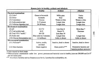
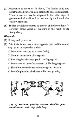
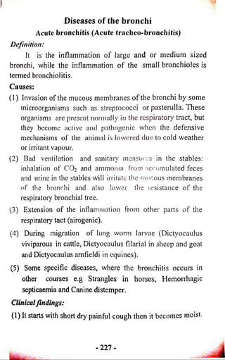
This document provides an overview of general systemic disturbances in farm animals, including dehydration, overhydration, and electrolyte imbalances. It discusses the physiology of body fluids and water, causes and pathogenesis of dehydration, clinical symptoms, diagnosis, and treatment. It also covers sodium imbalances like hypernatremia and hyponatremia, their causes and effects. The document contains detailed information on disturbances to body fluids and electrolytes.

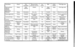
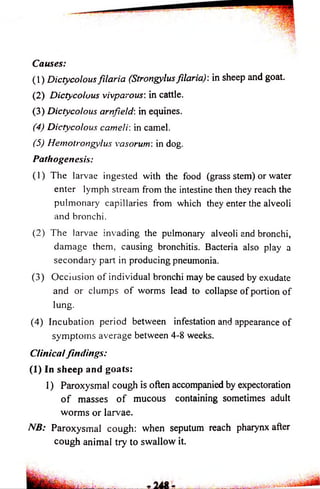
![Disturbances o f electrolytes
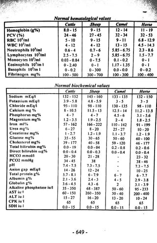
(1) The normal blood serum levels of Na+; K+ & CL" are 132-
152; 95-110 & 4-6 m.Eq/1, respectively.
(2) They are the most important electrolytes in the body. They
affected by certain diseases & body fluid disturbances.
(3) Sodium is the most abundant ion in ECF. It is responsible
for maintenance of ECF osmotic pressure.
Physiology of Sodium: Total body Na+ losses activate the
CNS to alter renal Na+ handling resulting in a decrease in renal
Na+ excretion. An increase in Na+ content in the ECF results in
changes opposite to those seen with Na+ loss.
[1] Sodium excess (Hypernatremia)
It is an elevation of sodium level in serum more than
normal. It arises from:
(1) Excessive IV saline solution or oral Na+ or salt poisoning
(2) Pure water loss or water deprivation.
[2] Sodium deficiency (Hyponatremia)
It is a decrease of Na+ level in serum or blood.
Causes:
(1) Excessive IV fluid therapy free from Na+.
(2) Low intake of Na+ with diuretics.
(3) Loss of sodium containing fluid in cases of diarrhea,
enteritis, excessive sweating & blood loss.
Pathogenesis:
Na+ depletion leads to increase renal losses of water to
maintain normal osmotic pressure resulting in withdraw of
- 11 -](https://image.slidesharecdn.com/vet-141021053329-conversion-gate02/85/Vet-internal-medicine-text-book-14-320.jpg)

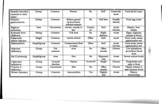
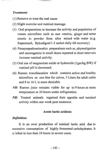
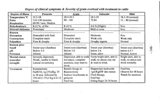
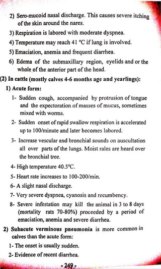
![Treatment:
By isotonic or hypertonic sodium (5%) solution. The
amount of m.Eq. of Na+ required in fluid therapy = serum Na+
level (Normal - Dehydrated) X BW / kg divided by 3.
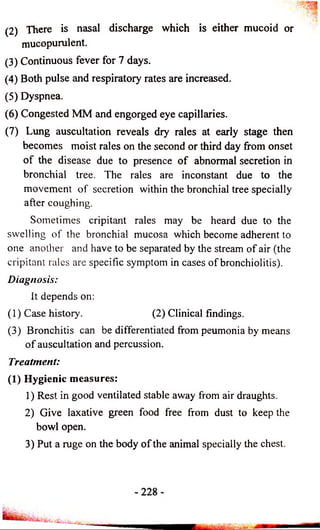
Physiology of potassium (K+): It is the main ICF cation. It
has an important role for potential difference of cell membrane
& excitation of cell so that excess or less K+ depress the heart
muscle, so it must not be injected IV.
[1] Potassium deficiency (Hypokalemia)
It is a decrease of serum K+ level.
Causes:
(1) Low K+ in diet (rare in grazing animal).
(2) Prolonged use of fluid therapy free from K+.
(3) Increase K+ excretion & loss by:
1) Excessive & rapid bicarbonate Injection.
2) Insulin & or glucose injection.
3) Injection of Na salt, high level of ACTH, aldosterone,
adreno-cortical hormones.
4) Vomiting or diarrhea. 5) Excessive sweating.
6 ) Prolonged anorexia. 7) Metabolic alkalosis.
(4) Acute gastric, abomasal or intestinal dilatation, impaction,
obstruction or torsion leading to excessive K+ loss and
excretion.
Symptoms:
K+ depletion is characterized by progressive muscular
weakness & decrease excitability of nerve:
- 13-](https://image.slidesharecdn.com/vet-141021053329-conversion-gate02/85/Vet-internal-medicine-text-book-16-320.jpg)
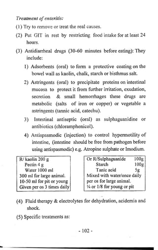
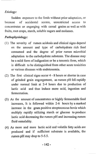
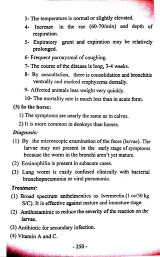
![(1) Weakness of myocardium lead to dilatation, arrhythmia,
heart failure
(2) Weakness of respiratory muscles lead to cyanosis, dyspnea,
and respiratory failure.
(3) Weakness of intestinal muscles leads to atony &
distension.
(4) Alkalosis occurs due to exchange of K+ (from urine to
blood) for EC (from blood to urine) in the renal tubular
fluid.
(5) Excessive K+ excretion in GIT lead to failure of gastric H+
& CL' to be reabsorbed by small intestine resulting in
hypokalemia, hypochloremia & metabolic alkalosis due
to retention of H* compensated by excessive K+
excretion.
Treatment:
(1) Remove causes.
(2) Oral K+ salt or fruit or milk or meat.
(3) IV injection of K+ may be very dangerous.
(4) IV saline solution to treat hypokalemia, hypochloremia &
alkalosis.
[2] Hyperkalemia
It is an elevation of serum K+ level than normal.
Causes:
(1) Increase K+ intake.
(2) Metabolic acidosis.
(3) Rapid infusion of K+ salts.
(4) Decrease renal elimination, oliguric acute renal failure, urethral
obstruction or terminal stages of chronic renal failure.
- 14 -](https://image.slidesharecdn.com/vet-141021053329-conversion-gate02/85/Vet-internal-medicine-text-book-17-320.jpg)
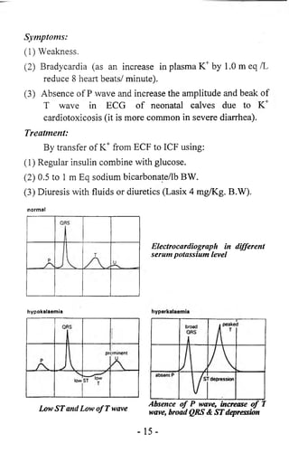
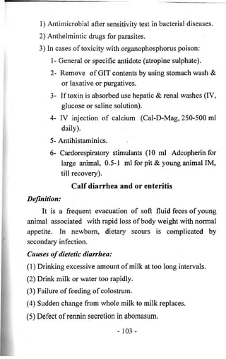
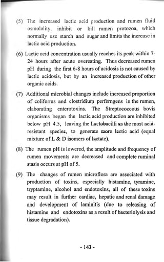
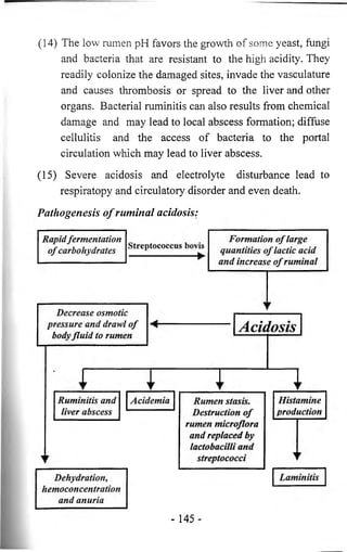
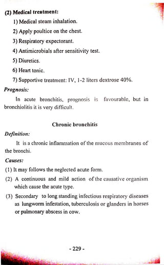
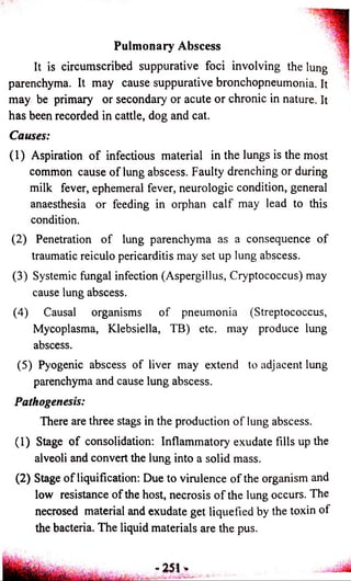
![Acidosis
Acidemia means shift of arterial blood pH toward the
acidity.
Acidosis refers to any acidotic process of body
(Formation, excretion and other pathogenesis even acidemia).
Causes:
[1] Metabolic acidosis :It occurs in cases of:
(1) Excessive loss of bicarbonate in enteritis, renal failure, urea
toxicity & damage of proximal tubules (less renal HCO3’
absorption).
(2) Excessive bicarbonate free fluids therapy in alkalosis.
(3) Over production of organic acids in:
1) Over feeding with carbohydrates.
2) Starvation, ketosis and pregnancy toxemia.
3) Colic with strangulated bowel.
4) Strangulated abomasal torsion.
5) Dehydration, bums, fever and shock.
6 ) Liver cirrhosis and hepatitis.
7) Abnormal gut flora in cases of diarrhea and GIT
disorders (Lactobacillus, etc).
8 ) Anaerobic tissue oxidation in dehydration and hypovolemia.
[2] Respiratory acidosis: It occurs in cases of:
(1) Depression of respiratory center.
(2) Defect in respiratory system that lead to poor pulmonary
ventilation & elevating C02, in cases of pneumonia,
pneumothorax, pleurisy, pulmonary edema, asphyxia Sc
severe emphysema.
- 18-](https://image.slidesharecdn.com/vet-141021053329-conversion-gate02/85/Vet-internal-medicine-text-book-21-320.jpg)
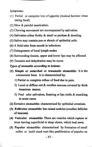
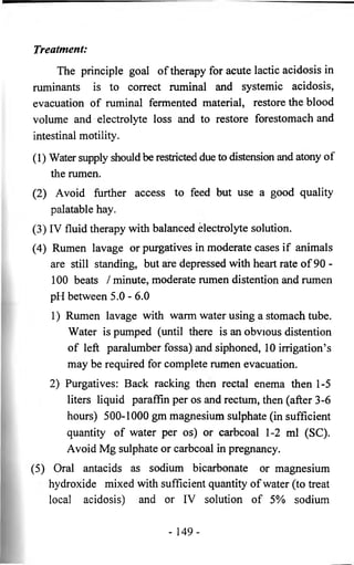
![Treatment:
(1) Complete rest, remove & treat the real cause.
(2) Antacid: By IV and or oral sodium bicarbonate (or acetate
or lactate) not more than lg/kg BW/day or by IV Ringer
lactate solution.
e.g. The bicarbonate deficit in horse with acute diarrhea &
acidosis = BW (kg) x 0.3 x (Normal level - observed
plasma bicarbonate) = 500 (Kg BW) x 0.3 x [26
(normal serum bicarbonate level in m Eq/liter) - 12
(Bicarbonate level in acidosis)]= 500 x 0.3 x 14 =
2100 m Eq/ Liter =2100 divided by 12= 175 gm Na H
C 03 (as 1 gm Na H C 0 3 yields 12 mEq HCO3 ).
(3) Saline solution to correct hyponatremia and hyperkalemia.
(4) In respiratory acidosis: Treat respiratory disorders firstly,
also use bronchodilator (Carbonic anhydrase inhibitor,
Diamox).
(5) In chronic respiratory acidosis use 40% 0 2 or use ventilator
in respiratory failure.
Required amount of sodium bicarbonate in g per 24 hrs given IV
in concentration of 2.6-5 or even 8.4% for calf40-50 kg BW.
Acidosis Na.bicarb. Calf state
Simple 5 g 1 -Can not sucking but can stand
Mild 10 g 1-No sucking 2- Need assistance to stand
Moderate 20 g Sternal recumbency +1, 2 (previous)
Severe 30 g 3- lateral recumbency + 1,2
4-Coldness o f extremities
Hyper
Severe
40 g 1,2,3,4 + opened mouth, dropped tongue, cardiac
arrythemia, hypoventilation, coma, then death.
-21 -](https://image.slidesharecdn.com/vet-141021053329-conversion-gate02/85/Vet-internal-medicine-text-book-24-320.jpg)

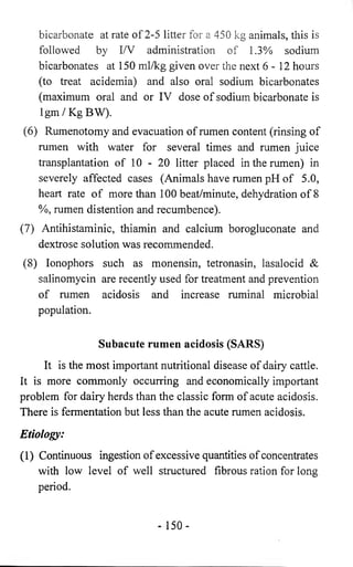
![Shock
Definition:
It is an inadequate blood flow to vital organs or even
body tissue causing inadequate tissue perfusion to meet the
nutritional requirements of the cells & remove the waste
product of metabolism.
Clinical signs:
Includes hypotension, weak pulse, tachycardia, cold
extremities, depression, pale mucous membranes, increase
capillary refill time and fever (in early stages of septic shock).
Types o f shock:
[1] Hypovolemic shock :It is a pathological decrease in
capillary perfusion due to decrease of blood volume.
Causes:
(1) Blood loss in cases of severe injury & hemorrhage.
(2) Plasma loss in cases of burns & or sepsis.
(3) Body water & electrolyte loss in case of dehydration.
Symptoms:
(1) Mild shock (up to 20% blood volume loss): It is a decrease
in perfusion of non vital organs & tissues (skin, fat,
skeletal muscle & bone). It is manifested by a pale & cool
skin.
(2) Moderate shock (20-40% blood volume loss): It is a
decrease in perfusion of vital organs (as liver, gut and
kidneys). It is manifested by oliguria to anuria & drop in
blood pressure.
- 43 -](https://image.slidesharecdn.com/vet-141021053329-conversion-gate02/85/Vet-internal-medicine-text-book-46-320.jpg)
![(3) Severe shock (40% or more blood volume loss): It is a
decrease in perfusions of heart & brain. It is manifested by
restlessness, agitation, coma, cardiac irregularities, ECG
abnormalities & cardiac arrest.
Treatment:
Shock is an acute emergency:
( 1 ) Complete rest in an airway.
(2) Try to treat & remove the real cause.
(3) IV fluid to restore blood volume by one or more of the
following:
1 ) Crystalloid as iso or hypertonic sodium chloride in
balanced salt solution (Ringer’s lactate or Ringer’s
acetate) with strictly use of sodium bicarbonate in
acidosis (to avoid alkalosis)
2) Colloids including whole blood & albumin:
1- Use bloods if shock persists after infusion two
liters of crystalloid.
2- Albumin solutions (25-50 gm in liter saline or
Ringer) in prolonged severe shock.
NB: Measuring of arterial blood pressure, central venous
pressure, pulse rate & hematocrit are essential for
diagnosis &prognosis.
NB: Prognosis is good by an increase in cardiac output, blood
pressure, urine flow & decrease in pulse rate.
[2] Septic shock: It is a failure of cells of vital organs to
oxygen utilization & to perform normal metabolic
function despite availability of oxygen.
- 4 4 -](https://image.slidesharecdn.com/vet-141021053329-conversion-gate02/85/Vet-internal-medicine-text-book-47-320.jpg)
![Causes:
(1) Gram negative septicemia: mainly from gastrointestinal or
genitourinary tract that increase capillary permeability &
increase fluid loss from vascular space producing
hypovolemia as well as direct toxic effect on heart with
depression of myocardial function
(2) Gram positive septicemia: it is less severe than G. negative.
It is limited to area of infection.
Symptoms:
(1) Infection, confusion & restlessness.
(2) Skin and exterimities are dry and warm.
(3) Increase heart beats.
(4) Pulmonary hypertension & hyperventilation.
(5) Urine output is normal at first, then it slows rapidly.
Treatment:
(1) Volume replacement: Balance salt solutions.
(2) Specific antibiotics & sulfonamides after sensitivity test or
mixing 5,000,000IU crystaline penicilline and 2-5mg/lb
dexamethasone with IV fluid.
(3) Surgical drainage for abscess or the focus of infection.
(4) Supportive measures for heart & lung.
[3] Neurogenic shock: It occurs due to a failure of arterial
resistance from nervous or psychic stimulation (sudden
pain or fright), vasodilator drugs (nitrites), spinal
anesthesia or spinal trauma.
- 45 -](https://image.slidesharecdn.com/vet-141021053329-conversion-gate02/85/Vet-internal-medicine-text-book-48-320.jpg)
![Symptoms:
(1) Dyspnea.
(3) Cardiac arrythemia.
(2) Pulmonary rales.
(4) Myocardial infarction.
(5) Later on hypovolemia
Treatment:
By correcting central venous pressure.
[5] Anaphylaxis&anaphylactic shock :It is a server condition
of acute antibody antigen reactions
Causes:
(1) Repeat blood transfusions from same donor.
(2) Repeat injection of same vaccine as FMD & Rabies
vaccine.
(3) After first injection of some drug as penicillin.
(4) In certain cattle, sudden stop of milk secretion.
(5) Killing or death of subcutaneous hypodermal larvae.
(6 ) Ingestion of certain protein at pasture or feedlot.
(7) Sensitivity to a protein substance entering the blood stream
& a second exposure to the same substance.
Pathogenesis:
Antigen and antibodies (Neutrophil, basophil, mast cells
or some specific tissues or cells) causing liberation of
anaphylactic mediators such as histamine, serotonine and
catecholamines resulting in:
(1) Systemic hypotension.
(2) Pulmonary congestion & edema then pulmonary hypertension.
- 4 7 -](https://image.slidesharecdn.com/vet-141021053329-conversion-gate02/85/Vet-internal-medicine-text-book-50-320.jpg)
![[6 ] Other types of shock:
(1) Electrical shock.
(2) Emotional shock
(3) Traumatic shock after severe trauma.
(4) Obstructive shock: It is a cardiogenic cause involves
obstruction of blood flow as severe heart worm disease,
aortic thromboembolism & tumor.
- 4 9 -](https://image.slidesharecdn.com/vet-141021053329-conversion-gate02/85/Vet-internal-medicine-text-book-52-320.jpg)
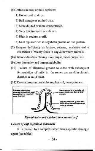
![e.g. defect in lactase activity in neonates resulting in
indigested lactose leading to hyperosmotic effect followed
by diarrhea .
(3) Digestive dysfunction:
It depends on motor and secretary function and, in
herbivores, on activity of the microflora which inhibit the
forestomachs of ruminants, or cecum and colon of equines.
1) Microbial digestion that digest carbohydrate & cellulose
in fore stomach of ruminant to volatile fatty acids &
convert nitrogenous substance to ammonia & protein.
2) Ruminal protozoa, bacteria, yeast may affected by oral
drugs & feeding (as acidosis or alkalosis).
(4) Absorptive dysfunction:
It is an increase irritability of GIT mucous will increase
its motility & passage of lumen content so less affected with
gut enzyme led to maldigestion & malabsorption.
(5) Autointoxication:
Toxic amines, phenols & cresols produced by
putrefaction of protein in the large intestine but normally
detoxified in the bowel wall could, if regurgitated into the
small intestine, be absorbed and cause depression, anorexia &
weakness.
Manifestation o f alimentary tract dysfunction:
[1] Abnormal prehension: It may be interfere with:
(1) Paralysis of muscles of jaw or tongue.
(2) Defect of incisor teeth or jaws.
(3) Stomatitis & oral foreign bodies.
-51 -](https://image.slidesharecdn.com/vet-141021053329-conversion-gate02/85/Vet-internal-medicine-text-book-54-320.jpg)
![(4) Congenital abnormalities of teeth, gum, tongue, lips or
jaws. In all cases not in systemic disease, the animal is
hungry & attempts to feed but can not do so.
(5) Lockjaw in tetanus.
[2] Abnormal mastication: It may be:
(1) Slow jaw movements in cases of bad teeth
(2) Painful mastication in cases of painful stomatitis (complete
refusal to chew).
(3) Incompleted mastication with drop of food from mouth
during mastication in cases of sharp teeth.
(4) Passage of food particles in feces in cases of bad teeth.
[3] Abnormal swallowing: It may be from:
(1) Dysphagia (it means a difficulty in swallowing): It
manifested by forceful attempts to swallow, accompanied
by extension of the head at first, followed by forceful
flexion & violent contraction of the muscles of neck &
abdomen.
(2) Defect in nervous control of the reflex or a narrowing of
the lumen of the pharynx or esophagus may interfere with
swallowing.
(3) Lesions in the pharynx cause regurgitation through the
nostril or coughing up of the material.
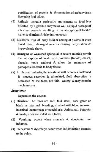
[4j Diarrhea :It is a frequent evacuation of soft feces, more in
bulky than normal. It occurs in enteritis, incomplete
digestion with passage of excessive fiber.
[5] Constipation: It is a dry, hard & of small bulk feces which
are passed at infrequent intervals. It occurs when the
period of food passage through GIT is prolonged &
52](https://image.slidesharecdn.com/vet-141021053329-conversion-gate02/85/Vet-internal-medicine-text-book-55-320.jpg)
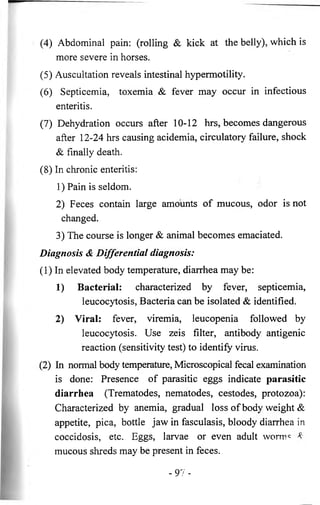
![intestinal activity is reduced. Constipation may cause
severe debility, dehydration, painful conditions in anus &
paralytic ileus (loss of intestinal movement).
NB: The food journey in the gut is 12-35 hours (ruminants), 1-
4 days (horses) & 2-4 days (dog, cat).
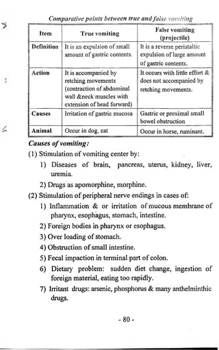
[6] Vomiting :It is a protective mechanism in the form of
reverse peristaltic movement with the function of
removing excessive quantities of ingesta or toxic materials
from stomach. It is not common in animals. Vomiting
occurs in two forms: as true or projectile vomiting.
[7] Alimentary tract hemorrhage: It occurs in cases of:
( 1 ) Stomach or intestinal ulceration with erosion of blood
vessels.
(2) Severe hemorrhagic enteritis.
(3) Structural lesions of intestinal wall as tumor .
(4) Infestation with helminthes(Blood sucking nematodes as
Bunostomiasis) or protozoa (Coccidia or Cryptosporidia).
(5) Local vascular engorgement or obstruction as in
intussusception, verminous thrombosis.
Alimentary tract hemorrhage
Blood origin Character of blood & GIT contents
Stomach Vomits are dark brow color like coffee grounds
due to formation o f acid haematin, feces are black
or very black brown tarry appearance (Melena).
Small intestine Feces are brown black
Colon or caecum Blood is unchanged giving red colored feces
Lower colon or
Clots o f whole blood are voided
rectum
- 5 3 -](https://image.slidesharecdn.com/vet-141021053329-conversion-gate02/85/Vet-internal-medicine-text-book-56-320.jpg)
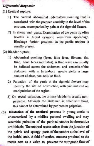
![[8 ] Abdominal pain: Different degree of abdominal pain
r occurs (Severity of pain varies with the nature of the
lesions). The pain varies with the species but pain
associated with diseases of abdominal viscera causes
similar signs irrespective of the organ involved:
(1) Acute pain of horses occurs in cases of colic including
gastric dilatation, intestinal obstruction, enteritis and colitis.
(2) Subacute pain of horses in cases of thromboembolic colic,
impaction of large intestine and ileal hypertrophy.
(3) Acute pain of cattle in cases of intestinal obstruction and
poisoning.
(4) Subacute pain of cattle occurs in cases of traumatic
reticuloperitonitis and general peritonitis.
[9] Grunting which may be
(1) Expiratory caused by pulmonary or thoracic diseases
including severe pulmonary emphysema, severe
pneumonia, pleuritis and or severe hydropericardium
(2) Non expiratory caused by:
1) Peritonitis (acute local or diffuse or chronic).
2) Distended organ includes bloat, abomasum, omasum,
severe hepatomegaly and or acute intestinal
obstruction.
3) Severe pain in genitourinary tract (vaginitis, vulvitis
and urolithiasis).
[10] Dehydration & shock: Dehydration is common in acute
diseases but shock may result from hyperacute one .The
acute rapid distension of stomach & or intestine may
- 5 4 -](https://image.slidesharecdn.com/vet-141021053329-conversion-gate02/85/Vet-internal-medicine-text-book-57-320.jpg)


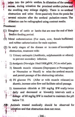
![Affection o f the stomach
The stomach is the first area affected by irritating
materials which are ingested, so the case history is the most
important part for a suspected gastric disorders. Affection of
the stomach includes:
[1] Vomiting (Emesis). [2] Gastric ulcer.
[3] Gastritis
Vomiting (Emesis)
Definition:
It is a forcible expulsion of stomach contents through
nose or mouth. It is not a disease but a symptom.
It is controlled by vomiting center in the medulla
oblongata (M.O.).
Vomiting may be:
(1) Central vomiting due to direct stimulation of the vomiting
center in M.O. due to diseases of the brain, uremia,
apomorphine poisoning.
(2) Reflex or indirect vomiting due to irritation in various
organs as esophagus, stomach, intestine, kidney, uterus.
1) The horse & ruminant seldom vomit. It occurs with
great difficulty & always with very grave or even fatal
signs.
2) Vomitus flows through the nose in horse (due to
elongated soft palate), through the mouth in other
species.](https://image.slidesharecdn.com/vet-141021053329-conversion-gate02/85/Vet-internal-medicine-text-book-82-320.jpg)
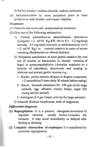

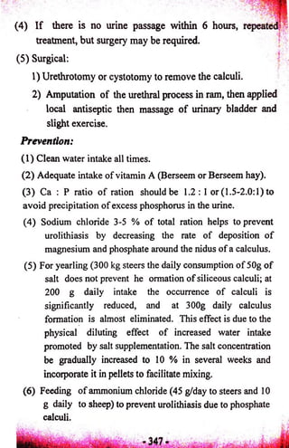
![Colic due to impaction of the intestine
It occurs when the large intestine remains impacted with
undigested food material causing partial obstruction, colic,
depression & anorexia.
Etiology:
(1) Overfeeding of grains or coarse food rich in cellulose and
bran for long period.
(2) Ingesting food materials, which contain large amounts of
mud or sand.
(3) Sluggish intestinal peristalsis especially in old debilitated
& or draught horses.
(4) Greedy feeding & defective teeth.
(5) Obstruction of the intestine or natural opening by large
foreign bodies or parasite.
(6) Inadequate water intake or green food.
(7) Enterolith, fiber balls, hairball.
(8) Encephalitic (equine rectal paralysis).
Colic may be due to:
(1) Impaction of the small intestine.
(2) Impaction of the colon.
[1] Impaction of the small intestine:
It occurs due to accumulation of sand in the small
intestine (sandy colic) or large number of parasites (Ascaris).
Symptoms:
(1) The symptoms varies according to the location of
impaction, when the duodenum is affected, the symptoms
occurs after feeding within few hours, when the ileum is
affected symptoms appear after several hours.
- 123 -](https://image.slidesharecdn.com/vet-141021053329-conversion-gate02/85/Vet-internal-medicine-text-book-126-320.jpg)
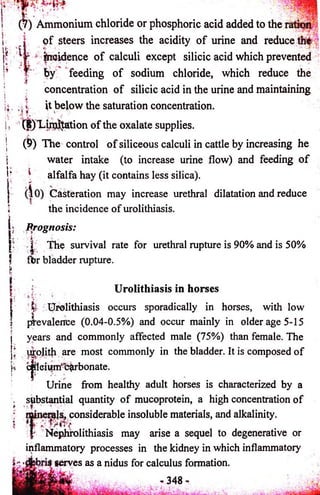
![(2) Restlessness and beats the ground with the forelimbs.
(3) In severe attacks the animal lies on the ground and rolls
with quick pulse & continuous pain during attack.
(4) During urination the animal throw the hindlegs more
backward and outward and urine comes out at intervals.
[2]: Impaction of the colon:
It is due to accumulation of undigested materials in the
colon. The large colon is the most common seat of impaction
in horse.
Symptoms:
(1) Subacute colic which occurs slowly, started with dullness
and abdominal discomfort, the animal looks at the flank
and kicking its belly.
(2) Constipation, Feces are passed in small amounts, hard in
consistency and covered with thick & sticky mucous.
(3) Intestinal sound are absent or much decreased in intensity.
(4) Moderate increase in pulse but the temperature &
respiration are normal.
(5) Rectal palpation revealed balloon shape colon impacted
with fecal mass.
(6) Dehydration, thirst, anorexia occur with constant effort to
urinate.
Diagnosis and differential diagnosis:
(1) In cases of sandy colic:
1) Sand is present in feces.
2) Sedimentation of sand in the feces.
- 124-](https://image.slidesharecdn.com/vet-141021053329-conversion-gate02/85/Vet-internal-medicine-text-book-127-320.jpg)

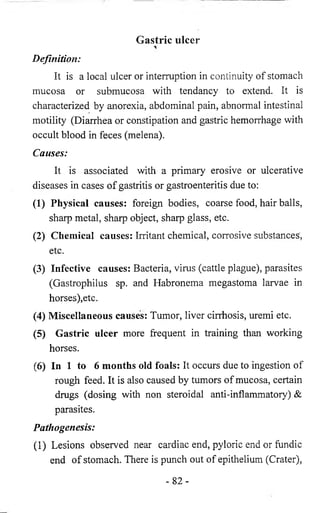
![Acute intestinal obstruction (specific colic)
[1] Embolic colic (Verminous aneurism):
It is disorder in the intestine due to the presence of larvae
of strongylus vulgaris in the anterior mesenteric artery of the
horse, causing aneurisms, emboli and thrombi of the
mesenteric artery and its branches.
It is characterized by intermittent and attacks of colic
which occurs suddenly during work as in spasmodic colic,
beside that the feces are bloody ¶sitic eggs are present in
fecal examination. Atoxyl solution 3 % or anthelminitic may
be used in treatment.
[2] Intestinal torsion (Volvulus) or Gut tie or Twist of the
intestine:
Volvulus is an intestinal obstruction due to rotation of
segment of the intestine around its mesenteric axis. It is either
partial or complete. Volvulus is common in small intestine
while torsion is common in large intestine in which the bowel
twists on its own or long axis.
Causes:
(1) Severe attack of colic, violent movements, rolling, jumping
or sudden fall of the animal during colic.
(2) Injections of large dose of carbacoal which lead to sudden
increase in the peristaltic movements.
(3) Heavy infestation with parasite (Ascaris) cause irregularity
in peristaltic movement of the intestine resulted in
torsion.
- 126-](https://image.slidesharecdn.com/vet-141021053329-conversion-gate02/85/Vet-internal-medicine-text-book-129-320.jpg)
![Treatment:
(1) Sedative.
(2) In partial torsion, give large doses of liquid paraffin or lin
seed oil & make rectal enema.
(3) Surgical removal of obstruction.
Prognosis:
(1) Complete torsion is unfavorable (death within 12-24
hours).
(2) In partial twist, the course & prognosis depends upon the
severity of case & cause.
[3] Intestinal strangulation:
It is the occlusion of the intestinal lumen by pressure
from out side. It occurs when a lope of the intestine passes
through a natural or artificial opening in the peritoneum and
held there as in case of inguinal hernia in stallion. Also in case
of pedunculated tumour which cause strangulation.
Symptoms: as torsion.
Treatment: Surgical.
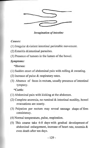
[4] Invagination (Intussusception, telescoping):
This is a form of acute intestinal obstruction caused by
telescoping of a section of the bowel into a portion
immediately behind it especially in ileocecal junction. The
affected part form a sausage shaped, painful swelling
composed of three segments (outer, middle & inner) as the
ileum is always invaginated into cecum or colon. It occurs
frequently in cattle & dog, seldom in sheep & horse.
- 128-](https://image.slidesharecdn.com/vet-141021053329-conversion-gate02/85/Vet-internal-medicine-text-book-131-320.jpg)
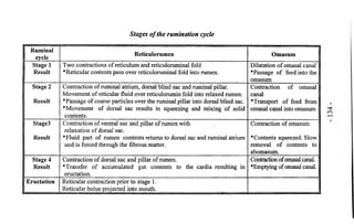
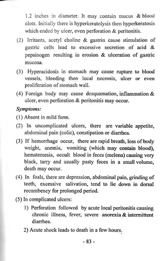
![Indigestion
It is a general term for a group of diseases characterized
by dysfunction of the reticulorumen, decrease its motility,
discontinuous grazing and abnormal feces. It is usually results
in anorexia, decrease in ruminal contraction, ruminal
distension, mild bloat, decrease milk production, sometimes
abdominal pain, diarrhea, recumbency and death.
The general causes o f rumen dysfunction:
(1) Inadequate quantity of feed
(2) Improper ratio of nutrient elements.
(3) Infrequent and irregular feeding.
(4) Too much feed.
(5) Sudden changes in feed.
(6) Infrequent and inadequate water intake.
(7) Spoilage or moldy feeds.
(8) Fever.
(9) Internal or external parasites.
(10) Prolonged or heavy oral dosing with sulpha drugs or
antibiotics.
Classification o f indigestion:
[1] Primary Indigestion:
(1) Reticuloruminal fermentative (Microbial /biochemical)
disorders.
1) Simple indigestion.
2) Acute rumen lactic acidosis.
3) Subacute rumen lactic acidosis.
4) Rumen alkalosis.
5) Chronic indigestion in calves.
- 135 -](https://image.slidesharecdn.com/vet-141021053329-conversion-gate02/85/Vet-internal-medicine-text-book-138-320.jpg)
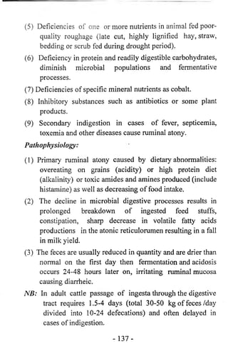
![(2) Reticuloruminal motor disorders/Diseases of the ruminal
wall.
1) Traumatic reticuloperitonitis.
2) Frothy bloat and free gas bloat.
3) Vagal indigestion.
[2] Secondary:
(1) Secondary reticuloruminal motor inactivity.
(2) Secondary reticuloruminal microflora inactivity
Reticuloruminal fermentative disorders
Simple indigestion
Definition:
It is the inability of animal to digest feed stuff due to an
abrupt change in the ration, where the rumen microflora are
not metabolically adapted with nutrient substrates or produce
inhibitory substances to decrease fermentation.
Etiology:
(1) Indigestible and damage foods include moldy or overheated
feeds, frosted forages and partly fermented spoiled or sour
silages. One or several animals on the same ration may
have signs.
(2) Indigestable substances as placenta, balls of hair or wool,
heavily contaminated roughage with sand, mud and/or
dust.
(3) Sudden change of food from green to dry.
(4) Less water intake especially during dry season.
- 136-](https://image.slidesharecdn.com/vet-141021053329-conversion-gate02/85/Vet-internal-medicine-text-book-139-320.jpg)
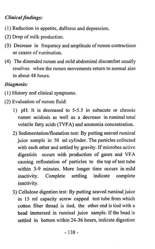
![Abomasum displacement and volvulus
Definition:
Abomasal displacement occurs either to the right or to the
left side of the abdomen when gas accumulates inside the
abomasum. Left displacement of the abomasum is most often
encountered (75%) than the right cases (25%). The highest
incidence in adult dairy cattle in the early postpartum period.
Etiology:
(1) Atony of the abomasum caused by an abnormally high
volatile fatty acid (VFA) concentration and continued
microbial fermentation of ingesta lead to gas
accumulation and resultant distention.
(2) Displaced abomasums was associated with nutrition-related
risk factors, use of minerals and sodium chloride,
inadequate concentrates feeding. Moreover hypocalcemia
with decreased abomasal smooth muscle tone may also
contribute to atony.
(3) Diets high in starch or deficient in roughage are commonly
associated with abomasal displacement.
[1] Left displacement of the abomasum (LDA):
Abomasal displacement occurs to the left side of the
abdomen, where the abomasum located between the rumen
and left abdominal wall.
Risk factors:
(1) Cows in early lactation are at greatest risk of developing
LDA, (occurred in the first 30 days after calving).
- 175 -](https://image.slidesharecdn.com/vet-141021053329-conversion-gate02/85/Vet-internal-medicine-text-book-178-320.jpg)
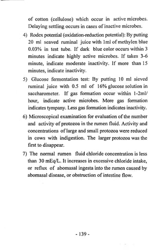
![[2] Right Displacement of the Abomasum (RDA):
Abomasal displacement occurs to the right side of the
abdomen, where the abomasum located between the liver and
right abdominal wall. RDA occurs at about 10% to 15% the
frequency of LDA. The predisposing causes, pathophysiologic
mechanisms, clinical pathologic conditions, and epidemiologic
characteristics are the same as LDA.
Clinical findings:
(1) The general systemic state of the cow with RDA is the
same as in LDA.
(2) An area of tympanitic resonant is heard on the right side
with auscultation and percussion.
(3) The condition must be differentiated from other causes of
right-sided pings, such as cecal distention (with or
without volvulus), gas in the spiral colon, pneumorectum
after rectal examination, pneumoperitoneum, physometra
(gas in the uterus), and abomasal volvulus.
(4) The ping usually is confined to an area under the last five
ribs in the upper half of the abdomen. Cecal and rectal
pings usually are detectable in a linear pattern just below
the transverse processes of the lumbar vertebrae
extending to the tuber coxae.
(5) The rectal examination identifies the gas-filled structure of
abomasums and the spiral colon may be palpated laterally
flattened, mildly distended. Abomasal volvulus in an
early case is the most difficult to differentiate from RDA.
With time the cow becomes progressively more
dehydrated and more severely ill with volvulus than is
usual with RDA. Later on a ping caused by the fluid level
in the abomasums occurs.
- 180-](https://image.slidesharecdn.com/vet-141021053329-conversion-gate02/85/Vet-internal-medicine-text-book-183-320.jpg)
![Treatment:
(1) The surgical treatment.
(2) Rolling for nonsurgical correction is contraindicated
because of the risk of creating abomasal volvulus from a
RDA.
(3) The prognosis for a successful recovery after surgery is
comparable to that for LDA.
[3] Abomasal volvulus (Right torsion of the abomasum):
Abomasal volvulus is a sporadic disease that proceeds by
RDA. Right torsion of the abomasum, (RTA), leads to
complete obstruction of the flow of ingesta through the
duodenum.
Pathphysiology:
(1) Risk factors predisposing to LDA or RDA probably
contribute to the pathogenesis of RTA. Whether true
RDA precedes RTA is not known.
(2) Dehydration and cardiovascular collapse occur in more
prolonged cases.
(3) Earlier cases have acid-base and electrolyte abnormalities
(hypochloremic, metabolic alkalosis, hypokalemia) as in
LDA but more marked
(4) In cases of severe distention of the abomasum and omasum
with vascular compromise, systemic cardiovascular
insufficiency develops.
(5) The rotation probably occurs most frequently at the
reticuloomasai junction.](https://image.slidesharecdn.com/vet-141021053329-conversion-gate02/85/Vet-internal-medicine-text-book-184-320.jpg)
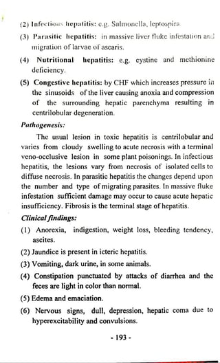
![Manifestations (Principles) o f liver dysfunction:
[I] Jaundice:
It is the most important clinical sign associated with liver
diseases, in which bile pigments accumulates in blood
(bilirubinemia) and then partly excreted by the kidney
(bilirubinuria) and partly deposited in the tissue such as
mucous membrane (of conjunctiva, nasal & mouth) and
unpigmented part of the skin. The sweat, milk and exudates
also contain bile.
Causes o f jaundice are classified as:
(1) Pre-hepatic (Intravenous hemolytic) jaundice:
1) Bacterial infection e.g. bacillary hemoglobinuria and
leptospirosis.
2) Viruses infection (Equine infectious influenza).
3) Protozoa e.g. babesiosis, anaplasma and infectious
equine anemia.
4) Hypophosphatemia.
5) Poisoning e.g. chronic copper poisoning: Arsenic;
Phosphorous; lead poisoning.
6) Isoimmune hemolytic anema especially in newborn.
Symptoms o f hemolytic jaundice is characterized by:
(1) Hemoglobinuria in severe cases.
(2) Anemia.
(3) Yellow mucosa (moderate degree).
(4) Increase urobilinogen & absence of bilirubin in urine.
- 186-](https://image.slidesharecdn.com/vet-141021053329-conversion-gate02/85/Vet-internal-medicine-text-book-189-320.jpg)
![(2) In constipation, direct cholagogues & laxative are used, for
horse give: Mag. sulphate 60, sod. bicarbonate 30 &
sod.citrate 10 gm dissolved in sufficient quantity of water
given as drench for 5 days.
(3) Oral and IV injection of glucose, calcium, polyvitamines
daily.
[2] Nervous signs:
Hyperexcitability, convulsions, terminal coma, muscle
tremor & weakness may be occur due to hypoglycemia & or
failure of hepatic detoxication which resulted in accumulation
of excess amino acids and ammonia. Inability to work,
drowsiness & yawning occurred with more slowly liver
damage & persistent hypoglycemic encephalopathy (decrease
of brain glucose).
[3] Diarrhea and constipation:
In hepatitis and hepatic fibrosis, the partial or complete
absence of bile salts from the alimentary tract deprives bile
salts from their laxative and mild disinfectant effect resulting
in anorexia & vomiting, in some species and constipation
punctuated by diarrhea with pale feces.
[4] Edema and emaciation:
Failure of the liver to anabolic amino acids and protein
during hepatic insufficiency is manifested by tissue wasting
and fall in the plasma protein, which lower the osmotic
pressure of the plasma lead to edema as Bottle Jaw. Edema is
much more severe & is limited to the abdominal cavity in
cases of obstruction of the portal circulation or hepatic
fibrosis.
- 188-](https://image.slidesharecdn.com/vet-141021053329-conversion-gate02/85/Vet-internal-medicine-text-book-191-320.jpg)
![[5] Photosensitization:
Most photosensitizing substances including phyllo-erythrin
(the normal breakdown product of the chlorophyll in
the alimentary tract) are excreted in the bile. In hepatic or
biliary insufficiency, excretion of these substances is retarted
and photosensitization occurs.
[6] Hemorrhagic diathesis:
(1) In severe diffuse diseases of the liver, there is a
deficiency in prothrombin formation, which prolonged
the clotting time of the blood.
(2) Absence of bile salts from the intestine retards the
absorption of the fat & fat soluble vitamins especially
vitamin K formation which is essential for
prothrombin, fibrinogen & thromboplastin formation.
[7] Abdominal pain:
It is caused by:
(1) Distension of liver with increased tension of the
capsule (due to liver engorgement with blood in acute
inflammation or CHF).
(2) The lesion of the capsule, beneath the capsule or in
parenchyma, causes local irritation to its pain end
organs. Pain may be included arched back, disinclined
to move, tenseness of abdomen, even pain on deep
hepatic palpation.
[8] Alteration in size of the liver:
It is seen in advanced congestion of the liver due to CHF
and when multiple neoplastic metastasis occurs. In acute
hepatitis the swelling is not sufficiently large to be detected
clinically. If fibrosis occurs, the liver becomes smaller.
- 189-](https://image.slidesharecdn.com/vet-141021053329-conversion-gate02/85/Vet-internal-medicine-text-book-192-320.jpg)
![[9] Hepatic coma:
It is usually seen in chronic than acute hepatic failure due
to hypoglycemia and ammonia toxicity which is caused by
breakdown of protein & urea by intestinal bacteria which
increase endogenous urea formation. It causes metabolic
encephalopathy & hepatic coma.
[10] Endocrine abnormalites:
Due to the role of liver in endocrine metabolism.
[11] Nutritional and metabolic abnormalities:
For protein, fat & carbohydrate. Fat soluble hypovitaminosis
may also be occurred.
[12] Blood & serum abnormalities:
Especially serum GPT, GOT, alkaline phosphate, etc.
Principles o f treatment in diseases o f liver:
(1) Rest, try to treat the real cause.
(2) Diet free from fat, rich in carbohydrate, protein of high
biological value, calcium & vitamins.
(3) Easily digested food, with mild laxative.
(4) Oral & or injected glucose 5% (hepatic wash), calcium
guanidate (to reduce intoxication, easily excreted),
vitamins A, C, K & B complex, diuretics, liver extract or
oral liver preparation.
(5) Specific antimicrobial drugs.
(6) In chronic diffuse hepatitis, fibrous tissue replacement
causes compression of the sinusoids, which is irreversible
except in the very early stages, where removal of fat from
the liver by administration of lypotrophic factors
including choline associated with diet low in fat and
protein.
- 190-](https://image.slidesharecdn.com/vet-141021053329-conversion-gate02/85/Vet-internal-medicine-text-book-193-320.jpg)

![(5) Microbes cause peritonitis, exudate formation which
coagulate causing adhesion o f abdominal organs.
(6) Inflammation of peritoneum, irritating nerve ending
causing continuos pain & reflexly cause rigidity o f
abdominal wall & arched back.
Symptoms:
[1] Peracute difuse peritonitis:
(1) Toxemia occurs in cows after calving or GIT rupture.
(2) Severe weakness, depression, circulatory failure,
recumbent, coma & subnormal temperature.
(3) Death occurs within 1-7 days in severe toxemia.
[2] Acute diffuse peritonitis:
(1) Animal grunts when move ,eat, urinate, defecate, lie
down.
(2) Animal walks with caution, when forced to do.
(3) Elevated temperature (39.5-41.5°C), pulse (double) &
respiration (with dyspnea & absence of abdominal
movement).
(4) Enlarged abdomen, tenderness o f abdominal wall,
muscle rigidity & abdominal pain which is more severe
by palpation & percussion in horse, dog, less in cattle.
(5) Pain is clearer in horse, It includes bellowing, grunting
& grinding of teeth.
(6) Horse tries to lie down while cattle remains standing
with great care to move or lie down & walks with short
steps.
(7) Animal stands with arched back, muscular rigidity and
closed feet under the body with lowering o f head &
neck downward.
199-](https://image.slidesharecdn.com/vet-141021053329-conversion-gate02/85/Vet-internal-medicine-text-book-208-320.jpg)
![(8) GIT motility (rumen or cecum) is reduced or absent. It
is observed by palpation or auscultation.
(9) Feces are hard, dark with mucous & foul odor causing
rectal tensemus & constipation, later on tympany may
occur. Rectal examination may be negative or only
mucous is present.
( 10) Bilateral lacrimation, tearing, purulent discharge may
be occur.
(11) In toxemia, severe weakness, depression, circulatory
failure & death may be occur, within 24-48 hours in
acute, 4-7 days in less acute, 2-15 hours in peracute.
[3] Acute local peritonitis:
(1) Similar, but less severe, to acute diffuse peritonitis.
(2) Pain is localized in small area.
(3) Arching back, disincline to move.
(4) Temperature & pulse are slightly affected.
[4] Chronic peritonitis:
(1) It takes a long course (some months).
(2) Loss o f appetite, slight rise of temperature & mild
colic.
(3) Emaciation & tenderness of abdomen.
(4) Rectal examination reveals signs o f visceral adhesion.
(5) Distended abdomen, accumulation of fluid in abdominal
cavity.
Clinical pathology:
(1) Leucopenia (2000 to 3000 leucocytes per c /mm) in peracute
cases.
(2) Neutrophilia in acute diffuse cases.
-2 0 0 -](https://image.slidesharecdn.com/vet-141021053329-conversion-gate02/85/Vet-internal-medicine-text-book-209-320.jpg)
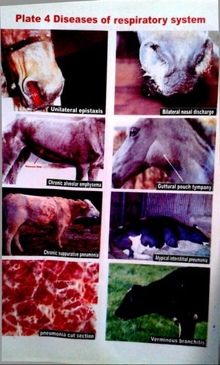
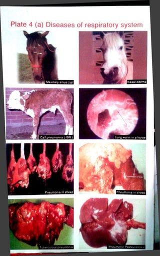
![Diseases o f the respiratory system
1 he function efficiency of the respiratory system depends
on its ability to oxygenate blood and remove carbon dioxide
from the blood in the respiratory circulation. Interference with
these functions can occur in a number o f ways but the final
defect in all instances is lack of adequate oxygen supply to the
tissues. The anoxia of respiratory insufficiency is responsible
for most of the clinical signs of respiratory diseases and
respiratory failure.
Principles o f respiratory insufficiency:
[1] Anoxia
It is a failure of the tissue to receive adequate oxygen.
In certain pulmonary diseases, gaseous exchange between
oxygen and carbon dioxide is impaired. This results in increase
o f depth of the respiratory movements and an increase in heart
rate and stroke volume.
Types o f anoxia:
(1) Anoxic anoxia: It occurs when there is defective
oxygenation of blood in the pulmonary circulation due to
respiratory diseases, such as pneumonia, pneumo-thorax,
pulmonary edema and pulmonary congestion.
(2) Anemic anoxia: It occurs when oxygen capacity of the
blood is reduced in cases o f anemia due to blood
parasites, copper poisoning and nitrite poisoning, etc.
(3) Stagnant anoxia: It occurs due to decrease the rate of
blood in the capillaries in case o f CHF, peripheral
circulatory faulty and local venous obstruction.](https://image.slidesharecdn.com/vet-141021053329-conversion-gate02/85/Vet-internal-medicine-text-book-214-320.jpg)
![(4) Histotoxic anoxia: It occurs due to failure of tissue
oxidation system in cases o f cyanide poisoning only. It
inhibits cytochrome oxidase so inhibit tissue oxidation.
[2] Respiratory failure:
Normal respiratory movements are involuntary and
stimulated by respiratory centers in medulla oblongata (MO)
as well as pH, 0 2 and C 0 2 tensions of the cranial arterial blood
supply.
Respiratory failure is the terminal stage of respiratory
insufficiency in which the activity of the respiratory centers
diminishes to the point where movements of respiratory
muscles ceases.
[3] Hypercapnia
It is the retention of C 0 2 in blood and tissues due to
faulty in elmination of C 0 2 during respiratory insufficiency,
which stimulate respiratory center in MO.
General manifestations o f respiratory insufficiency:
It may be includes:
(1) Respiratory noises:
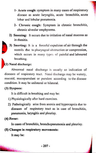
1) Cough: It is a sudden expulsion of air proceeded by
deep inspiration. It is caused by irritation of the
respiratory mucosa of the air passages by inhalation of
foreign body or dust. It indicates the presence of
disease in the respiratory system. Cough is classified
into:](https://image.slidesharecdn.com/vet-141021053329-conversion-gate02/85/Vet-internal-medicine-text-book-215-320.jpg)
![Acute Lobular Pneumonia
(Catarrhal or bronchopneumonia)
Definition:
It is a catarrhal inflammation o f bronchi, bronchioles as
well as alveoli (Broncho-pneumonia). It affects small group o f
lobules in one or both lobes o f the lung. It differs from acute
lobar pneumonia in the following points:
(1) Recurrent fever (Specific symptom): It shoots hig h and
remain for 3 days then drop suddenly to normal and stay
for few days, then shoots high again and so on, this is due
to appearance o f a new foci o f infection in the lungs.
(2) Percussion and auscultation on the lung area reveal all
sounds o f different stages o f pneumonia in different areas
at the same time, in addition to the resonant and vesicular
sound as well as rales.
(3) Compensatory emphysema can be detected by percussion
and auscultation around the affected areas specially the red
consolidation stage.
Comparison between acute lobar and lobular pneumonia
1 — Lobar pneumonia Lobular pneumonia j
] Onset Sudden --------- ---—A-----------------------------------j
Gradual and preceded by j
bronchitis. 1
I Temperature Continuos Recurrent i
| Course 1-7 days, By crisis 3-4 weeks. By lysis
[Cough
Short Painful, weak and
£
frequent
Sputum Rusty Frothy and mucopurulent
Percussion Differ according to
different stages
Scattered areas of
dullness in both lungs.
Auscultation According to stage Consildatin and captation
over different areas of
lungs. _
P.M. One lobe or part
affected
Scattered areas oF lobe or
lobes](https://image.slidesharecdn.com/vet-141021053329-conversion-gate02/85/Vet-internal-medicine-text-book-246-320.jpg)

![Diseases of the pleura
[1] Pleurisy - Pleuritis
The pleura o f the thorax is composed o f visceral and
parietal pleura.
(1) The visceral pleura covers the lung surface, lackes
specific pain receptors.
(2) The parietal pleura lines the chest wall, diaphragm, and
mediastinum, contains pain receptors, thus, when the
pleural lining is inflamed, it can be a source o f significant
pain for the animal.
Definition:
Pleurisy is an acute inflammation of the pleura causes
pain during respiration.
Causes:
(1) An extension of infection, inflammation, rupture abscesses
from the respiratory tract.
(2) Traumatic perforation o f the thoracic wall or sequle of
traumatic reticulo-peritonitis or rupture o f the thoracic
part of esophagus.
(3) Infectious: such as contagious bovine pleuro-pneumonia
and caprine pleuro-pneumonia o f sheep and goat,
infectious equine pneumonia and strangles.
Pathogenesis:
(1) First stage (acute dry stage): In early affection the
contact and frictional movement between the partial and
visceral pleurae cause pain due to stimulation of pain and
organ in the pleura.
- 2 5 9 -](https://image.slidesharecdn.com/vet-141021053329-conversion-gate02/85/Vet-internal-medicine-text-book-270-320.jpg)
![(7) Signs o f toxemia with loss o f appetite, dullness antfi
depression.
(8) Percussion on the chest causes severe pain, while in the 2I^ |
exudative stages where there is a formation o f exudate
which goes downwards by gravity to the floor o f the
pleural sacs will give rise to line o f demarcation
(pleuretic line) which is horizontal and at which!
percussion on and below this line gives a dull sound. The
pleuretic line will be changed as you change the positon
o f the animal (It is diagnostic method to differentiate
pleurisy from bronchopneumonia)
(9) Auscultation on the chest give friction sound in the first ]
stage while in the 2nd stage give normal vesicular sound
above the pleuretic line and no sound or muffled sound ;
below it.
(10) Exiensiun of the inflammation may take place to the
pericardium and death may occur in any time due to
combination of toxemia and anoxia.
NB: Chronic pleurisy which occur in tubeieuiosis is usually
symptomless, no inflammatory signs and no fluid
exudation. Weight loss is one o f the most common sign.
Diagnosis and differential diagnosis:
(1) History and symptoms.
(2) Thoracic puncture to obtain a sample from the inflammatory
fluid for bacteriological examination.
(3) Differential from pneumonia, emphysema and hydrothorax.](https://image.slidesharecdn.com/vet-141021053329-conversion-gate02/85/Vet-internal-medicine-text-book-272-320.jpg)
![[3] Hydropericardium
It is an excessive formation o f serous fluid in pericardial
sac. It is common in dog & cat. It is a secondary disease
usually accompanies:
(1) Chronic disease o f heart & lung such as myocardosis &
pericarditis as well as ascites & liver fibrosis.
(2) Worm infestation.
(3) Hydremic diseases in chronic nephritis, anemia.
Clinical findings:
(1) Increase in cardiac dullness as in pericarditis.
(2) Cardiac tone is weak & is not sharply detected with a small
rapid pulse
(3) Dyspnea.
(4) Cyanosis o f mucosa.
(5) Filling o f capillaries with blood.
(6) There is no fever or cardinal signs.
Diagnosis:
(1) Symptoms.
(2) Increase area o f cardiac dullness without pain.
(3) Puncture o f pericardial sac gives clear yellow transudate.
(4) Jaundice occurs in liver cirrhosis.
Treatment:
(1) Complete rest, treat the real cause.
(2) Diet free from salt & restriction o f water supply.
(3) Cardiac stimulant as adcopherine & cardiac tonics as digitalis.
(4) Diuretics & laxative.
(5) Slowly IV injection o f 10% calcium chloride.
- 2 8 4 -](https://image.slidesharecdn.com/vet-141021053329-conversion-gate02/85/Vet-internal-medicine-text-book-295-320.jpg)
![[4] Pneumo-pericardium
Causes:
(1) Stomach gases, following the perforation by foreign
bodies.
(2) As a complication of putrefactive purulent pericarditis.
Clinical findings:
Percussion over heart reveals loud & tympanic sound.
Treatment:
As pericarditis and surgical interference.
[5] Endocarditis
It is the Inflammation of the endocardium which may
interfere with blood flow from the heart by causing
insufficiency or stenosis of the valves, murmurs sounds are
clearly audible from heart and if interference with blood flow
is sufficiently severe, CHF develops.
Causes:
(1) Bacterial infection (whether by direct adhesion to
undamaged endothelium or through the valvular surfaces
or by hematogenous spread). In cattle, Alpha-hemolytic
streptococci & Coryne bacterium pyogenes may be a
cause.
(2) The infection may be emboli from suppurative lesions in
other sites especially traumatic reticulitis, metritis and
mastitis.](https://image.slidesharecdn.com/vet-141021053329-conversion-gate02/85/Vet-internal-medicine-text-book-296-320.jpg)
![Diagnosis: A
(1) Failure to observe a valvular murmur may result in
confusion between endocarditis and pericarditis or other
causes o f CHF.
(2) The commonest error in cattle is in differentiation o f the
disease from lymphomatosis which respond to treatment
with penicillin.
(3) Blood culture may reveal the causative organism.
Treatment:
(1) Specific antibiotics after sensitivity test.
(2) Penicillin is the antibiotic of choice, effective in large
doses, 4-6 million units daily for a 1200 lb cow should be
given for 3 days.
(3) Cardiac tonics.
Diseases characterized by abnormalities o f the cellular
elements o f the blood:
These include: [I] Anemia, [II] Leukosis, [III] Leucopenia
Anemia
It is a reduction in the number of erythrocytes,
hemoglobin (Hb) or both in circulating blood.
Causes:
(1) Excessive loss o f blood by hemorrhage.
(2) Insufficient production or excessive destruction of
erythrocytes.](https://image.slidesharecdn.com/vet-141021053329-conversion-gate02/85/Vet-internal-medicine-text-book-298-320.jpg)
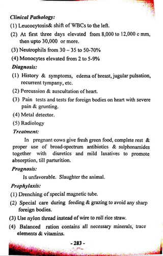
![Etiological classification o f anemia:
[1] Hemorrhagic or blood loss anemia:
(1) Acute in cases of trauma, surgery or defects o f the
coagulin mechanism.
(2) Simple chronic or secondary anemia in cases of:
1) Internal parasites as hook worms, stomach worms,
coccidia, nodular worm, liver fluke, strongylosis.
2) External parasites as ticks, some flea, blood suckling
parasites.
3) GIT lesions as ulcer, hemorrhagic gastritis, enteritis.
4) Urogenital tract bleeding as nephritis, uremia,
excessive esterus hemorrhage.
[2] Hemolytic anemia:
Due to destruction of red blood cells or shortened life
span of erythrocytes in cases of:
(1) Blood parasites: Anaplasma, piroplasma, babesia,
trypansoma.
(2) Bacterial infection: leptospirosis, bacillary hemoglobinuria.
(3) Viral infections: Equine infectious anemia.
(4) Chemical agents: Poisoning with copper, lead,
phenothiazine & methylene blue.
(5) Poisonous plants: Caster bean, wild onion, kale.
(6) Hemolytic reptile poison: snake poison.
(7) Metabolic: Post parturient hemoglobinuria or
hypophosphatemia, common in cattle which feed on diet
rich in calcium &poor in phosphorus (Alfa alfa, berseem).
- 2 8 8 -](https://image.slidesharecdn.com/vet-141021053329-conversion-gate02/85/Vet-internal-medicine-text-book-299-320.jpg)
![(8) Auto immune hemolytic anemia.
(9) Immune hemolytic anemia o f the newborn due to mother &
fetus antibodies incompatibility or due to hemolysin in the
colostrum.
(10) Isoimmunization following multiple blood transfusions
between animals.
[3] Hypoplastic anemia
Due to depression of bone marrow caused by:
(1) Physical agents: Irradiation from x-rays, radium, and
radioactive isotopes.
(2) Chemical agents as estrogens, chloramphenical,
sulphaguanidine, copper, lead, mercury.
(3) Biological toxin produced in course of chronic suppurative
processes.
(4) Parasitic toxins produced by intestinal parasites (Taenia &
Ascaris) or blood protozoa (Anaplasm, Theilaria,
Trypansoma).
(5) Tumors o f leukemic cells in the bone marrow or osteolytic
tissue.
[4] Nutritional deficiency anemia:
In cases of:
(1) Iron deficiency in diet or pasture or malabsorption or
severe chronic hemorrhage.
(2) Copper deficiency in diet or pasture or excessive
molybdenum intake.
-2 8 9 -](https://image.slidesharecdn.com/vet-141021053329-conversion-gate02/85/Vet-internal-medicine-text-book-300-320.jpg)
![(3) Cobalt deficiency with decreased synthesis o f vitamin B12?
or diet or pasture deficient on cobalt (Vitamin B12 is V,
erythrocyte maturing factor).
(4) Vitamins deficiency: Folic acid, riboflavin, pyridoxine,
nicotic acid, and vitamin C.
5) Protein deficiency due to insufficient intake or digestion o f
protein or marked serum protein loss.
Morphological & etiological classification o f anemia
Serial
Morphological classification
Etiological Size of classification
RBC Hb content
111
121
Macrocytic
Macrocytic
Normochromic
Hypochromic
Cobalt & B12 deficiency
Anaplasma, Piroplasma,
leptospira, Bacillary Hb uria,
parturent Hb uria.
[3]
or (4]
or [5]
Normocytic
Normocytic
Microcytic
Normochromic
Hypochromic
Normochromic
! - Acute blood loss
2- Nephritis with terminal
uremia jj
3- Subacute or chronic |
inflammatory diseases ||
4- Stomach worm infection j
(except Hemonchus causes |
blood loss).
5- Leukemia
6- Hypoplastic anemia,
radiation, injury or soybean
meal poisoning
I6J Microcytic Hypochromic 1 - Deficiency of iron
2- Defect in utilization of
iron stores of body in copper
deficiency or molybdenum
poisoning. ___.](https://image.slidesharecdn.com/vet-141021053329-conversion-gate02/85/Vet-internal-medicine-text-book-301-320.jpg)

![[2] Decreased plasma osmotic pressure-Hypoproteinemia:
(1) Renal diseases causing continuous loss o f protein in urine
(albumin urine), occurs in anasarca in the anterior part o f
the body (head, eye lids, neck).
(2) Parasites as Fascula sp & Haemonchus sp in ruminants,
Strongylus in horses, Hook worm in dogs causing loss o f
osmotic due to protein loss.
(3) Malnutrition due to:
1) Defect in digestion, absorption, metabolism, utilization
o f protein & plasma protein.
2) Decrease protein level in diet.
3) Impairement o f liver function.
4) Vitamin A deficiency causes edema of legs specially in
calves.
5) Copper deficiency in sheep.
(4) Liver damage in heavy parasitic infestation or malnutrition
or bacterial, viral infection or toxicity. They causing
failure o f protein synthesis.
[3] Obstruction o f blood or lymph or portal circulation due to
tumor, fibrosis, surgical, congential obstruction in calf,
ulcerative lymphangitis in horse or parasites as filaria.
[4] Allergic condition: In which allerge & histamin like
substance are released causing local liberation o f
vasodilators increasing capillary permeability, dilatation,
vascular damage o f small vessels and hydrostatic pressure
increasing fluid & protein passage to interstitial space than
that reabsorbed by lymphatic fluid causing angioedema,
urticaria, wheels or purpura hemorrhagica.
- 296-](https://image.slidesharecdn.com/vet-141021053329-conversion-gate02/85/Vet-internal-medicine-text-book-307-320.jpg)
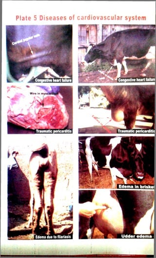
![[5J Toxines in the course of some infectious diseases as
Anthrax, Black leg, Malignant edema, Pasteurlosis,
Filariasis, Edematous skin disease as well as equine infectious
anemia, viral arteritis, infectious rhinopneumonia.
Etiological classification o f edema:
(1) Physiological or mammary edema:
(2) Cardiac edema: It occurs in CHF
(3) Renal edema
(4) Hepatic edema.
(5) Pulmonary edema: due to disturbance of circulatory &
plumonary circulation, nervous system, together with
physiochemical factors regulating fluid exchange in
tissues.
(6) Obstructive edema: due to obstruction of blood or lymph or
portal circulation.
(7) Allergic edema.
(8) Nutrional edema.
Pathogenesis:
[1] In normal state:
(1) The absorbed water reaches blood to enter intravascular
space (vascular water contains more protein), interstitial
space & intracelllar.
(2) There are a constant flow (to & fro) between vascular &
interstitial water which occur between capillary arterial
end (due to higher hydrostatic pressure & lower osmotic
pressure) & capillary venous end (due to lower
hydrostatic pressure & higher osmotic pressure) & carries
nutrient & metabolites to body tissue.
(3) At capillary venous end the reverse occurs.
- 297-](https://image.slidesharecdn.com/vet-141021053329-conversion-gate02/85/Vet-internal-medicine-text-book-308-320.jpg)
![[2] In diseased condition:
When the hydrostatic pressure increased & osmotic
pressure decreased leads to:
(1) An excessive fluid tends to pass into tissue space at the
capillary arterial end as the hydrostatic pressure o f the
blood is sufficient to overcome its osmotic pressure.
(2) An excessive fluid tends to pass into tissue space (instead
o f returning to the vascular system) at the capillary
venous end as the position is reversed.
(3) Failure o f fluid to return to the capillaries re su ltin g in
accumulation o f fluid into tissue space or e sc ap e into
serous cavity forming edema.
Clinical symptoms (Symptomatical classification o f edema):
(1) Anasarcas: SC formation o f transudate in abdominal floor,
sternum, brisket, intermandibular space,pharyngeai and
perineal region. This edema is soft, painless and pit on
pressure.
(2) Ascites: Transudate formation in peritoneal cavity causing
enlarged abdomen with pear shape. Percussion o f the fluid,
thrill is seen and can be detected on the other side and at
top line o f fluid.
(3) Hydropsy: Fluid formation in uterus.
(4) Cerebral edema is manifested by nervous symptoms.
(5) Hydropericardium: fluid in pericardium causing restriction
o f cardiac movements and muffled heart sounds occur.
(6) Pulmonary edema: It is accompanied by re sp ira to iy
disorders, moist rales and frothy discharge from the nose.](https://image.slidesharecdn.com/vet-141021053329-conversion-gate02/85/Vet-internal-medicine-text-book-309-320.jpg)
![Common types of clinical edema (examples of associated
conditions):
(1) Intermandibular edema (parasitic).
(2) Thoraco- abdominal (parasitic/ heart).
(3) Supra-orbital fossa (south AF.H.S./ renal)
(4) Pharyngeal region (pharyngitis/hemorrhagic septicemia).
(5) Perineal area (urticaria).
(6) Buttock region (black leg).
(7) Udder region (physiological/mastitis).
(8) Limbs, whole or with demarcation (heart/renal).
(9) Brisk! region (heart).
(10) Dewlap (pericarditis).
(11) Head region (purpura hemorrhagica, swelled head in rams
& blue tongue).
(12) Generalized (systemic).
Edema may be:
[1] Non inflammatory (cold) edema:
(1) May be local or general edema.
(2) Cold and painless swelling contains transudate (serous
fluid).
(3) Causes: Increase in hydrostatic pressure & plasma colloid
osmotic pressure (PCOP).
[2] Inflammatory (hot) edema:
(1) Local edema.
(2) Hot (local or general fever), red (in unpigmented skin),
firm & painful swelling contains exudate.
(3) Caused by:](https://image.slidesharecdn.com/vet-141021053329-conversion-gate02/85/Vet-internal-medicine-text-book-310-320.jpg)
![' ' ' ; [2] Variations in daily urine flow
An increase or decrease in urine volume over 24 hours.
(1) Polyuria:
It is an increase in urine volume over 24 hours period,
diagnosed by determine o f specific gravity or osmolality,
blood urea and creatinine.
1) Extrarenal causes:
1- Diabetus insipidus.
2- Diuretic drugs including corticosteroids.
3- Transient: e.g. excessive water intake, Diet deficient in
Na Cl, Hyperglycemia, Fear or Emotion stress.
2) Renal cause:
1- Exceeded resorptive capacity of remaining tubules.
2- When the osmotic gradient in the renal medulla is not
adequate to produce concentrated urine.
(2) Oliguria and Anuria:
It is a reduction in the daily output (oliguria) and
complete absence of urine (anuria).
Oliguria is caused by dehydration, CHF, peripheral circulatory
failure and terminal stages of all forms of nephritis.
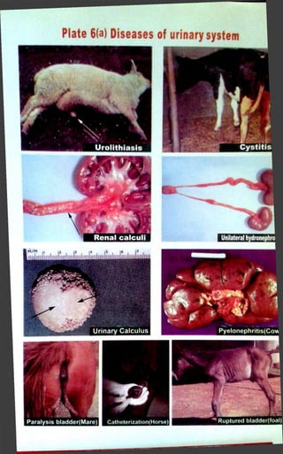
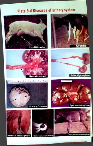
Anuria is caused by urolithiasis,
NB: It leads to retension of solutes and disturbances of acid-base
balance that contribute to uremia.
(3) Pollakiuria:
It is an abnormal frequent passage of urine. It may occur
nth or without an increase in the volume of urine excreted](https://image.slidesharecdn.com/vet-141021053329-conversion-gate02/85/Vet-internal-medicine-text-book-323-320.jpg)
![and: is commonly associated with disease o f the lower uritjip
Tract such as cystitis, the presence o f calculi in the bladder
'frri4tritis, and partial obstruction o f the urethra.
(4) Dribbling:
i : ' | 7 , i It is an intermittent passage o f small volumes o f urine du^.
", to lack o f sphincter control. Occurs in cases o f non-obstrucfr^l
urolithiasis and persistent urachus.
[3] Abnormal painful or difficult urination
(1) Jysuria or painful or difficult urination:
; It is accompanied by a frequent passage of small amounts
of urine. It occurs in cystitis, vesical calculus and urethritis.
Grunting and abdominal pain may occur with painful urination
and: the animal may remain in the typical posture after
urination is completed.
(2) Stranguria:
It is a slow and painful urination which accompanied by
strains to pass each drop of urine. It occurs in cystitis, vesical
calculus, urethral obstruction and urethritis.
(3) Urine scalding of the perineum or urinary burn:
lit is caused by frequent wetting of the skin with urine.
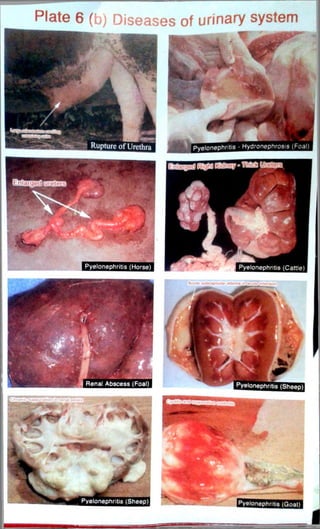
[4] Morphological abnormalities of urinary tracts
(1) Enlargement or decreased size of kidneys or ureters.
(2) Abnormalities of the bladder: Gross enlargement of the
bladder, rupture of the bladder, a shrunken bladder fol-
|lowing rupture.](https://image.slidesharecdn.com/vet-141021053329-conversion-gate02/85/Vet-internal-medicine-text-book-324-320.jpg)
![(3) Abnormalities o f the urethra include enlargement and ?
o f the pelvic urethra and its external aspects in male cattle
and ram with obstructive urolithiasis.
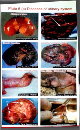
[5] Acute and chronic renal failure
The clinical findings o f urinary tract disease vary with the
rate o f development and stage o f the disease. In horses, mental
depression, colic and diarrhea are common with oliguria or
polyuria. Cattle with uremia are similar and in addition are
frequently recumbent and may have a bleeding diathesis. In
chronic renal disease of all species, there is a severe loss o f
body weight, anorexia, polyuria, polydipsia and ventral edema.
[6] Uremia
It is the systemic state that occurs in the terminal stages o f
renal insufficiency due to retention o f some solutes and
disturbances o f acid base balances. Anuria or oliguria may
occur with uremia.
Clinical signs:
(1) Depressed,anorexic with muscular weakness and tremor.
(2) In chronic uremia: Poor body condition (due to continued
loss o f protein in the urine) and dehydration may occur.
(3) The respiration is usually increased in rate and depth but is
not dyspenic; in the terminal stages it may become
periodic in character.
(4) The heart rate is markedly increased because o f terminal
dehydration and myocardial asthenia.](https://image.slidesharecdn.com/vet-141021053329-conversion-gate02/85/Vet-internal-medicine-text-book-325-320.jpg)

![Nervous Reflexes:
There are three examined reflexes o f CNS:
[1] Superficial reflexes:
(1) Conjunctival reflex:
They are the sensory nerve fibers in the ophthalmic and
maxillary branches of the fifth cranial nerve and the motor
fibres o f the seventh cranial nerve.
(2) Corneal Reflex:
They concerned sensory fibers in the ophthalmic branch
o f the fifth nerve and motor fibers in the seventh cranial nerve.
(3) Pupil Reflex:
The nerves involved are the second sensory nerve (optic)
and the third motor nerve (oculomotor).
(4) Perineal Reflex:
This reflex is useful in cattle and horses. In cases of
recumbency and failure to response by the perineal reflex,
must be regarded as a sign of serious import. This reflex is a
convenient means of testing the functional of the local spinal
reflexes. One of the folds of the skin radiating from the anus is
pinched between the forefinger and thumb; this should result
in a reflex contraction of the perineal musculature, causing the
skin of the region to become tense.
(5) Pedal Reflex:
This reflex is tested only in recumbent animals which
cannot rise or cannot stand when assisted to its feet. In cattle
and horses, this reflex may be tested by stimulating the skin of
the bulb of the heel with a pin. In dog and cats, one of the
- 357-](https://image.slidesharecdn.com/vet-141021053329-conversion-gate02/85/Vet-internal-medicine-text-book-373-320.jpg)
![folds o f skin between the bads is nipped between the ball o f
the forefinger and thumbnail, quick and active retraction o f the
leg should take place. In parablegic dogs, the movement may
be sluggish and limited if the spinal reflexes are still intact or
the response may be completely absent i f there is a breakdown
o f the reflex arc.
[2] Deeper Reflexes (Musclo-tendon reflexes):
These reflexes are confined to cases o f recumbent animal
to investigate the state o f the neuro-muscular mechanism, or
cases in which damage to the spinal cord is suspected.
(1) Patellar Reflex,
(2) Tarsal Reflex:
It is more convenient to perform on small animals. For
testing this reflex, the hock must be slightly flexes. If attempts
to flex the hock are countered by the animal vigoursly
extending the limb, so it is not necessary to perform the test.
When the hock has been flexed the tendo-achilles must be
struck a quick sharp blow. Avigorous contraction o f the
gastrocnemius muscle should result.
[3] Organic Reflexes:
(1) Respiration; stimulation o f the respiratory center may
be reflex by visceral pain or by increase blood carbon
dioxide.
(2) Deglutition reflex.
(3) Defecation reflex.
(4) Micturation reflex.](https://image.slidesharecdn.com/vet-141021053329-conversion-gate02/85/Vet-internal-medicine-text-book-374-320.jpg)
![(7) Vaccine or immunization:
Such as rabies & distemper vaccine.
(8) Metabolic diseases:
Such as hypocalcemia, hypomagnesemia and ketosis
(Nervous form) in cattle; pregnancy toxemia in sheep & goats.
Clinical approach of nervous system
[1] History of feeding & environment.
[2] Inspection:
It should be made from various angles. All the body parts
of the animals starting from anterior to posterior extremity
should be thoroughly observed. Behaviour of the animal
ranging from decline to aggressiveness should be noted.
Response of the animal to the external stimuli will give a clear
idea about the state of animal’s health. Clinical signs like
recumbency, enlarged head, dilated pupils, droopy ears and
eyelids often point to wards hydrocephalus. Spinal injury
points to peripheral nerve paralysis. Semiflex of foreleg directs
to radial paralysis.
[3] Posture:
Abnormal posture may arise from injury of
musculoskeletal & or nervous system. Animal adopt different
types of posture due to neurological disorder. When head is
twisted in one side and can be made straight manually it will
suggest middle ear infection but if it cannot be straightened, it
is indicative of inner ear involvement. Paresis or paralysis of
one leg is caused by peripheral nerve injury. Paraplegia is
indicative of spinal cord involvement. Hemiplegia suggests
brain Lesions.
-3 6 0 -](https://image.slidesharecdn.com/vet-141021053329-conversion-gate02/85/Vet-internal-medicine-text-book-376-320.jpg)
![[4] Gait:
Abnormalities in gait are observed when the animal walks
or run. Brain lesion may cause circling. Vestibular lesions
cause circling in closed circle whereas lesions o f thalamus
cause circling in wide circle. Cerebellar lesions cause
dysmetria characterized by uncoordinated gait with wide apart
legs (goose-stepping). Standing or walking in one side is seen
in small animals due to cerebral affection.
[5] Palpation:
A systemic palpation o f vertebral column and skull will
reveal about dislocation and fracture. The involved area
becomes hyper or hyposensitive to touch.
[6] Sensitivity:
Sensitivity disorders arise as a result of impaired
transmission o f excitation along sensory nerves from the
peripheral receptors and along afferent nerves to the cerebral
cortex. Sensitivity may be diminished (hypoaesthesia), lost
(anaesthesia) or increases (hyperaesthesia). Simple sensitivity
is divided into superficial (skin, mucosa), or deep (muscles,
joints, bones) or interoceptive (internal organs).
Sensory reflex
It includes analgesia, loss o f superficial reflex
hypersensitivity e.g.Acetonemia, hypomagnesemia, tetanus
show hyperaestheia skin to touch.](https://image.slidesharecdn.com/vet-141021053329-conversion-gate02/85/Vet-internal-medicine-text-book-377-320.jpg)
![3) Tarsal reflexes in dog & index, response is just to retract
the finger. It is indicated if recumbency accompanied
by trauma o f spinal cord or not.
General Manifestations o f diseases o f the nervous system:
HI Mental state:
This include mania and frenzy.
Mania occurs in the nervous form o f acetonaemia in
cattle,and liver insufficiency in horses poisoned by
certain plants.
Frenzy, occurs in rabies, acute lead poisoning.
Mania and frenzy are manifestations of general excitation
o f the cerebral cortex.
Depressive mental states include somnolence, lassitude,
syncope & coma. They are all manifestations of
depression of cerebral cortical function in various
degrees.
Head pressing a syndrome characterized by the animal
pushing its head against fixed objects may be due
to a combination o f headache & mania.
[2] Involuntary movements:
They include convulsions and tremor. Convulsions
always originate in the cerebral cortex but the primary cause
may be a dysfunction in a system other than the nervous
system. True tonic convulsions occur in strychnine poisoning
and in tetanus. Convulsions may occur in various intoxications
(e.g. lead, arsenic, mercurials & phosphates chlorinated
hydrocarbon, etc.).
- 3 6 4 -](https://image.slidesharecdn.com/vet-141021053329-conversion-gate02/85/Vet-internal-medicine-text-book-380-320.jpg)
![[3] Posture and gait:
Head pressing or rotation, dog sitting posture dropping o f
lips, eyelids & ears are examples.
[4] Paralysis:
Loss o f motor power or sensation or both in cases o f
nerve injury resulting in loss o f voluntary muscular
movements.
NB: Paresis (relaxation): The power of contraction is weaker
than normal. It may be local of general.
Symptomatic classification o f paralysis:
(1) Spastic paralysis means paralysis in contractile stage.
(2) Flaccid paralysis means paralysis in relaxation stage.
The type o f paralysis is often indicative of the site of the
lesion. Most paralysis seen in farm animals is flaccid and is
caused by lesions in the spinal cord.
Clinical classification o f paralysis:
(1) Monoplegia: Paralysis of one limb or one muscle.
(2) Diplegia: Similar bilateral paralysis.
(3) Hemiplegia: Paralysis of one side of the body.
(4) Quadriplegia: Paralysis of all four legs.
(5) Paraplegia: Paralysis of the posterior part of the body and
the hind legs due to spinal cord affection.
There are two types of paraplegia:
1) Paraplegia in extension due to hypertonicity of the
extensor group of muscles.
2) Paraplegia in flexion due to increased tone of the flexor
group of muscles.](https://image.slidesharecdn.com/vet-141021053329-conversion-gate02/85/Vet-internal-medicine-text-book-381-320.jpg)
![Peripheral nerve paralysis:
It includes the suprascapular, brachial plexus and radial
nerves o f the forelimbs as well as femoral, obturator, tibial,
peroneal and sciatic nerves o f the hindlimbs.
[5] Disturbances in sensation:
Lesions o f the peripheral sensory neurons cause hyper or
hyposensation o f the area supplied by the nerve.
Principles o f treatment in diseases o f the nervous system:
Treatment o f disease o f the nervous system have some
particular problems, because o f the failure o f nervous tissue in
the brain and spinal cord to regenerate and because o f the
impermeability o f the blood- brain barrier to many drugs.
When peripheral nerves are served regeneration occurs if the
damage is not extensive. When neurons are destroyed in the
brain and spinal cord no regeneration occurs and the provision
o f nervous stimulants have no effect on the loss o f function
that occurs. The emphasis in treatment o f diseases o f the
nervous system must be on prevention o f further damage.
(1) Control o f Infection:
By the broad spectrum antibiotic and chloramphenicol.
Treatment o f the diseases o f the brain is limited by the
existence o f the blood- brain barrier which prevents
penetration o f some substances into nervous tissue. The
cerebrospinal fluid is probably o f little importance as a mear
o f bringing antibacterial drugs to brain tissue.
(2) Decompression:
Increased intracranial pressure probably occurs in mos
cases of inflammation of the brain; acute cerebral oedema an<
hypovitaminosis A. In these circumstances, some treatmen
s h o u ld hp o iv p n tn n /ith d ro t i/ f lu id fr r m flip h ro in ticcup nni](https://image.slidesharecdn.com/vet-141021053329-conversion-gate02/85/Vet-internal-medicine-text-book-382-320.jpg)
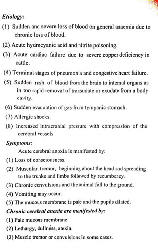
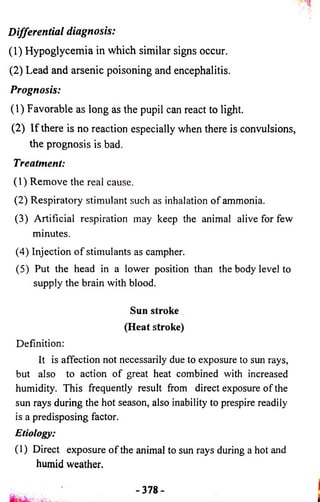
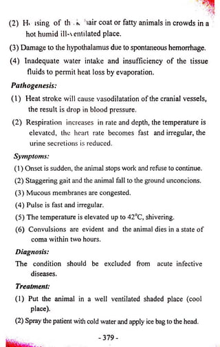
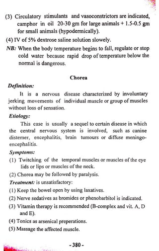
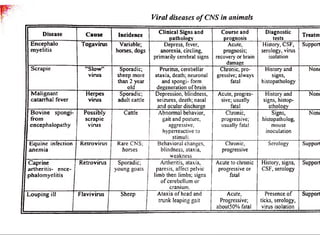
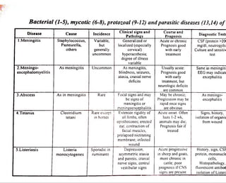
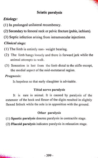
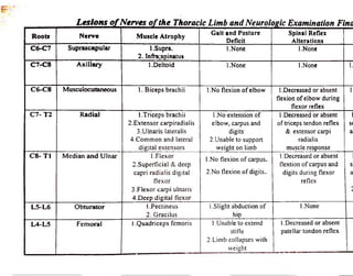
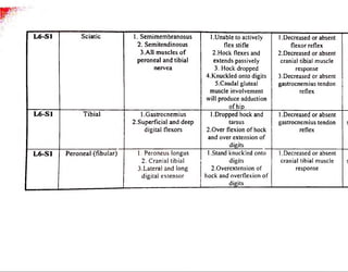
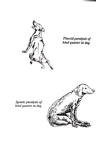
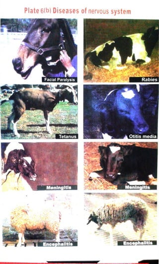
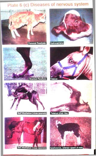
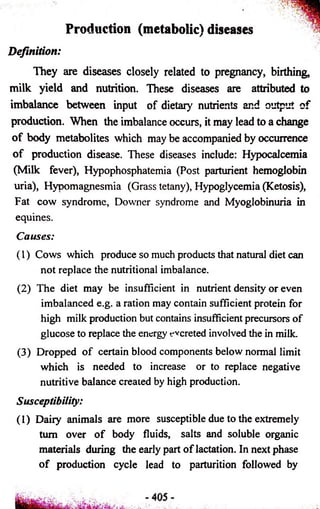
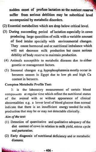
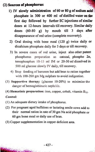
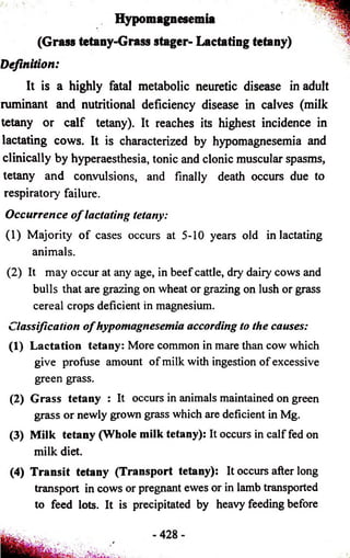
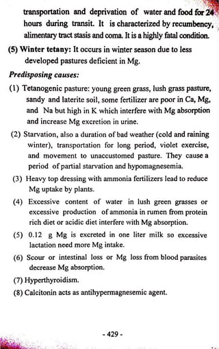
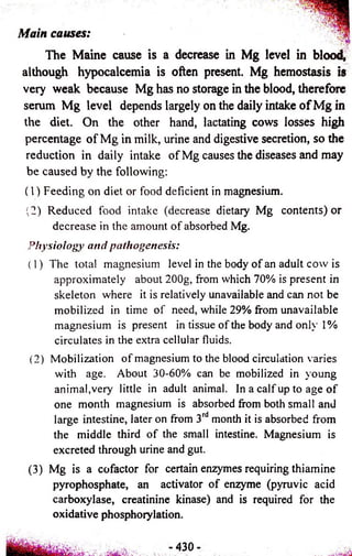
![(4) A decrease in Mg:Ca ratio stimulate secretion of acetyl
choline estrase (ACH) which is responsible for
hyperaesthesia, muscular tremors, convulsion, etc.
(5) Tetany may result from central effect of brain due to lower
of Mg level of CSF in hypomagnesemic animal.
Clinical findings:
The disease may be acute, subacute and chronic:
[1] Acute lactation tetany:
(1) The animal may be grazing and suddenly cease to graze and
appear uncomfortable with twitching of the muscles and
ears.
(2) Severe hyperaesthesia so that any slight disturbances such as
sudden noise or touch leads to attacks of continuos
bellowing and frenzied galloping.
(3) Staggering gait then fall down on the ground with obvious
tetany of the limbs which is rapidly followed by clonic
spasm and convulsions lasting for about a minute.
(4) During the convulsive episodes there is opithotonus (Back
head), nystagmus (rotation of eye ball), champing of the
jaws, frothing at the mouth, pricking of the ears, retardation
of the eye lids. Between episodes the animal lies quite but a
sudden noise or touch stimulate another attack.
(5) The temperature rises (40 to 40.5°C) after severe muscle
exertions.
(6) Death usually occurs (from respiratory failure) within a half
to one hour and the mortality rate is high.](https://image.slidesharecdn.com/vet-141021053329-conversion-gate02/85/Vet-internal-medicine-text-book-449-320.jpg)
![[2] Subacute lactation tetany:
(1) The onset is more gradual. It takes from 3 -4 days, there is
slight inappetence, wildness of the facial expression and
exaggerated limb movements.
(2) Stagger gait or stiffness of the limbs
(3) Loss of appetite which accompanies by rumen stasis and
decrease in milk yield.
(4) Spasmodic urination and frequent defecation.
(5) Anoxious, muscular tremors and mild tetany in the hind legs
and tail.
(6) Unsteady walking, then the animal falls down on and the
affected cow rolls on her side with legs stretched backward.
[3] Chronic lactation tetany:
(1) Low serum-magnesium levels with or without signs.
(2) A few animals do some vagus syndrome including dullness,
convulsion, unthriftiness and indifferent appetite.
(3) It may also occur in animals, which recover from the
subacute form of the disease.
Diagnosis:
(1) Case history: A bad weather or grazing green cereal crops or
luch grass.
(2) Clinical symptoms: Inco-ordination, hyperaethesia and
tetany.
(3) Clinical pathology:
1) Serum Mg level dropped from 1.7-3 (normal) to less
than 1.0-0.5 mg% (diseased).](https://image.slidesharecdn.com/vet-141021053329-conversion-gate02/85/Vet-internal-medicine-text-book-450-320.jpg)
![2) CSF Mg level dropped from 2 (normal) to l e s a l l i^ S
mg% (in tetany, even after 12 hours after death)*'
3) Serum Ca level may be reduced.
Differential diagnosis:
(1) Nervous fonn of ketosis: It is not usually accompanied b]
convulsions or letany, presence of ketonuria anc
hypoglycemia.
(2) Rabies: Presence of straining, ascending paralysis, anesthesia
and an absence of tetany.
(3) Acute lead poisoning: Blindness and mania, history of access
to lead.
(4) Enterotoxemia caused by Clostridium perfringes type D.
(5) Polio-encephalomalacia.
(6) Tetanus.
(7) Vitamin A deficiency.
(8) Meningitis.
Hygienic treatment:
Complete rest of the affected animal in quite place.
Medical treatment:
(1) Source of magnesium: By IV (200-300 ml of 5%)
solution of Mg sulfate. (It causes rapid rise in serum
Mg within 3-6 h) followed by SC injection of 200-300
ml of 20% solution of Mg sulfate.
N B :
1) The IV injection of magnesium salts may induce
cardiac arrythemia, or medullar depression may be
severe enough to cause respiratory failure and
death.](https://image.slidesharecdn.com/vet-141021053329-conversion-gate02/85/Vet-internal-medicine-text-book-451-320.jpg)
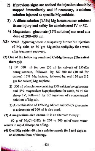
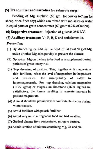
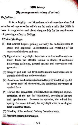
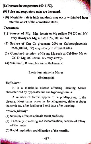
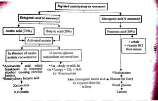
![Symptoms o f bovine ketosis:
There are five forms of bovine ketosis
[1] Subclinical form where the urine and blood contain ketone
bodies in excess amount but there is no obvious
symptoms o f ketosis
[2] Wasting or digestive form:
It is the most common form. It is manifested by:
(1) Gradual but moderate decrease in appetite and milk
production over a period of 2-4 days.
(2) Body weight is lost rapidly, from the decrease in appetite.
(3) Cows become woody (Woody cow) due to the apparent
wasting and loss of cutaneous elasticity.
(4) The feces are firm and dry due to ruminal atony.
(5) The cow is moderately depressed and disinclined to move
or eat may be due to abdominal pain.
(6) Temperature, pulse and respiration are normal.
(7) Ketone smell in breath, milk, sweat, urine and stable.
[3] Nervous form:
It occurs quite suddenly
(1) The animal walks incircles and crossing the fore legs.
(2) Pushing the head or lean it against objects.
(3) Apparent blindness, aimless movement and wandering.
(4) Hyperaethesia, isolated muscle twitches on shoulder and
flank, moderate tremors, tetany and stagar gait.
(5) Bellowing on being pinched or stroked.](https://image.slidesharecdn.com/vet-141021053329-conversion-gate02/85/Vet-internal-medicine-text-book-461-320.jpg)
![(6) Depraved appetite with excessive salivation, exaggerated
licking, abnormal chewing movements.
(7) Nervous signs occur in 1-2 hours and may recur in interval
of about 8-12 hours. Animal may injure itself during
episoides.
[4] Mixed form of wasting and nervous forms
[5] Milk fever like form:
It resembles milk fever disease but the muscular
twitching and hypersensation are constant symptoms. This
form responds to treatment with calcium and glucose therapy
due to presence of hypoglycemia and hypocalcemia.
Diagnosis o f bovine ketosis:
(1) History of calving or late pregnancy and winter season.
(2) Clinical symptoms of ketone odor, wasting and or nervous
form.
(3) Biochemical abnormality (See the table).
Rothers test fo r detection o f ketone bodies:
(For urine): To 5 ml urine add ammonium sulfate to
saturation (about 1 gm) and 2 or 3 drops of sodium
nitropursside solution; mix and cover with strong ammonium
hydroxide. A reddish purple ring is positive.
A positive test is considered as warming rather than a
sign of disease.
(For serum): Put 2 drops of plasma or serum in a test
tube and supersaturate with ammonium sulfate crystals by
shaking. Add 2 drops of approximately 5% sodium
nitropursside solution and shake. Let stand 3 minutes. A
permanganate color indicates a trace; a light blue, a moderate
amount; and a deep blue, a large amount of ketone bodies.
445-](https://image.slidesharecdn.com/vet-141021053329-conversion-gate02/85/Vet-internal-medicine-text-book-462-320.jpg)
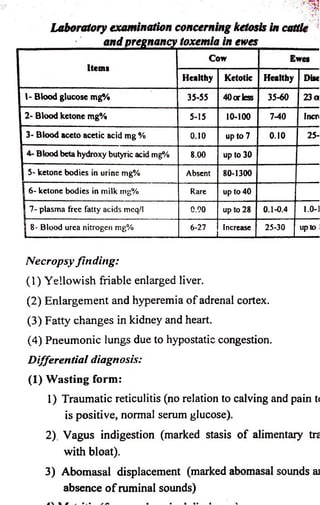
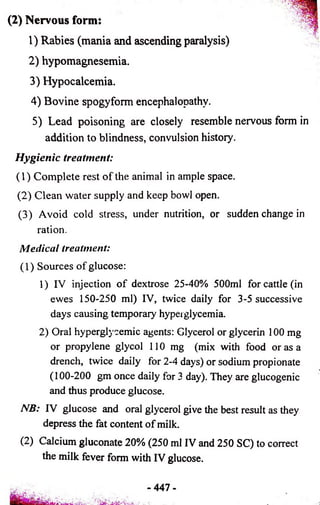
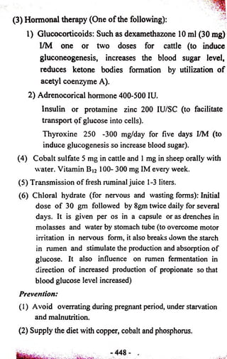

![Control: ■
• •
(1) Reduce the grain ration to half on rest day and when the '
horse is getting no exercise.
(2) Daily exercise to minimizes the disease.
Downer syndrome
Definition:
It is a recumbency of animal and unable to rise again.
Causes:
[11 Before parturition:
(1) Parturient paresis (hypocalcemia).
(2) Grass tetany.
(3) Ketosis.
(4) Severe torsion of uterus with possible rupture of uterine
artery.
(5) Hydrops of th* allantois.
(6) Septic metritis of pregnancy.
(7) Dislocation of hip or sacro-iliac joint, or fracture of a leg or
pelvis.
[2] During four days after calving:
(1) Parturient paresis.
(2) Grass tetany.
(3) Ketosis.
(4) Septic metritis with or without retained placenta.
(5) Septic mastitis.
(6) Rupture of uterus and diffuse peritonitis.](https://image.slidesharecdn.com/vet-141021053329-conversion-gate02/85/Vet-internal-medicine-text-book-476-320.jpg)
![(7) Traumatic gastritis with diffuse peritonitis.
(8) Obturator paralysis.
(9) Peroneal nerve paralysis.
(10) Dislocation or subluxation of the hip or sacro-iliac joints,
fracture of the leg, pelvis or spine.
(11) Rupture of gastrocnemius tendon.
(12) Toxic indigestion after hypocalcemia due to prolonged
suppression of intestinal activity.
(13) Degeneration of muscle.
(14) “Cast” with or without debility.
(15) Albuminuria due to primary nephritis (rare).
[3] During four to fourteen days after calving:
(1) Septic metritis with or without retained placenta.
(2) Septic mastitis.
(3) Traumatic gastritis with diffuse peritonitis.
(4) Debility.
(5) Obturator paralysis.
(6) Fracture or dislocation.
(7) Suppurative arthritis of knee or back hock and pyemia.
(8) Ketosis.
(9) Parturient paresis (hypocalcemia).
(10) Grass tetany or magnesium deficiency.
(11) Postparturient hemoglobinuria (possibly leptospirosis).
(12) Albuminuria (nephritis).
(13) Advanced severe pyelonephritis.
(14) Degeneration of muscle.](https://image.slidesharecdn.com/vet-141021053329-conversion-gate02/85/Vet-internal-medicine-text-book-477-320.jpg)
![[4] At any time: j |
(I) Metabolic disturbances and deficiencies of:
1) Hypocalcemia
2) Hypophosphatemia.
3) Hypomagnesmia.
4) Hypoglycemia.
5) Vitamin A: hypervitaminosis A in calves and lambs
(Downer calf or Downer lamb)
6) Vitamin E: hypovitaminosis E (white muscle disease of
calves or stiff lamb disease of lambs).
7) Ca, ph and vitamin D deficiency in Rickets (Downer
calf) or osteomalacia in adults (Cow and buffaloe).
8) Trace element deficiencies and electrolytes disturbances:
1- Selenium and vitamin E deficiency.
2- Potassium deficiency in hypokalemia particularly in
the so-called Creeper downer cows which are alert
and crawl but unable to rises.
3- Protein deficiency in hypoproteinemia particularly if
accompanied by unspecific digestive disturbances.
(2) Infectious diseases:
Generally the last stages of any acute febrile infectious
disease can cause recumbancy, for instance:
(1) Anthrax. (2) Black leg.
(3) Malignant edema. (4) Mucosal disease.
(5) Acute mastitis (due to anemia).
(6) Generalized T.B. (due to tuberculous meningitis).
(7) Last stage of tetanus (due to generalized tonic spasm).](https://image.slidesharecdn.com/vet-141021053329-conversion-gate02/85/Vet-internal-medicine-text-book-478-320.jpg)
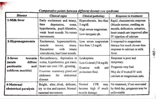
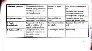
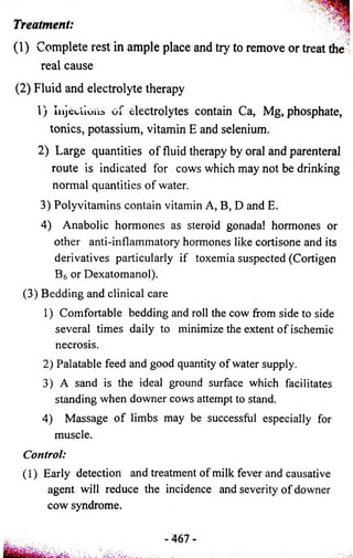
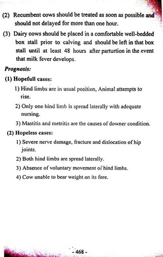
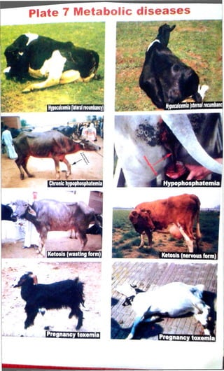
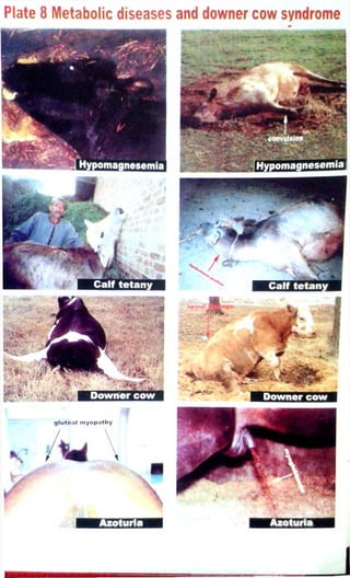
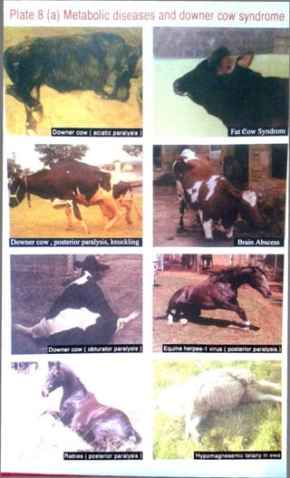
![Nutritional deficiency diseases
(1) Minerals:
(1) Major elements are essential for growth. Their deficiency
include: Phosphorous deficiency, Rickets, Osteomalacia
and Gi.i.eodystrophic fibrosis.
(2) Trace elements are essential for healthy growth. They
include: copper, iron, zinc, iodine, selenium, cobalt and
manganese.
(2) Vitamins:
(1) Fat soluble vitamins as vitamin A, D, E & K.
(2) Water soluble vitamins as Bb B2, B6, B|2 & C.
Etiology; o f the disease:
11] Evidence of existence of deficiency:
(1) Diet:
1) The type of the soil affects the plant growing on it.
2) Farming practices and marketing may affect the
presence of specific nutrients in live stock.
(2) Abnormal absorption:
Diet may contain adequate amounts or others substances
which can decrease the absorption such as:
1) Excess phosphate reduce calcium absorption.
2) Excess calcium reduce the absorption of iodine.
3) Absence of bile salts prevent absorption of fat soluble
vitamin.
4 | Chronic enteritis reduces dietary digestion and absorption.](https://image.slidesharecdn.com/vet-141021053329-conversion-gate02/85/Vet-internal-medicine-text-book-489-320.jpg)
![(3) Abnormal utilization of ingested nutrient:
Ingested certain material may effect on the development
of the deficiency e.g. molybdenum and sulphate reduce copper
absorption,metabolism and storage.
(4) Abnormal requirement:
Stimulation of the growth rate of animals by improved
nutrition of other practices may increase their requirement of
specific nutrient to the point where deficiency disease occurs.
[21 Evidence of a deficiency:
As to the cause of disease (clinical signs & PM): e.g
Night blindness indicate vitamin A deficiency. Level of
vitamins and minerals in the blood, urine, liver biopsy and
postmortem lesion help in diagnosis.
[3] Evidence based on cure or prevention by correction of
deficiency:
By observing the effects of specific addition to the ration
(diagnostic therapy).
Distribution of Calcium, phosphorus and magnesium:
(1) Ca, inorganic ph and Mg ions are all found ECF. Ca
and ph constitute the abundant cation after K in bone. Mg
is abundant anion after K in all ICF.
(2) Bone contains 99% of ca, 85% of ph and 75% of Mg of the
body. The main function of the three ions is in the bone
formation.
(3) Magnesium and to a lesser extend calcium are important
cofactor for many enzyme.](https://image.slidesharecdn.com/vet-141021053329-conversion-gate02/85/Vet-internal-medicine-text-book-490-320.jpg)
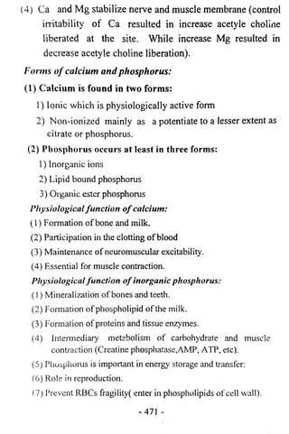
![(3) Poor calcification of diphyseal bone results in stresses and
bowing of long bones and enlargement of the joints.
Symptoms:
(1] Subclinical rickets:
The most important one and respond easily to treatment. It
is detected only by blood and serum analysis ofCa, ph and
vitamin D.
(2) Clinical rickets:
(1) Stiffness in gait.
(2) Enlargement of the limb joints especially in the forelegs
and costochondral junction (button like projection).
(3) The long bones show abnormal curvature usually forward
and outward at the knee.
(4) Lameness and tendency to lie down.
(5) Arching of the back
(6) Irregular and delayed eruption o f the teeth, poorly calcified
teeth with pitting grooving and pigmentation.
(7) Thickening and softening ofjaw bones.
(8) Dyspnea and chronic rumen tympany in severe cases.
(9) Finally hypersensitivity, tetany, fracture may occur
spontaneously, lameness, recumbency and death.
Clinical pathology:
Decrease in serum Ca level to 4-5 mg% (normal 10-12
g%) and serum ph level to l-2mg% (normal 6 mg%) or
imbalance between them.](https://image.slidesharecdn.com/vet-141021053329-conversion-gate02/85/Vet-internal-medicine-text-book-494-320.jpg)
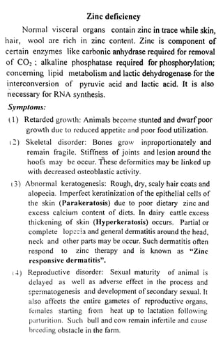
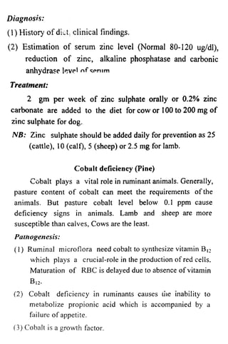
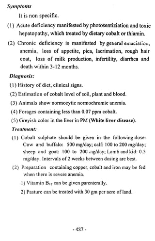
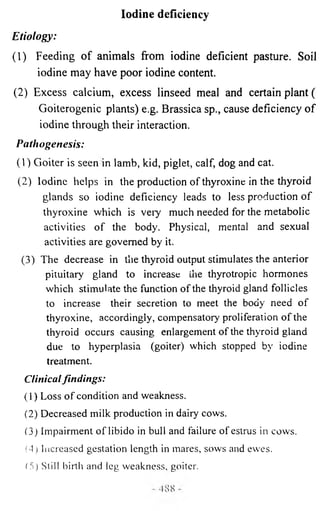
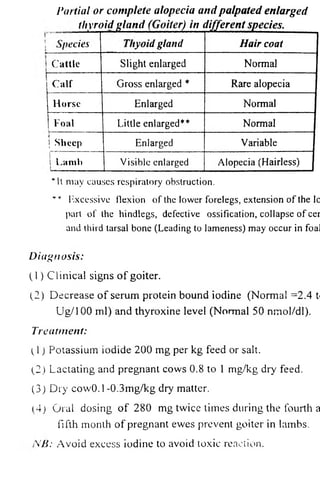
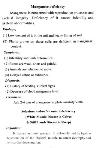

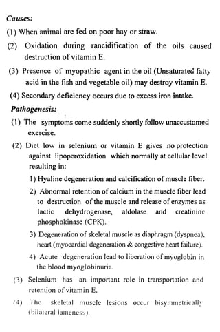
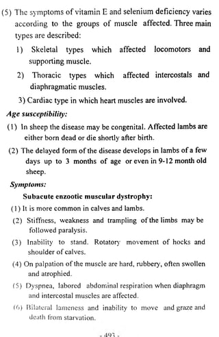
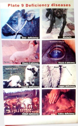
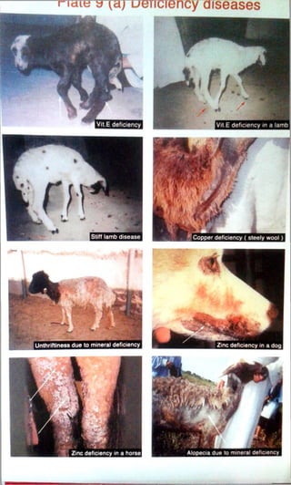
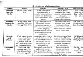
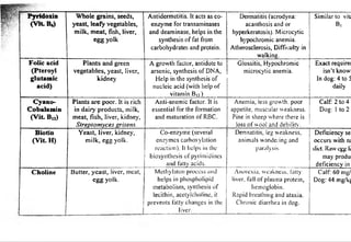
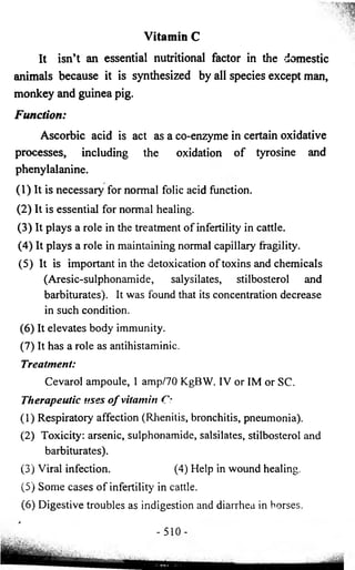
![Dermatology
(Skin Diseases)
The skin is a covering for the whole body so it is a mirror
that reflects many systemic and local diseases. The incidence of
skin diseases in Uutneslic animals is high. Many of the skin
diseases are contagious therefore quick and accurate diagnosis
is important in their control and eradication.
Function o f the skin:
(1) Protection from light, organisms and mechanical.
(2) Immunologic function.
(3) Vitamin D synthesis.
(4) Heat regulation.
(5) Secretary organ by sweating : e.g. wastes, urea.
(6 ) Water reserve.
(7) Sensory organs.
(8 ) Reflection of internal feelings.
Causes o f skin diseases:
[1] Infectious:
(1) Viral:
1) Contagious erythema.
2) Ulcerative dermatitis in sheep.
3) Lumpy skin diseases (in cattle).
4) Pox.
5) Vesicular papular dermatitis (in horse).
-511 -](https://image.slidesharecdn.com/vet-141021053329-conversion-gate02/85/Vet-internal-medicine-text-book-533-320.jpg)
![(2) Bacterial:
1) Bacterial dermatitis. 2) Tuberculosis. 3) Foot rot.
(3) Mycotic:
1) Ring worm. 2) Epizootic lymphangitis.
3) Spirocheta.
(4) Parasitic:
1) Protozoa e.g. Lishmaniasis.
2) Arthropodes as mange.
3) Helminthes as filariasis.
[2] Non infectious:
(1) Nutritional deficiency of zinc, cobalt, iodine and copper.
(2 ) Chemical injury as acids, alkalies and gases.
(3) Histamine production: urticaria.
(4) Lesions due to severe abrasion or friction.
(5) Disturbance in circulation such as erythema.
(6 ) Distribution in nervous tissue “itching”.
(7) Internal diseases (constipation, nephritis).
(8 ) Neoplasms (wastes, papilloma, carcinoma).
(9) Congenital defects (alopecia).
(10) Over feeding of carbohydrate without enough exercise.
(11) Traumatic (wounds).
(12) Thermal, excessive heat, sun rays.
(13) Long administration of some drugs as arsenic, mercury,
serum causing skin rashes.
(14) Endocrine disorders.
- 5 1 2 -](https://image.slidesharecdn.com/vet-141021053329-conversion-gate02/85/Vet-internal-medicine-text-book-534-320.jpg)
![Scientific Term used in Skin Diseases
[1] Primary lesions (less than 1 cm in size):
(1) Macule: It is a flat circumscribed discoloration o f the skin
or mucous membrane.
(2) Papule: A solid elevation of the skin or mucous membrane
extending deeper in the epidermis.
(3) Nodule: A solid elevation of the skin or mucous membrane
extending into the dermis (neoplastic or inflammatory).
(4) Vesicle: A fluid filled superficial circumscribed elevation
of skin or mucous membrane.
[2] Lesions more than 1 ml:
Such as cyst, tumor, granuloma, plaque.
[3] Primary lesions of varying sizes:
(1) Pustule: Vesicle of the skin or mucous membrane which
contain pus.
(2) Wheal: An irregularly shaped, elevated transient lesion of
the skin or mucous membrane due to edema.
[4] Secondary cutaneous lesions:
(1) Erosion: Shallow ulcer of the epidermis doesn’t penetrate
the entire epidermis.
(2) Erythema: A redness of skin due to capillary congestion.
(3) Hyperkeratosis: An increased thickening of the keratin
layer of the epidermis (normal in the foot pads, nose of dog
and cat).
(4) Scab or crust: A collected dried exudate on the surface of
the epidermis adherent to hairs.](https://image.slidesharecdn.com/vet-141021053329-conversion-gate02/85/Vet-internal-medicine-text-book-535-320.jpg)
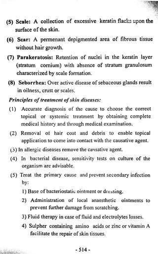
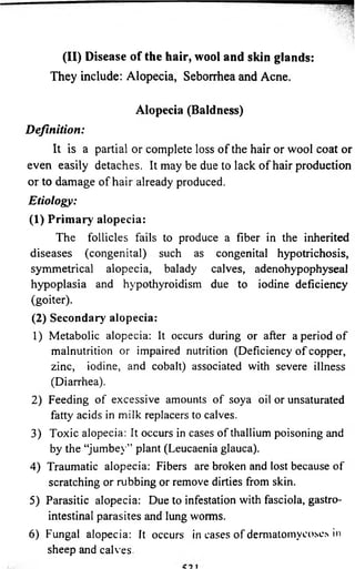
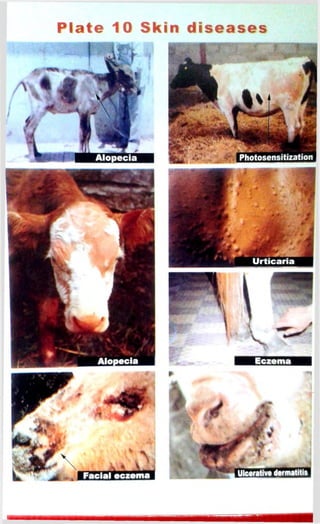
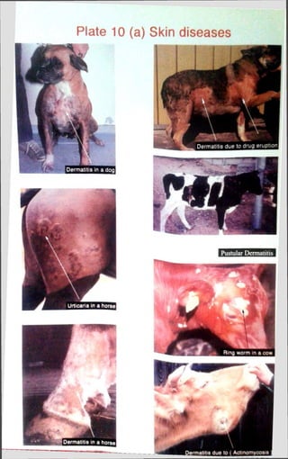
![Diseases of musculoskeletal system
Anatomy:
The musculoskeletal system consists o f muscles & their
attachments, bones & joints.
Physiology:
This system has an important role for movement gait,
posture & locomotion as well as respiration, mastication,
urination, and defecation protection certain vital organs.
Locomotor disorders & Lameness:
They may be involved in many diseases o f
musculoskeletal system (Bones, muscles, tendons &
ligaments) nervous system &even blood vascular system.
Clinical approach o f musculoskeletal system:
[1] History:
(1) Numbers o f animals showing similar condition.
(2) Age (e.g Rickets in young, osteomalacia in adult.
(3) Relation with pregnancy or production.
(4) Relation with unhygienic ground condition.
(5) History o f transportation, trauma, injury or accident.
(6) Injury, fracture or lameness due to overcrowding & or floor
soiled with urine, feces, mud & water.
(7) Duration o f illness.
(8) History o f gait, posture, movement, lameness.
(9) History o f diets as:
1) Copper deficient diet inhabit osteoblast activity
2) High magnesium & low calcium cause poor bone growth.](https://image.slidesharecdn.com/vet-141021053329-conversion-gate02/85/Vet-internal-medicine-text-book-566-320.jpg)
![3) Low magnesium produces muscle incoordination^
poor bone development.
4) Low zinc causes poor ostoblastic activity & osteopenia.
5) Low vitamin A level impairs osteoblastic activity&
fragile bone
6) Low vit.D level may bring rickets.
7) Low protein level may reduce osteoid formation.
[2] Inspection:
(1) Posture:
It is the state o f animal during standing or recumbency.
Abnormal posture may include:
(1) Cow sits on sternum, head rest on flank: occurs in case o f
milk fever.
(2) Sheep or goat lies in frog-like posture: occurs in case o f
parturient hypocalcemia.
(3) A calf in lateral recumbency with head hold in
opisthotonus along with tonic clonic contraction o f limbs:
occurs in casp o f bypomagnesemic tetany.
(4) A cow keeping on the carpal joints & unable to stand: in
case o f calcinosis condition o f nutritional origin.
(5) A horse shifting its weights from one limb to other limb
continuously: in Laminitis, osteodystrophia fibrosa.
(6) A cow stands with crossing o f forelimbs: occurs in cases o f
ketosis, painfull fracture o f third phalanx.
(7) A cow in sternal recumbency with hindlimbs extended
behind (frog like posture): in bilateral hip dysplasia.](https://image.slidesharecdn.com/vet-141021053329-conversion-gate02/85/Vet-internal-medicine-text-book-567-320.jpg)
![[3] Local examination:
Using palpation, pressure test, percussion, probang &
compression to determine pain, swelling, thermal response
(hot or cold), crepitation, mobility & movement o f limbs &
joints.
(1) Passive movements of limbs to identify:
1) Fracture
3) Pain
2) Dislocations
(2 ) P a lp a tion o f muscles for evidence of:
1) Enlargement 2) Atrophy
3) Pain test for evidence o f paralysis or inflammation.
S p e c ia l examination o f musculoskeletal system:
(1) Radiography: for diagnosis the diseases o f bones, joints &
soft tissue swelling o f limbs.
(2) Muscle biopsy: for microscopic & histochemical evaluations.
(3) Arthroscopy: using special endoscope for visceral
inspection o f jo in t cavity & articular surface .
(4) Arthrocentensis: collection of joint fluid for cellular,
biochemical & inflammatory conditions.
(5) Ultrasonography: for soft tissue examination.
(6) Electromyography: to detect the activity of muscle & nerve.
(7) Clinical pathology: for synovia, blood, urine, tissue
specially serum level o f Ca,P & vitamin D for bone
diseases; Creatine phosphokinase (CPK), Aspartate
amino-transferase (AST) & Alkaline phosphatase enzyme
for muscle diseases.](https://image.slidesharecdn.com/vet-141021053329-conversion-gate02/85/Vet-internal-medicine-text-book-569-320.jpg)
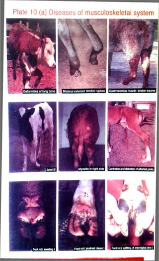
![Lameness above the fetlock origin
It may be caused by:
[1] Nerve paralyses:
Of the forelimb include the suprascapular, brachial plexus
and radial. The main hindlimb involvement is with the
femoral, obturator, tibial, peroneal and sciatic. The usual cause
is trauma. The treatment in all forms involves careful nursing
and supportive care (e.g. oral fluids).
[2] Contracted flexor tendons:
Symptoms:
Arthrogryposis or articular rigidity.
Etiology:
(1) May be congenital in cattle, usually in flexion.
(2) Dystokia.
(3) Akabane virus d-Ingestion of various toxic plants.
Clinical signs and diagnosis:
(1) Mild cases:
1) May be self-correcting as the calf moves around to an
increasing extent.
2) The mild cases show about 10-20 excessive flexion of
the carpus and forelocks. Forced extension discloses
the tautness in flexor tendons and suspensory ligament.
3) Contracted tendons usually affect both deep and
superficial flexors & sometimes the suspensory
ligaments. Sometimes the hindlimbs have abnormal
hock extensor rigidity and fetlock flexion.](https://image.slidesharecdn.com/vet-141021053329-conversion-gate02/85/Vet-internal-medicine-text-book-592-320.jpg)
![Lameness above the fetlock origin
It may be caused by:
[1] Nerve paralyses:
Of the forelimb include the suprascapular, brachial plexus
and radial. The main hindlimb involvement is with the
femoral, obturator, tibial, peroneal and sciatic. The usual cause
is trauma. The treatment in all forms involves careful nursing
and supportive care (e.g. oral fluids).
[21 Contracted flexor tendons:
Symptoms:
Arthrogryposis or articular rigidity.
Etiology:
(1) May be congenital in cattle, usually in flexion.
(2) Dystokia.
(3) Akabane virus d-Ingestion of various toxic plants.
Clinical signs and diagnosis:
(1) Mild cases:
1) May be self-correcting as the calf moves around to an
increasing extent.
2) The mild cases show about 10-20 excessive flexion of
the carpus and forelocks. Forced extension discloses
the tautness in flexor tendons and suspensory ligament.
3) Contracted tendons usually affect both deep and
superficial flexors & sometimes the suspensory
ligaments. Sometimes the hindlimbs have abnormal
hock extensor rigidity and fetlock flexion.](https://image.slidesharecdn.com/vet-141021053329-conversion-gate02/85/Vet-internal-medicine-text-book-594-320.jpg)
![(3) The hind limbs are hyperextended caudally in a fixed • ■
manner that may persist for several minutes. Spasms may 4i
extend forward to the forelimbs and neck muscles.
(4) The head is extended and the forefeet advanced with the
back arched. If then forced to move, the gait is stiff, and
episodic raising of hind limbs may occur during forward
movement.
(5) Animals may recumbent when the condition is advanced.
Treatment:
The spastic syndrome is incurable. Vitamin D, TriB, E,
bone meal and sedatives can be used.
[4] Lameness of iatrogenic:
Iatrogenic damage may be a result of treatment for
diseased or injury. It occur in cases of:
(1) Radial paralysis form prologed lateral recumbency during
anesthesia.
(2) Neonatal long bone fracture & physical separation
resulting from excessive traction in dustokia.
IM injection, result in a massive abscess, which cause
Lameness that require surgical interference.](https://image.slidesharecdn.com/vet-141021053329-conversion-gate02/85/Vet-internal-medicine-text-book-595-320.jpg)
![Diseases of the newborn
Newborn diseases have a great economic importance
among dairy farms. These diseases cause a high mortality rate
among newborn lambs (20%) and calves (10-30 %). These
diseases may be:
[1] Physical cause: (Hyper or hypothermia)
[2] Parturient injury from dystokia
[3] Congenital Defects
[4] Poisonous plants: May cause teratogenic effects.
[5] Fetal hypoxia (neonatal hypoxia)
[6] Neonatal hypothermia.
[7] Hypoglycemia
[8] Failure of passive transfer (FPT)
[9] Sepicemia
[10] Umbilical abnormalities
Congenital Defects
These defects have a maternal causes. They include:
(1) Virus infections (2) Nutritional Deficiency
1) Blue virus: in lambs. 1) Iodine: goiter in all species.
2) Bovine viral diarrhea virus. 2) Copper deficiency in lambs.
3) Akabane virus. 3) Cobalt.](https://image.slidesharecdn.com/vet-141021053329-conversion-gate02/85/Vet-internal-medicine-text-book-596-320.jpg)
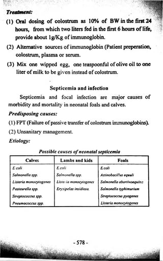
![, - v - ■ . ; : Y • Y / . - y y
'* ;» • v Y:' ■ ' . Y ' .. 'I
2) Appropriate and early therapeutic intervention
with in uteroacquired infections often results
favorable outcome: survival rates greater than 75%.
(2) The long-term prognosis for future pedormance is guarded
if multifocal bone or joint disease is diagnosed.
Prevention:
(1) Good hygenic sanitory condition.
(2) Sanitory management o f the dam specially udder and
genital system
Umbilical abnormalities
[1] Umbilical infections
Definition:
It is inflammations which may be involve the urachus,
umbilical veins (omphalophlebitis), one or both umbilical
arteries (omphaloarteritis), or many structures (Omphalitis,
umbilical abscess).
Pathogenesis:
Infection may originate following contamination o f the
external umbilicus fter irth r esult rom ther ites uring
periods o f septicemia. Bacteria may localize in the umbilical
vessels, urachus, bladder, or interstitial tissues and the
infection may extend into the peritoneal cavity or progress to a
generalized septicemia. Urachal abscessation can cause the
previously closed urachus to become patent externally or allow
urine to leak subcutaneously or into the peritoneal cavity.
-5S5-](https://image.slidesharecdn.com/vet-141021053329-conversion-gate02/85/Vet-internal-medicine-text-book-610-320.jpg)
![[2] Patent urachus
Etiology
(1) Congenital patent urachus caused by excessive torsion o f
the umbilical cord in utero causes urachal obstruction,
urine retention in the bladder, distention of the proximal
urachus and interference with urachal involution.
(2) Acquired patent urachus: Reestablishment of urine flow
after a normal urachal closure at birth by any insult that
causes tension on the abdominal wall (e.g., prolonged
recumbency, tenesmus, abdominal distention) r
umbilical inflammation (e.g., omphalitis) may lead to the
development of an acquired patent urachus.
Clinical findings:
(1) Urine is dribbling or flowing from the umbilical stump.
(2) Dermatitis may develop on the hindlimbs and ventral
abdomen from urine scalding.
(3) Fever, purulent discharge, and pain on palpation of the
umbilicus
Treatment:
(1) Sanitary management.
(2) Local therapy and removal of predisposing conditions.
(3) Cauterization of the urachus with silver nitrate sticks, 2%
iodine, or local procaine penicillin.
(4) Surgical management.
Prevention:
(1) Sanitary enviroment.
-587-](https://image.slidesharecdn.com/vet-141021053329-conversion-gate02/85/Vet-internal-medicine-text-book-612-320.jpg)
![(2) The umbilicus should be allowed to rupture spontan
(3) Critically ill neonates should be restrained to prevent f
excessive tension on the ventral abdomen.
[3] Excessive bleeding from the umbilicus
Bleeding from the umbilicus may occur, particularly if it
was cut or ligated. Occasionally, hemorrhage is severe enough
to necessitate a blood transfusion. Rarely, hemoperitoneum
will result from hemorrhage from an intra-abdominal umbilical
vessel
588](https://image.slidesharecdn.com/vet-141021053329-conversion-gate02/85/Vet-internal-medicine-text-book-613-320.jpg)
![Fluid therapy
[1] Parenteral fluid therapy:
1) Must be injected slowly IV at a rate o f 10 ml / min.
2) Avoid sudden & or excessive dilution of blood.
3) Must be within normal body temperature.
4) Similar to blood viscosity, pH, osmotic pressure.
5) Complete excretion or metabolized with long duration
circulation.
6) Clean, sterile, no turbidity or precipitate.
7) Non toxic, non antibody formation.
(1)Electrolytes:
Sodium replaces Na loss & increase body water uptake.
Potassium replace K loss & act as antacids, e.g.: Saline;
Ringer; Ringer’s lactate solution.
(1) Saline solution (0.9% sodium chloride).
(2) Ringer solution (contains Na, K, Cl & Ca at dose of 130,
40, 11& 3 m. Eq/ L, respectively).
(3) Ringer’s lactate solution (as Ringer solution plus 28 m.
Eq./ L, Lactate).
(2) Glucose or Dextrose:
For energy supply. It increases Na & body water uptake.
(1) Glucose 5% is used as fluid therapy.
(2) 10 to 25 % as a source of energy, renal & hepatic wash.
(3) 25 to 40% in cases of hypoglycemia (ketosis).
(4) 40% should not exceed 20 ml/Kg In 24hrs period.
(5) Glucose 50% is used as diuretic in renal failure.](https://image.slidesharecdn.com/vet-141021053329-conversion-gate02/85/Vet-internal-medicine-text-book-619-320.jpg)
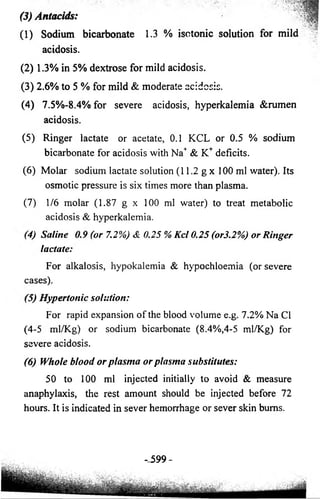
![Blood transmission:
Must be from same species, breed, and age or from the
dam (of the same blood group).
Blood transfusion:
Collect 500 ml of whole blood from donar animal o f the
same species in a sterile autoclaved saline bottle containing an
anticoagulant like sodium citrate solution (3.85%) 10 ml for
100 ml of blood. Blood is generally collected from the jugular
vein using a wide bore needle or a canula. The blood can be
collected at the rate of 10-15 ml/kg BW from the donar animal
in the inner wall of the bottle to prevent frothing. Filter the
blood through the sterile gauze if froth is formed. Blood can be
stored in freeze for a week. Cattle blood could be preserved for
22 days for a safe transfusion.
[2] Oral fluid therapy formation:
(1) Oral electrolytes as the intestinal absorption of sodium
replaces Na loss & increase body water uptake.
Potassium replaces K loss & antacids e.g. Saline; Ringer.
(2) Oral glucose or dextrose for energy supplies increase Na &
body water uptake. Glucose sodium ratio should be 1:1 to
3:1 mixed with milk.
(3) Amino acid, glycine & alanine for nitrogen source increase
Na & water uptake; glutamine may facilitate repair of
damaged intestinal epithelium; acetate &propionate also
increase intestinal Na & water uptake. These preparation
as well as acetate containing fluid can be fed with milk as
acetate does not raise abomasal pH or inhibit milk clot.
- 600-](https://image.slidesharecdn.com/vet-141021053329-conversion-gate02/85/Vet-internal-medicine-text-book-621-320.jpg)
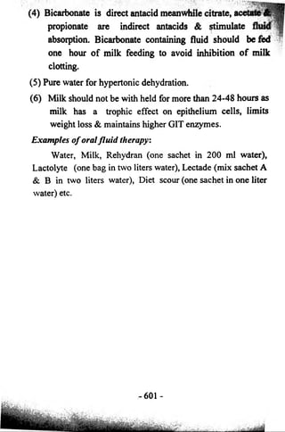
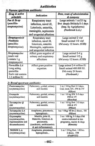
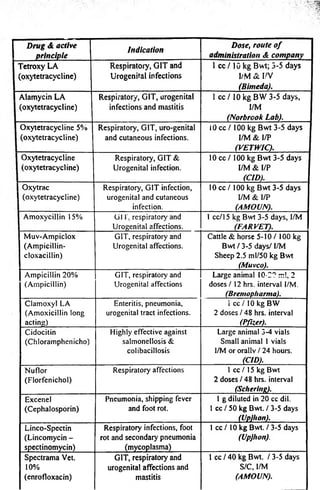

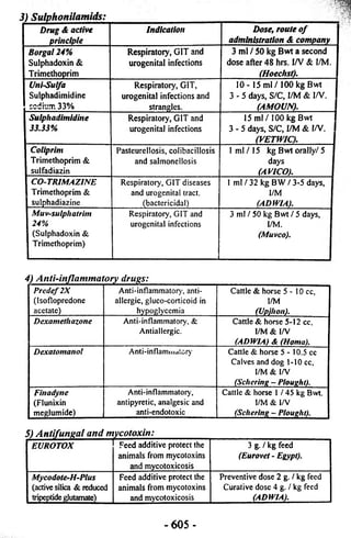
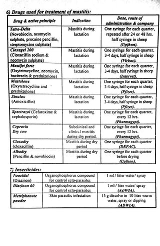

![Drugs A active
principle Indication Dose, route o f - 3 |
administration A companr
Levamisoie Broad spcct. anthelmentics
against round worm of
GIT, lung of cattle, sheep
and goat.
S ml/15 kg oral drench
(Pharma-SweaJ)I
Hapadex GIT nematodes, cestodes,
fasciola hepaticaand
paramphistomum
15 ml/100 kg Bwt
paramphist. 40 ml/100 kg
(Sobering- plough). ]
Vaibazine sus
(Albendazole)
GIT nematodes, round,
tape worms and fasciola
4ml/10kgBwt orally for GIT
nematodes&aduit fascioltasis
(Pfizer).
Vaibazine tab
(Albendazole)
GIT nematodes, round,
tape worms and fasciola
1 tablet/80 kg Bwt orally
(Pfizer).
Piperazine
Citrate 50%
Ascaridia and oxyuris in
cattle and horses
40 g / 100 kg Bwt. Orally
(Uccma).
Piperazine
DHC 52%
Ascaridia and oxyuris in
cattle and horses
11 g / 100 kg Bwt. Orally
(ADWIA).
Yomesan
(Niclozamid)
Tenia spp. and
paramphistomum.
Cattle, sheep & dog 1
tablet / 10 kg Bwt Orally
(Byer).
Equvalan
(Ivermectin)
Internal parasites of horse Past syring / 600 kg Bwt
on the base of the tongue
(MSDAgvet).
9) Drugs used for treatment of blood parasites:
Berenil
Babesiosis,
(Diminazine &
trypanosomiasis and
antipyrin)
theiieria
Prepared 7% aqueous solution, 3.5
mg/kg Bwt; vial 1.05g. dis. 12.5
ml water for 300 kg Cattle, I/M
(Hoechst).
Imizol
(Imidocarb
diproionate 12%)
Babesiosis and
anaplasmosis.
Cattle & sheep 1 ml /100 kg Bwt as
one d^e. I/M & S/C Horse 2 ml/100
kg Bwt two doses 48 hrs. Intervals.
Dog 0.5 ml / 10 kg Bwt.
(Schering-Plough).
Butalex
(Buparvaquone)
Thieleriosis Cattle 1 m l/20 kg/l/M
(Pitman moors).
Naganol Trypanosomiasis 1 g / 50 kg Bwt dissolved in
distilled water 10%soln; I/V
maximum dose in Camel 8 g.
(Byer).
Arsina! 10 %
(Na Methyl arsenate)
Thieleriasis and
rumen stimulant.
Cattle 8 -12 cc S/C & l/M
(VETWIC).
-608](https://image.slidesharecdn.com/vet-141021053329-conversion-gate02/85/Vet-internal-medicine-text-book-629-320.jpg)
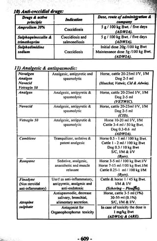
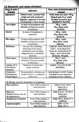
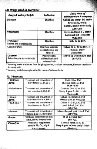
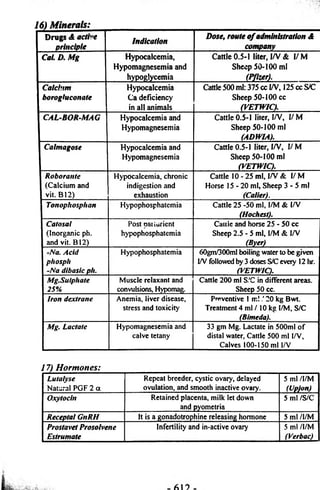
![18) Fluid therapy:
1 Drugs A active
I principle
Indication Dose, route of 1
administration A company 1
I Adwla Electrolytes C m ] electrolyte for calves in
cases of dehydration,
diarrhea, hyponatremia
And acidosis.
50- 100 g./calves or j
Iambs, dissolve in 2 liters of I
w a te r o r m ilk . I
(ADWIA). |
1 Lactolyte Oral electrolyte for calves in
(A, f 4 I a • « wuocj w l u w itju ia tiu ii,
d ia rrh e a , h y p o n a tre m ia a n d
a c id o s is .
1 b a g ( 9 0 ) d is s o lv e d in 1
w o rm w a te r ( 2 lit e r ) , tw ic e I
tim e s d a ily 2 - 3 d a y s . 1
(Vlrbac). I
I Lectade R e h y d ra te s s c o u rin g in
c a lv e s ,
M ix s a c h e t A a n d B tw o J
lit e r o f w o rm w a te r, 2 - 3 1
tim e s d a ily . j
(Beech am). j
1 Diet scour
I ( a n tib io tic , v ita m in s
I & e le c tro ly te s )
P re v e n tio n a n d tre a tm e n t o f
s c o u r in Iam b s a n d c a lve s .
1 00 g d is s o lv e d in o n e - lite r I
w a te r, 2 -3 tim e s d a ily . J
(Virbac). |
1 Rehydran O ra l re h y d ra tio n s o lu tio n E a c h s a c h e t d is s o lv e d in }
2 0 0 m l w a te r, tw ic e d a ily . I
(CID). 1
1 Sodium chloride
0 .9 , 5 ,7 . 2 , 1 1 .2%
D e h y d ra tio n , h y p o n a tre m ia
a n d a lk a lo s is
A c c o r d in g to d e g re e o f j
d e h y d ra tio n .
(ADWIA). I
1 Ringer
(N a C l, K C I, C a C l)
It is u se d fo r re h y d ra tio n a n d
as s o u rc e o f c a lo rie s
A c c o rd in g to d e g re e o f
d e h y d ra tio n .
(ADWIAA
1 Ringer lactate
(N a C l, K C I, C a C l)
It is u sed fo r re h y d ra tio n a nd
as s o u rc e o f c a lo rie s
A c c o rd in g to d e g re e o f
d e h y d ra tio n .
(ADWIAj.
1 Glucose or Dextrose
5 %
I t is u sed fo r re h y d ra tio n a nd
as s o u rc e o f c a lo rie s
A c c o rd in g to d e g re e o f
d e h y d ra tio n .
(ADWIA).
I Dextrose 25% It is used as so u rce o f
c a lo rie s , as o sm o tic d iu re tic
A c c o rd in g to d e g re e o f
d e h y d ra tio n .
(ADWIA).
I Dextrose 50% It is used as s o u rc e o f
c a lo rie s as o sm o tic d iu re tic
A c c o rd in g to d e g re e o f
d e h y d ra tio n .
(ADWIA).
1 A fa bicarbonate
1.3%
In case o f m ild a c id o s is . A c c o rd in g to d e g re e o f
a c id o s is .
(ADWIA).
I Na bicarbonate
3 -5 , 5 , 8 .4 %
H y p e rto n ic s o lu tio n fo r
se ve re a c id e m ia .
A c c o rd in g to d e g re e o f
a c id o s is .
(ADWIA).](https://image.slidesharecdn.com/vet-141021053329-conversion-gate02/85/Vet-internal-medicine-text-book-634-320.jpg)
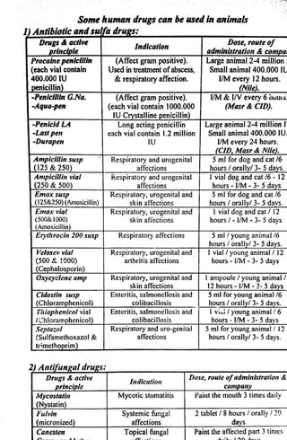
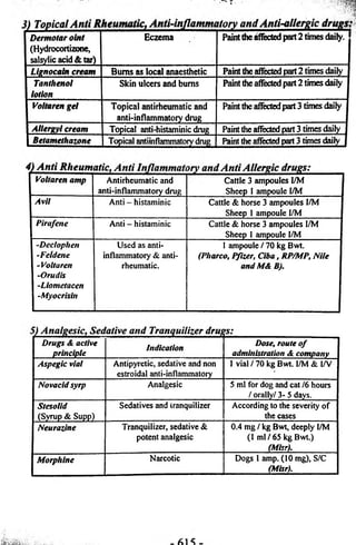
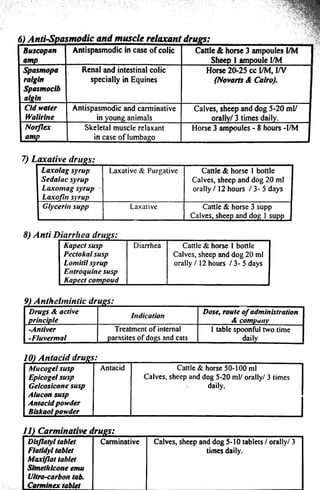
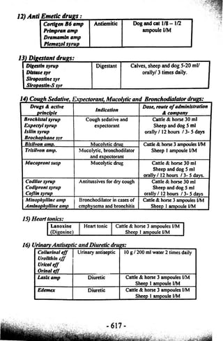
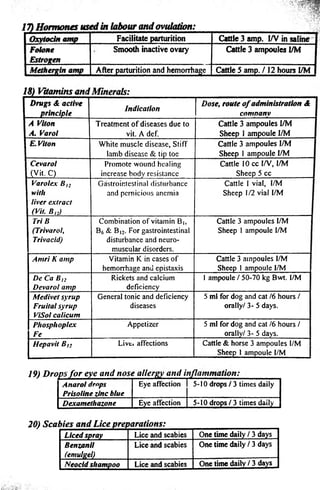
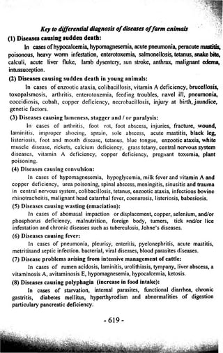
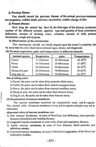
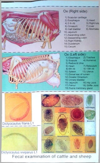
![Laboratory Diagnosis
[1] Fecal examination:
The fecal sample (5-10 g) is collected directly from the animal in a clean dry
glass container. The feces are kept in a refrigerator at 4°C in delay examination.
Methods of fecal examination:
1) Direct method:
Place a drop of distilled water in the middle of a clean dry glass slide, add
small amount of feces, mix and place a cover slide. Examine it under microscope
for the presence of parasitic ova. If no parasitic ova is detected it should be
examined by qualitative method.
2) Qualitative concentration method:
Feces are mixed with either of the saturated sugar, saturated salt solution or
41% magnesium sulfate solution. The parasitic ova, being lighter float on the top of
fluid and can be concentrated for examination.
(1) Simple flotation method: 1 g of feces is mixed with few ml of distilled water,
filtered through a fine sieve, mixed with 4-5 ml of saturated salt solution, then
placed in a tube or cylinder and filled up to the top with solution, cover the
tube with glass slide and left it 30-60 minutes at room temperature. Remove
the cover slide and examine under the microscope.
(2) Concentration flotation method: 1 g of feces mixed with few ml of distilled
water, filtered through a fine sieve. The filtrate is mixed with saturated suger
solution in a ratio of 1:3 in a test tube, mix the contents and centrifuge at 1500
rpm / 5 minutes. Transfer the small amount of superficial contents of tube on a
clean and dry glass slide and examine for the presence of parasitic ova. The
sediment can be examined for eggs of trematodes.
(3) Baermen,s technique in cattle and horse: Put a Small amount of feces in guaze
inside funnle filled with worm water, examine the first few drops to detect
the larva after 2 hours.
(4) Vida technique in sheep: Pellet of feces is mixed with warm water in a
petredish for 10 minutes then crushed by forceps and examine after 10
minutes.
[2] Urine examination:
Urine samples can be collected
in cows and sheep either by
stimulation of the urethra through
valva or by catheterization. In delay
exam, store the urine in refrigerator
at 4°C.
Suburethral
diverticulum
Urinary
bladder
External
urethral orifice
- 6 2 7 -](https://image.slidesharecdn.com/vet-141021053329-conversion-gate02/85/Vet-internal-medicine-text-book-649-320.jpg)
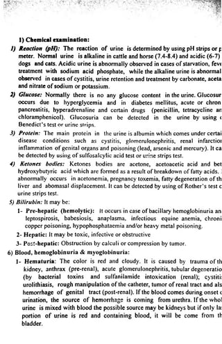
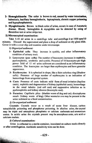
![[3] Skin scraping examination:
The scrapings must be collected deeply from the most affected parts o f the
skin, it is better to moist the affected area with mineral oil before scrabing.
1) Direct method: The skin scrapings are placed on a clean and diy glass slide
with one drop 10% sodium hydroxide, cover it with cover slip and examined
under low power o f microscope.
2) Sedimentation methods: The skin scrapings are kept in 10% potassium or
sodium hydroxide, 2-4 hours then transfer to a centrifuge tube and centrifuged
at 3000 rpm/10 minutes. The supernatant is discarded and one drop o f the
sediment examine under microscope.
3) Examination of skin scrapings for fungi:
/- Examination by Wood’s lamp: The Wood’s lamp has UV light, which is
directed on the skin or scrapings collected in petri dishes. If the microsporum
fungi are present, it will give a yellow green fluorescence, while no
fluorescence in negative infection.
2- Microscopic examination: Collection o f skin scrapings should be from the
center as well as from the periphery o f the lesion. Swab the lesion with 95%
alcohol to remove any saprophytic organism. The skin scrapings are
collected in sterilized petri dishes containing 10% sodium hydroxide or
potassium hydroxide. Put one drop on a clean slide, cover it and apply
Vaseline around the rim o f cover slip. The slide is warmed gently for few
seconds, then examine for the presence o f chains o f hyphae and spores.
[4] Examination of milk:
Collection o f the samples: The udder o f the animal should be cleaned with
water and antiseptic solution like potassium permanganate (1:1000). The hands o f
this examiner should be cleaned with soap and antiseptics. Disinfect the teats with
alcohol 70%. collect the milk sample from each teat in a separate tube (5-10 ml)
and discard the first 3-4 streams o f milk, all tubes should be stoppered and
transported to in an ice bag for the laboratory examination.
(1) Physical examination of milk:
1) Color: The normal color o f milk is white, but in acute mastitis it may become
reddish (presence o f blood) or Yellow which occur during colostral period,
feeding o f carrots and/or due to tetracycline therapy.
2) Reaction: The pH o f normal milk is 6.4 to 6.6 but in mastitis it becomes alkaline
up to 7.4 due to the presence o f sodium bicarbonate in the milk. The reaction
can be determined by using pH strips or pH meter.](https://image.slidesharecdn.com/vet-141021053329-conversion-gate02/85/Vet-internal-medicine-text-book-652-320.jpg)
![3) Odor: The normal color o f milk is pleasant but in mastitis due to actinomyces /,
pyogenes it has an offensive odor. In ketosis the odor o f milk becomes sweet ?
and fruity (acetone smell).
4) Consistency: The colostrum is normally viscous in character. In acute and
subacute mastitis the milk contains fine and large flakes, while it becomes
watery in case o f poor feeding and chronic mastitis.
(2) Chemical examination of milk:
1) White slide test: Milk (4-5 drops) are placed on a clean dry slide, add a drop o f
4% sodium hydroxide and mix with a glass rode. Thickness and flakes appear
in mastitis.
2) California mastitis test: This test is based on an increase in the number o f
leucocytes and increase alkalinity in mastitic milk. Take 0.5 ml milk from each
quarter in plastic peddle cups and add equal quantity o f California reagent,
mix well by circular movement o f paddle on a horizontal plane.
1- Liquid milk with no streaks or precipitation is negative for mastitis.
2- Streaky milk is weak positive (+).
3- Slimy is strong positive (++).
4- Gelatinous is very strong positive (+++).
3) Leucocytic count: Mark the area on central portion o f the slide (1 square cm).
Put 0.1 ml, spread the milk sample by bacteriological loop in this area, dry the
smear and dissolve the fat by rinsing in xylene for 2-5 minute. Fix the smear
with alcohol 4-5 minutes and stain with methylene blue for 1 minute. Remove
the excess stain by immersing the slide in alcohol. Count the leukocyte under
oil immersion lens. The cell o f 10 field are counted and averaged and
multiplied by 500.000 to get the total number o f leukocytes in the milk.
(3) Bacteriological examination of milk:
1) Direct microscopical examination.
2) Cultural examination: The milk is collected in sterilized vials, stored in a
refrigerator and send to the laboratory, for isolation o f the organism and
antibiotic sensitivity test.
[5] Rumen juice examination:
Examination o f rumen juice (RJ) gives rapid diagnostic test for monitoring
the function o f the rumen as well as the nutritional health o f the animals. The
rumen juice is collected from animals by using a stomach tube that was introduced
through the mouth, then moved to and fro to obtain a representative sample from
different areas o f the rumen. The pH o f the rumen juice, ammonia concentration,
and volatile fatty acids must be measured as soon as possible.
- 631 -](https://image.slidesharecdn.com/vet-141021053329-conversion-gate02/85/Vet-internal-medicine-text-book-653-320.jpg)