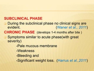
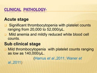
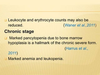
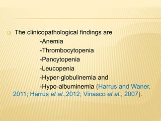
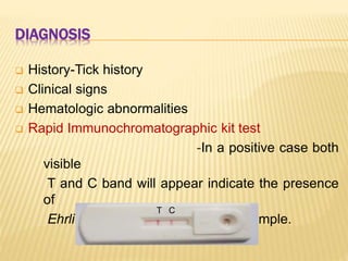
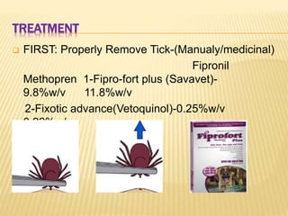
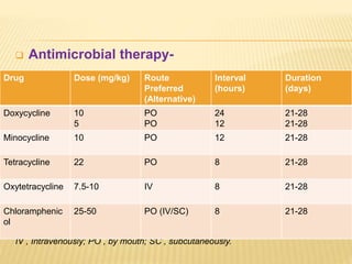
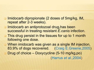
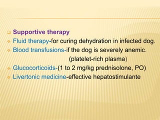
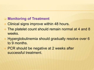
Department of Veterinary clinical Medicine,ethics and Jurisprudence
College of Veterinary and Animal Sciences,Bikaner
(Rajasthan University of Veterinary and Animal Sciences,Bikaner)
A seminar On
Canine Monocytotropic
Ehrlichiosis
PRESENTED BY
Dr.RAM CHANDER Tiwari
M.V.Sc. Scholar
OUTLINE
 Introduction
 Etiology
 Epidemiology
 Pathogenesis
 Clinical findings
 Clinical pathology
 Diagnosis
 Treatment
 Prevention and Control
INTRODUCTION
 Synonyms –
- Tracker dog disease
- Canine monocytic ehrlichiosis
- Tropical canine thrombocytopenia
- Canine haemorragic fever
- Canine rickettsiosis
 The disease was initially identified by Donatien and
Lestoquard in Algeria in 1935.
(Donatien and Lestoquard,
1937)
 In 1970s a large number of American military dogs,
died during the Vietnam War from this rickettsial
disease.
(Groves et al.,
1975)
 In India, E.canis was first reported in Madras by
Mudaliar 1944.
 Infection occurs mainly during the warm season
when the vector tick is active.
ETIOLOGY
 A number of Ehrlichia spp. infections have been
reported in dogs including
-E. canis
-E. chaffeensis
-E. ewingii
(Qurollo et al, 2013)
 Ehrlichiosis is a rickettsial disease of dogs caused
by Ehrlichia spp., principally transmitted by nymphs
and adults of Rhipicephalus sanguineus.
(Moraes-Filho et al.,
2015)
ORGANISM MORPHOLOGY
 Coccobacilli
-Small, pleomorphic shape
-Gram negative
-Obligate intracellular (Rikihisa et al., 2001)
 Three intracytoplasmic forms
-Initial body
-Elementary body
-Morulae
 It consists of a single circular chromosome
containing 1,315,030 nucleotides.
(Mavromatis et al.,
Ehrlichia canis morulae within the
cytoplasm of a monocyte as seen
in blood smears (photographed at × 1000).
The brown dog tick, Rhipicephalus . A
male (left) and engorged female
(right).
Species Name of
disease
Common
natural
host
Cells
mostly
infected
Primary
vector
Distribution
E. canis Canine
monocytic
ehrlichiosis
(CME)
Canids Primarily
monocytes
and
lymphocyte
s
Rhipicephalus
sanguineus,
Dermacentor
variabilis
Worldwide,
tropical,
subtropical,
& temperate
E.
chaffeensi
s
Human
monocytic
ehrlichiosis
(HME)
Humans,
dogs,
goats,
lemurs
in captivity
Monocytes,
macrophag
es
Amblyomma
americanum,
Dermacentor
variabilis
USA,
Europe,
Africa, South
and Central
America,
E. ewingi Canine
granulocyti
c
ehrlichiosis,
human
granulocytic
ehrlichiosis
Dogs,
humans
Primarily
neutrophils
and
eosinophils
Amblyomma
americanum,
Otobius
megnini
USA, Africa,
Korea
E.
ruminantiu
Heartwater
disease
Ruminants Endothelial
cells
Amblyomma
spp.
Africa,
Caribbean
EPIDEMIOLOGY
 Worldwide distribution.
 Free of E. canis infection - Australia.
 Vertebrate hosts - family Canidae.
 Arthropod vector -brown dog tick
(Rhipicephalus sanguineus)
-American dog tick
(Dermacentor variabilis )
(Dumler et al.,
2001)
 German shepherd dog -most susceptible breed,
higher morbidity and mortality compared to other
breeds . (Nelson and Couto,
2003)
 Ehrlichia canis is transmitted transstadially by
Rhipicephalus sanguineus ticks.
(Bremer et al.,
2005)
 Also transmitted by blood transfusion.
TRANSMISSION
 Rhipicephalus sanguineus is the primary vector of
E. canis, which is the cause of monocytic
ehrlichiosis.
(Groves et al.,
1975)
 Rhipicephalus sanguineus is the vector of tick-
borne pathogens affecting dogs and occasionally
humans.
(Dantas-Torres,
2010)
 E. canis is currently not considered an organism
.
Life cycle of Ehrlichia canis. The organism is transmitted only transstadially
(from larva to nymph to adult) within the tick.
PATHOGENESIS
 The incubation period is followed by three
consecutive stages:
-Acute phase
-Subclinical phase
-Chronic phase
(Skotarczak,
2003).
Variable
Reductio
n in Hb,
RBC,WBC
Splenomegaly,
Lymphedinomeg
aly
Hyper-
globulinem
ia
Resolved
fever,clinically
healthy,Persistent
thrombocytopenia
Hemorrhagic
Tendencies
Fever, Anorexia, Meningitis,
Mentaldepression,Hypoalbumine
mia
Retinal
detachment
Pancytopenia
Vector/Blood borne
Infection
Mononuclear cell
Mononuclear phagocytosis
(Liver,Spleen,Lymph nodes)
Lymphocytic perivascular cuff
(Lung,Kidney,Meninges,Eye)
Altered Cell mediated immunity
Thrombocytopenia
Lymphocyte & Plasma
cell proliferation
Recoverd
Hyperviscos
ity
ACUTE
2-4weeks
SUB-CLINICAL
months-year
CHRONIC
months8-20 days
CLINICAL FINDINGS
 ACUTE PHASE (develop 1-3 weeks after the bite)
- High Fever
- Depression
- Lethargy
- Anorexia
- Lymphadenomegaly
- Splenomegaly and
- Hemorrhagic Tendencies
- Dermal petechiae
- Ecchymoses
- Epistaxis.
`
 Ocular Signs-
-Hyphema
- Uveitis
- Scleral bleeding
- Retinal detachment. (Komnenou et al.,
2007)
 Neuromuscular Signs-(CNS symptom)
-Seizures
-Cerebellar Dysfunction
-Anisocoria
-Hyperesthesia
Ventral abdominal skin petechiation due
to thrombocytopenia in a bitch
Mucosal petechiae
Epistaxis in a dog due to Ehrlichia canis
infection.
Ocular Signs
Hyphema in a dog Scleral bleeding in a
dog
SUBCLINICAL PHASE
 During the subclinical phase no clinical signs are
evident. (Waner et al., 2011)
CHRONIC PHASE (develops 1-4 months after bite )
 Symptoms similar to acute phase(with great
severity)
-Pale mucous membrane
-Weakness
-Bleeding and
-Significant weight loss. (Harrus et al.,2011)
CLINICAL PATHOLOGY-
Acute stage
 Significant thrombocytopenia with platelet counts
ranging from 20,000 to 52,000/μL.
 Mild anemia and mildly reduced white blood cell
counts.
Sub clinical stage
 Mild thrombocytopenia with platelet counts ranging
as low as 140,000/μL.
(Harrus et al.,2011; Waner et
al.,2011)
 Leukocyte and erythrocyte counts may also be
reduced. (Waner et al.,2011)
Chronic stage
 Marked pancytopenia due to bone marrow
hypoplasia is a hallmark of the chronic severe form.
(Harrus et al.,
2011)
 Marked anemia and leukopenia.
 The clinicopathological findings are
-Anemia
-Thrombocytopenia
-Pancytopenia
-Leucopenia
-Hyper-globulinemia and
-Hypo-albuminemia (Harrus and Waner,
2011; Harrus et al.,2012; Vinasco et al., 2007).
DIAGNOSIS
 History-Tick history
 Clinical signs
 Hematologic abnormalities
 Rapid Immunochromatographic kit test
-In a positive case both
visible
T and C band will appear indicate the presence
of
Ehrlichia Canis antibodies in the sample.
CT
 Buffy coat smears- For demonstration of
morulae in monocytes.
-Morulae can also be seen in bone marrow cells
and cerebrospinal fluid.
 PCR - PCR has been shown to be a sensitive
method for detecting acute E. canis infection in
dogs.
(Harrus et al., 2004; Mylonakis et al.,
2004)
 IFA-(‘gold standard test’) An indirect fluorescent
antibody (IFA) test is usually performed to detect
antibodies in dogs infected by Ehrlichia canis.
(McBride et al., 2003)
 ELISA-Enzyme-linked immunosorbent assays
(ELISA) have been useful in the diagnosis E.canis.
(Harrus et al.,
2002)
 Organism Cultivation-this method is expensive and
not routinely available
 USG- Multiorgan dysfunction with liver and spleen
involvement is common in clinical cases of canine
monocytic ehrlichiosis.
(Ganguly and
Mukhopadhayay, 2008)
TREATMENT
 FIRST: Properly Remove Tick-(Manualy/medicinal)
Fipronil
Methopren 1-Fipro-fort plus (Savavet)-
9.8%w/v 11.8%w/v
2-Fixotic advance(Vetoquinol)-0.25%w/v
0.22%w/v
 Antimicrobial therapy-
IV , Intravenously; PO , by mouth; SC , subcutaneously.
Drug Dose (mg/kg) Route
Preferred
(Alternative)
Interval
(hours)
Duration
(days)
Doxycycline 10
5
PO
PO
24
12
21-28
21-28
Minocycline 10 PO 12 21-28
Tetracycline 22 PO 8 21-28
Oxytetracycline 7.5-10 IV 8 21-28
Chloramphenic
ol
25-50 PO (IV/SC) 8 21-28
 Imidocarb dipropionate (2 doses of 5mg/kg, IM,
repeat after 2-3 weeks).
 Imidocarb an antiprotozoal drug,has been
successful in treating resistant E.canis infection.
 This drug persist in the tissues for up to 1 month
following one dose.
 When imidocarb was given as a single IM injection,
83.9% of dogs recovered. (Craig E.Greene,2005)
 Drug of choice – Doxycycline (5-10 mg/kg,po)
(Harrus et al.,2004)
 Supportive therapy
 Fluid therapy-for curing dehydration in infected dog.
 Blood transfusions-if the dog is severely anemic.
(platelet-rich plasma)
 Glucocorticoids-(1 to 2 mg/kg prednisolone, PO)
 Livertonic medicine-effective hepatostimulante
 Monitoring of Treatment
 Clinical signs improve within 48 hours.
 The platelet count should remain normal at 4 and 8
weeks.
 Hyperglobulinemia should gradually resolve over 6
to 9 months.
 PCR should be negative at 2 weeks after
successful treatment.
PREVENTION
 A vaccine is not currently available.
 Minimizing tick exposure.
 Use prophylactic drug Doxycycline (3mg/kg PO
q24h) in highly endemic areas.
 All newly introduced dog into a kennel should be
serotested, treated for ticks.
 Whole blood should be tested before transfusion.
REFERENCES
 Abeygunawardena IS, Kakoma I, Smith RD. 1990. Pathophysiology of canine ehrlichiosis,
pp 79-92. In Williams JC, Kakoma I (eds): Ehrlichiosis. Kluwer, Dordrecht, The
Netherlands.
 Anderson BE, Greene CE, Jones DC, et al. 1992. Ehrlichia ewingii sp. nov., the etiologic
agent of canine granulocytic ehrlichiosis. Int J Syst Bacteriol 42:299-302.
 Anderson BE, Dawson JE, Jones DC, et al. 1991. Ehrlichia chaffeensis, a new species
associated with human ehrlichiosis. J Clin Microbiol 29:2838-2842.
 Anziani OS, Ewing SA, Barker RW. 1990. Experimental transmission of a granulocytic
form of the tribe Ehrlichieae by Dermacentor variabilis and Amblyomma americanum to
dogs. Am J Vet Res51:929-931.
 Harrus, S., Kenny, M., Miara, L., Aisenberg, I., Waner, T., Shaw, S., 2004. Comparison of
simultaneous splenic sample PCR with blood sample PCR for diagnosis and treatment of
experimental Ehrlichia canis infection. Antimicrobial Agents and Chemotherapy 48, 4488–
4490.
 Yu, X.J., McBride, J.W., Walker, D.H., 1999. Genetic diversity of the 28-kilodaltonouter
membrane protein gene in human isolates of Ehrlichia chaffeensis. J. Clin.Microbiol. 37,
1137–1143.
Thank You

Canine ehrlichiosis

  • 1.
    Department of Veterinaryclinical Medicine,ethics and Jurisprudence College of Veterinary and Animal Sciences,Bikaner (Rajasthan University of Veterinary and Animal Sciences,Bikaner) A seminar On Canine Monocytotropic Ehrlichiosis PRESENTED BY Dr.RAM CHANDER Tiwari M.V.Sc. Scholar
  • 2.
    OUTLINE  Introduction  Etiology Epidemiology  Pathogenesis  Clinical findings  Clinical pathology  Diagnosis  Treatment  Prevention and Control
  • 3.
    INTRODUCTION  Synonyms – -Tracker dog disease - Canine monocytic ehrlichiosis - Tropical canine thrombocytopenia - Canine haemorragic fever - Canine rickettsiosis  The disease was initially identified by Donatien and Lestoquard in Algeria in 1935. (Donatien and Lestoquard, 1937)
  • 4.
     In 1970sa large number of American military dogs, died during the Vietnam War from this rickettsial disease. (Groves et al., 1975)  In India, E.canis was first reported in Madras by Mudaliar 1944.  Infection occurs mainly during the warm season when the vector tick is active.
  • 5.
    ETIOLOGY  A numberof Ehrlichia spp. infections have been reported in dogs including -E. canis -E. chaffeensis -E. ewingii (Qurollo et al, 2013)  Ehrlichiosis is a rickettsial disease of dogs caused by Ehrlichia spp., principally transmitted by nymphs and adults of Rhipicephalus sanguineus. (Moraes-Filho et al., 2015)
  • 6.
    ORGANISM MORPHOLOGY  Coccobacilli -Small,pleomorphic shape -Gram negative -Obligate intracellular (Rikihisa et al., 2001)  Three intracytoplasmic forms -Initial body -Elementary body -Morulae  It consists of a single circular chromosome containing 1,315,030 nucleotides. (Mavromatis et al.,
  • 7.
    Ehrlichia canis morulaewithin the cytoplasm of a monocyte as seen in blood smears (photographed at × 1000). The brown dog tick, Rhipicephalus . A male (left) and engorged female (right).
  • 8.
    Species Name of disease Common natural host Cells mostly infected Primary vector Distribution E.canis Canine monocytic ehrlichiosis (CME) Canids Primarily monocytes and lymphocyte s Rhipicephalus sanguineus, Dermacentor variabilis Worldwide, tropical, subtropical, & temperate E. chaffeensi s Human monocytic ehrlichiosis (HME) Humans, dogs, goats, lemurs in captivity Monocytes, macrophag es Amblyomma americanum, Dermacentor variabilis USA, Europe, Africa, South and Central America, E. ewingi Canine granulocyti c ehrlichiosis, human granulocytic ehrlichiosis Dogs, humans Primarily neutrophils and eosinophils Amblyomma americanum, Otobius megnini USA, Africa, Korea E. ruminantiu Heartwater disease Ruminants Endothelial cells Amblyomma spp. Africa, Caribbean
  • 9.
    EPIDEMIOLOGY  Worldwide distribution. Free of E. canis infection - Australia.  Vertebrate hosts - family Canidae.  Arthropod vector -brown dog tick (Rhipicephalus sanguineus) -American dog tick (Dermacentor variabilis ) (Dumler et al., 2001)
  • 10.
     German shepherddog -most susceptible breed, higher morbidity and mortality compared to other breeds . (Nelson and Couto, 2003)  Ehrlichia canis is transmitted transstadially by Rhipicephalus sanguineus ticks. (Bremer et al., 2005)  Also transmitted by blood transfusion.
  • 11.
    TRANSMISSION  Rhipicephalus sanguineusis the primary vector of E. canis, which is the cause of monocytic ehrlichiosis. (Groves et al., 1975)  Rhipicephalus sanguineus is the vector of tick- borne pathogens affecting dogs and occasionally humans. (Dantas-Torres, 2010)  E. canis is currently not considered an organism
  • 12.
    . Life cycle ofEhrlichia canis. The organism is transmitted only transstadially (from larva to nymph to adult) within the tick.
  • 13.
    PATHOGENESIS  The incubationperiod is followed by three consecutive stages: -Acute phase -Subclinical phase -Chronic phase (Skotarczak, 2003).
  • 14.
    Variable Reductio n in Hb, RBC,WBC Splenomegaly, Lymphedinomeg aly Hyper- globulinem ia Resolved fever,clinically healthy,Persistent thrombocytopenia Hemorrhagic Tendencies Fever,Anorexia, Meningitis, Mentaldepression,Hypoalbumine mia Retinal detachment Pancytopenia Vector/Blood borne Infection Mononuclear cell Mononuclear phagocytosis (Liver,Spleen,Lymph nodes) Lymphocytic perivascular cuff (Lung,Kidney,Meninges,Eye) Altered Cell mediated immunity Thrombocytopenia Lymphocyte & Plasma cell proliferation Recoverd Hyperviscos ity ACUTE 2-4weeks SUB-CLINICAL months-year CHRONIC months8-20 days
  • 15.
    CLINICAL FINDINGS  ACUTEPHASE (develop 1-3 weeks after the bite) - High Fever - Depression - Lethargy - Anorexia - Lymphadenomegaly - Splenomegaly and - Hemorrhagic Tendencies - Dermal petechiae - Ecchymoses - Epistaxis.
  • 16.
    `  Ocular Signs- -Hyphema -Uveitis - Scleral bleeding - Retinal detachment. (Komnenou et al., 2007)  Neuromuscular Signs-(CNS symptom) -Seizures -Cerebellar Dysfunction -Anisocoria -Hyperesthesia
  • 17.
    Ventral abdominal skinpetechiation due to thrombocytopenia in a bitch Mucosal petechiae
  • 18.
    Epistaxis in adog due to Ehrlichia canis infection.
  • 19.
    Ocular Signs Hyphema ina dog Scleral bleeding in a dog
  • 20.
    SUBCLINICAL PHASE  Duringthe subclinical phase no clinical signs are evident. (Waner et al., 2011) CHRONIC PHASE (develops 1-4 months after bite )  Symptoms similar to acute phase(with great severity) -Pale mucous membrane -Weakness -Bleeding and -Significant weight loss. (Harrus et al.,2011)
  • 21.
    CLINICAL PATHOLOGY- Acute stage Significant thrombocytopenia with platelet counts ranging from 20,000 to 52,000/μL.  Mild anemia and mildly reduced white blood cell counts. Sub clinical stage  Mild thrombocytopenia with platelet counts ranging as low as 140,000/μL. (Harrus et al.,2011; Waner et al.,2011)
  • 22.
     Leukocyte anderythrocyte counts may also be reduced. (Waner et al.,2011) Chronic stage  Marked pancytopenia due to bone marrow hypoplasia is a hallmark of the chronic severe form. (Harrus et al., 2011)  Marked anemia and leukopenia.
  • 23.
     The clinicopathologicalfindings are -Anemia -Thrombocytopenia -Pancytopenia -Leucopenia -Hyper-globulinemia and -Hypo-albuminemia (Harrus and Waner, 2011; Harrus et al.,2012; Vinasco et al., 2007).
  • 24.
    DIAGNOSIS  History-Tick history Clinical signs  Hematologic abnormalities  Rapid Immunochromatographic kit test -In a positive case both visible T and C band will appear indicate the presence of Ehrlichia Canis antibodies in the sample. CT
  • 25.
     Buffy coatsmears- For demonstration of morulae in monocytes. -Morulae can also be seen in bone marrow cells and cerebrospinal fluid.  PCR - PCR has been shown to be a sensitive method for detecting acute E. canis infection in dogs. (Harrus et al., 2004; Mylonakis et al., 2004)  IFA-(‘gold standard test’) An indirect fluorescent antibody (IFA) test is usually performed to detect antibodies in dogs infected by Ehrlichia canis. (McBride et al., 2003)
  • 26.
     ELISA-Enzyme-linked immunosorbentassays (ELISA) have been useful in the diagnosis E.canis. (Harrus et al., 2002)  Organism Cultivation-this method is expensive and not routinely available  USG- Multiorgan dysfunction with liver and spleen involvement is common in clinical cases of canine monocytic ehrlichiosis. (Ganguly and Mukhopadhayay, 2008)
  • 27.
    TREATMENT  FIRST: ProperlyRemove Tick-(Manualy/medicinal) Fipronil Methopren 1-Fipro-fort plus (Savavet)- 9.8%w/v 11.8%w/v 2-Fixotic advance(Vetoquinol)-0.25%w/v 0.22%w/v
  • 28.
     Antimicrobial therapy- IV, Intravenously; PO , by mouth; SC , subcutaneously. Drug Dose (mg/kg) Route Preferred (Alternative) Interval (hours) Duration (days) Doxycycline 10 5 PO PO 24 12 21-28 21-28 Minocycline 10 PO 12 21-28 Tetracycline 22 PO 8 21-28 Oxytetracycline 7.5-10 IV 8 21-28 Chloramphenic ol 25-50 PO (IV/SC) 8 21-28
  • 29.
     Imidocarb dipropionate(2 doses of 5mg/kg, IM, repeat after 2-3 weeks).  Imidocarb an antiprotozoal drug,has been successful in treating resistant E.canis infection.  This drug persist in the tissues for up to 1 month following one dose.  When imidocarb was given as a single IM injection, 83.9% of dogs recovered. (Craig E.Greene,2005)  Drug of choice – Doxycycline (5-10 mg/kg,po) (Harrus et al.,2004)
  • 30.
     Supportive therapy Fluid therapy-for curing dehydration in infected dog.  Blood transfusions-if the dog is severely anemic. (platelet-rich plasma)  Glucocorticoids-(1 to 2 mg/kg prednisolone, PO)  Livertonic medicine-effective hepatostimulante
  • 31.
     Monitoring ofTreatment  Clinical signs improve within 48 hours.  The platelet count should remain normal at 4 and 8 weeks.  Hyperglobulinemia should gradually resolve over 6 to 9 months.  PCR should be negative at 2 weeks after successful treatment.
  • 32.
    PREVENTION  A vaccineis not currently available.  Minimizing tick exposure.  Use prophylactic drug Doxycycline (3mg/kg PO q24h) in highly endemic areas.  All newly introduced dog into a kennel should be serotested, treated for ticks.  Whole blood should be tested before transfusion.
  • 33.
    REFERENCES  Abeygunawardena IS,Kakoma I, Smith RD. 1990. Pathophysiology of canine ehrlichiosis, pp 79-92. In Williams JC, Kakoma I (eds): Ehrlichiosis. Kluwer, Dordrecht, The Netherlands.  Anderson BE, Greene CE, Jones DC, et al. 1992. Ehrlichia ewingii sp. nov., the etiologic agent of canine granulocytic ehrlichiosis. Int J Syst Bacteriol 42:299-302.  Anderson BE, Dawson JE, Jones DC, et al. 1991. Ehrlichia chaffeensis, a new species associated with human ehrlichiosis. J Clin Microbiol 29:2838-2842.  Anziani OS, Ewing SA, Barker RW. 1990. Experimental transmission of a granulocytic form of the tribe Ehrlichieae by Dermacentor variabilis and Amblyomma americanum to dogs. Am J Vet Res51:929-931.  Harrus, S., Kenny, M., Miara, L., Aisenberg, I., Waner, T., Shaw, S., 2004. Comparison of simultaneous splenic sample PCR with blood sample PCR for diagnosis and treatment of experimental Ehrlichia canis infection. Antimicrobial Agents and Chemotherapy 48, 4488– 4490.  Yu, X.J., McBride, J.W., Walker, D.H., 1999. Genetic diversity of the 28-kilodaltonouter membrane protein gene in human isolates of Ehrlichia chaffeensis. J. Clin.Microbiol. 37, 1137–1143.
  • 34.

Editor's Notes

  • #17 Uvea-iris, ciliary body and choroid,Anisocoria- an unequal size of the eyes pupils,  Hyperesthesia-abnormal increase in sensitivity to stimuli of the sense
  • #27 hypo echogenicity of liver
  • #29 Chloramphenicol has been recommended for puppies younger than 5 month to avoid yellow discoloration of erupting teeth from tetracycline.
  • #31 Immune mediated mechanism-suppression by pred