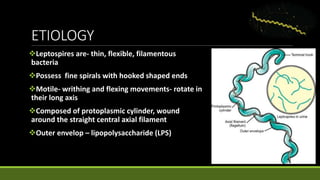
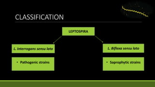
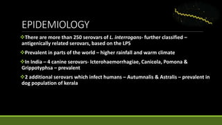
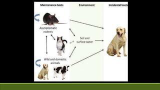
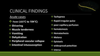
Prevalence of canine leptospirosis has increased in recent years.
As many as 8.2% of dogs are shedding leptospires, some asymptomatically.49
Weather changes, population growth, and habitat encroachment have all increased human and canine exposure to pathogens and their carriers.
Transmission of leptospirosis can occur through direct contact or indirectly through environmental exposure.
Leptospires enter the body through mucous membranes in the mouth, eyes, or nose, or through abraded or water-softened skin.
Leptospires multiply in a host animal's bloodstream.
Leptospires move from the bloodstream to the kidneys and other tissues to continue reproducing.
Leptospires pass from the kidneys into the urine; then are shed back into the environment.
Other dogs, wild animals, or people can become infected through direct or indirect contact.
clinical signs
Fever
Lethargy
Weight loss
Anorexia
Depression
Acute renal failure
Jaundice
Abdominal discomfort
Vomiting and diarrhea
Blood in urine is uncommon, but may occur
Respiratory distress
Dogs at risk
Dogs at risk for developing leptospirosis include those with
Access to ponds, lakes, streams, or standing water
Exposure to urine from other infected animals, including:
Other dogs in shelters or other pet care facilities
Wildlife (e.g. rodents, racoons, opossum, deer), either through direct contact with urine or through contaminated water
Morbidity threats
As leptospirosis progresses, it can result in
Leptospiremia
Leptospires can multiply in the bloodstream and spread to many tissues and organs
Vascular damage/thrombocytopenia
Can lead to kidney failure and interfere with liver function
Contributes to coagulatory abnormalities and hemorrhages
Severe kidney and liver damage
Acute renal failure occurs in dogs with severe clinical signs
Acute hepatic dysfunction or chronic hepatitis have been caused by specific serovars
etiology, local names, definition, transmission, source of infection, epidemiology, pathogenesis, clinical signs, diagnosis, differential diagnosis, treatment prevention and control
Babesiosis is the diseased state caused by the protozoal (single celled) parasites of the genus Babesia. Infection in a dog may occur by tick transmission, direct transmission via blood transfer from dog bites, blood transfusions, or transplacental transmission.
Paratuberculosis is a contagious, chronic and sometimes fatal infection that primarily affects the small intestine of ruminants.
It is caused by the bacterium Mycobacterium avium subspecies paratuberculosis.
Infections normally affect ruminants (mammals that have four compartments of their stomachs, of which the rumen is one),
but have also been seen in a variety of non ruminant species, including rabbits, foxes, and birds. Horses, dogs, and nonhuman primates have been infected experimentally.
Paratuberculosis is found worldwide
www.petsgroomingtips.com is one of the front-runners in providing complete digital information to the pet owners, which would guide theme through various process of grooming their beloved kids. A shabby puppy or kitten not only appears dirty but soon infested with disease if not treated properly. Our various tutorials and free PDF guides cover every aspect of the grooming process.
Rinderpest virus is a Morbillivirus, closely related to the viruses causing peste des petits ruminants, canine distemper and measles.Rinderpest virus is shed in nasal and ocular secretions and can be transmitted during the incubation period (1–2 days before onset of fever). Transmission required direct or close indirect contact between susceptible animals and sick animals shedding the virus.
Malignant catarrhal fever (MCF) is an infectious systemic disease that presents as a variable complex of lesions affecting mainly ruminants and rarely swine. It is principally a disease of domestic cattle, water buffalo, Bali cattle (banteng), American bison, and deer. In addition to these farmed animals, MCF has been described in a variety of captive ruminants in mixed zoologic collections.MCF results from infection by one of several members of a group of closely related ruminant gammaherpesviruses of the Rhadinovirus genus.
Leptospirosis: Its Epidemiology, Diagnosis and Control Chandrani Goswami
Leptospirosis is a zoonosis caused by pathogenic spirochetes of the genus Leptospira.
Disease was first described by Adolf Weil in 1886
In 1908, a Japanese research group led by Ryokichi Inada and Yutaka to first identified the bacterium as the causative agent of leptospirosis and noted its presence in rats in 1916
Generally it is transmitted by the infected urine of rodents.
Leptospirosis is in the group of 17 neglected tropical diseases, categorized by WHO.
Leptospirosis is an underreported disease, and there are no reliable global incidence figures (WHO, 2015)
Synonyms: Weil's Syndrome, Weil-Vasiliev disease, Swineherd's disease, Rice-field fever, Waterborne fever, Nanukayami fever, Cane-cutter fever, Swamp fever, Mud fever, Stuttgart disease, and Canicola fever.
Tuberculosis- International Perspectives on Epidemiology, diagnosis and ControlsRanjini Manuel
Tuberculosis (TB) is caused by bacteria (Mycobacterium tuberculosis) that most often affect the lungs. Tuberculosis is curable and preventable.
TB is spread from person to person through the air. When people with lung TB cough, sneeze or spit, they propel the TB germs into the air. A person needs to inhale only a few of these germs to become infected.
About one-quarter of the world's population has latent TB, which means people have been infected by TB bacteria but are not (yet) ill with the disease and cannot transmit the disease.
People infected with TB bacteria have a 5–15% lifetime risk of falling ill with TB. Persons with compromised immune systems, such as people living with HIV, malnutrition or diabetes, or people who use tobacco, have a higher risk of falling ill.
Tropical theileriosis or Mediterranean coast fever Ranjini Manuel
heileriases are a group of tickborne diseases caused by Theileria spp. A large number of Theileria spp are found in domestic and wild animals in tick-infested areas of the Old World. The most important species affecting cattle are T parva and T annulata, which cause widespread death in tropical and subtropical areas of the Old World. T lestoquardi, T luwenshuni, and T uilenbergi are important causes of mortality in sheep.
Taenia multiceps multiceps is an intestinal parasite of canids (especially dogs, foxes, and jackals) and occasionally people. Its intermediate hosts include sheep, goats, deer, antelope, chamois, rabbits, hares, horses, and less commonly cattle, which acquire this tapeworm's eggs while grazing. After ingestion, some oncospheres hatch and reach the brain, developing by endogenous budding into a metacestode (larval) stage known as Coenurus cerebralis. Initial invasion and development of the oncospheres may be responsible for acute suppurative meningoencephalitis.
Bovine herpesvirus 1 (BHV-1) is associated with several diseases in cattle: infectious bovine rhinotracheitis (IBR), infectious pustular vulvovaginitis (IPV), balanoposthitis, conjunctivitis, abortion, encephalomyelitis, and mastitis.
Bluetongue is an infectious arthropod-borne viral disease primarily of domestic and wild ruminants. Infection with bluetongue virus (BTV) is common in a broad band across the world, which until recently stretched from ~35°S to 40°–50°N. Bluetongue virus is the type-species of the genus Orbivirus in the family Reoviridae. The geographic restriction is in part related to the climatic and environmental conditions necessary to support the Culicoides vectors.
Guinea pigs are generally hardy, healthy animals but are susceptible to certain problems and diseases. Here are the common bacterial infections of guinea pigs.
Infectious laryngotracheitis (ILT) is an economically important respiratory disease of poultry. This highly contagious disease is caused by Gallid alpha herpesvirus type 1 (GaHV-1), commonly known as infectious laryngotracheitis virus (ILTV). The virus can be easily transmitted by infected birds and fomites. Lax biosecurity, transportation of infected birds, and spread of contaminated litter facilitates spread of the virus. Clinical signs of respiratory disease are not pathognomonic. Diagnosis is by real-time PCR and histopathology . Implementation of biosecurity is necessary for prevention, but vaccination is commonly used for control of the disease in endemic regions worldwide.
Infectious coryza is an acute respiratory disease of chickens characterized by nasal discharge, sneezing, and swelling of the face under the eyes. It is found worldwide. The disease is seen only in chickens; reports of the disease in quail and pheasants probably describe a similar disease that is caused by a different etiologic agent.
Pseudorabies is an acute, frequently fatal disease with a worldwide distribution that affects swine primarily and other domestic and wild animals incidentally. The pseudorabies virus has emerged as a significant pathogen in the USA since the 1960s, probably because of the increase in confinement swine housing or perhaps because of the emergence of more virulent strains. Clinical signs in nonporcine animals are similar to those of rabies, hence the name “mad itch” (pigs do not display this sign). Pseudorabies is a reportable disease and has been successfully eradicated from the vast majority of the USA.
Special staining techniques for microorganismsRanjini Manuel
cells and microorganisms that we observe under the microscope lack color and contrast. This makes it difficult, if not impossible, to detect important cellular structures and their distinguishing characteristics without artificially treating specimens. We have already alluded to certain techniques involving stains and fluorescent dyes, and in this section we will discuss specific techniques for sample preparation in greater detail. Indeed, numerous methods have been developed to identify specific microbes, cellular structures, DNA sequences, or indicators of infection in tissue samples, under the microscope.
Canine parvovirus (CPV) is a highly contagious and relatively common cause of acute, infectious GI illness in young dogs. Although its exact origin is unknown, it is believed to have arisen from feline panleukopenia virus or a related parvovirus of nondomestic animals. It is a nonenveloped, single-stranded DNA virus, resistant to many common detergents and disinfectants, as well as to changes in temperature and pH. Infectious CPV can persist indoors at room temperature for at least 2 mo; outdoors, if protected from sunlight and desiccation, it can persist for many months and possibly years.
This presentation includes
Etiology- Echinococcus granulosus- hydatid cyst
distribution
Transmission cycle
Life cycle & pathogenesis
clinical findings
Associated Necropsy findings
Diagnosis
Control
This presenation includes
Introduction
Etiology- structure, various species which comes under the genus lyssa virus
Epidemiology
Distribution
Transmission
Pathogenesis
Clinical signs
Diagnosis
prevention and control
Resistance to physical and chemical action
Prophylaxis
How to Split Bills in the Odoo 17 POS ModuleCeline George
Bills have a main role in point of sale procedure. It will help to track sales, handling payments and giving receipts to customers. Bill splitting also has an important role in POS. For example, If some friends come together for dinner and if they want to divide the bill then it is possible by POS bill splitting. This slide will show how to split bills in odoo 17 POS.
Palestine last event orientationfvgnh .pptxRaedMohamed3
An EFL lesson about the current events in Palestine. It is intended to be for intermediate students who wish to increase their listening skills through a short lesson in power point.
Welcome to TechSoup New Member Orientation and Q&A (May 2024).pdfTechSoup
In this webinar you will learn how your organization can access TechSoup's wide variety of product discount and donation programs. From hardware to software, we'll give you a tour of the tools available to help your nonprofit with productivity, collaboration, financial management, donor tracking, security, and more.
We all have good and bad thoughts from time to time and situation to situation. We are bombarded daily with spiraling thoughts(both negative and positive) creating all-consuming feel , making us difficult to manage with associated suffering. Good thoughts are like our Mob Signal (Positive thought) amidst noise(negative thought) in the atmosphere. Negative thoughts like noise outweigh positive thoughts. These thoughts often create unwanted confusion, trouble, stress and frustration in our mind as well as chaos in our physical world. Negative thoughts are also known as “distorted thinking”.
2024.06.01 Introducing a competency framework for languag learning materials ...Sandy Millin
http://sandymillin.wordpress.com/iateflwebinar2024
Published classroom materials form the basis of syllabuses, drive teacher professional development, and have a potentially huge influence on learners, teachers and education systems. All teachers also create their own materials, whether a few sentences on a blackboard, a highly-structured fully-realised online course, or anything in between. Despite this, the knowledge and skills needed to create effective language learning materials are rarely part of teacher training, and are mostly learnt by trial and error.
Knowledge and skills frameworks, generally called competency frameworks, for ELT teachers, trainers and managers have existed for a few years now. However, until I created one for my MA dissertation, there wasn’t one drawing together what we need to know and do to be able to effectively produce language learning materials.
This webinar will introduce you to my framework, highlighting the key competencies I identified from my research. It will also show how anybody involved in language teaching (any language, not just English!), teacher training, managing schools or developing language learning materials can benefit from using the framework.
Students, digital devices and success - Andreas Schleicher - 27 May 2024..pptxEduSkills OECD
Andreas Schleicher presents at the OECD webinar ‘Digital devices in schools: detrimental distraction or secret to success?’ on 27 May 2024. The presentation was based on findings from PISA 2022 results and the webinar helped launch the PISA in Focus ‘Managing screen time: How to protect and equip students against distraction’ https://www.oecd-ilibrary.org/education/managing-screen-time_7c225af4-en and the OECD Education Policy Perspective ‘Students, digital devices and success’ can be found here - https://oe.cd/il/5yV
The French Revolution, which began in 1789, was a period of radical social and political upheaval in France. It marked the decline of absolute monarchies, the rise of secular and democratic republics, and the eventual rise of Napoleon Bonaparte. This revolutionary period is crucial in understanding the transition from feudalism to modernity in Europe.
For more information, visit-www.vavaclasses.com
2. INTRODUCTION
1st reported by Adolf Weil in Heidelberg in 1886
Leptospirosis is a zoonotic disease of world wide
significance that affects many animal species
Caused by the infection with –leptospiral spirochetes of
the species- Leptospira interrogans sensu lato
3. ETIOLOGY
Leptospires are- thin, flexible, filamentous
bacteria
Possess fine spirals with hooked shaped ends
Motile- writhing and flexing movements- rotate in
their long axis
Composed of protoplasmic cylinder, wound
around the straight central axial filament
Outer envelop – lipopolysaccharide (LPS)
5. EPIDEMIOLOGY
There are more than 250 serovars of L. interrogans- further classified –
antigenically related serovars, based on the LPS
Prevalent in parts of the world – higher rainfall and warm climate
In India – 4 canine serovars- Icterohaemorrhagiae, Canicola, Pomona &
Grippotyphsa – prevalent
2 additional serovars which infect humans – Autumnalis & Astralis – prevalent in
dog population of kerala
6. EPIDEMIOLOGY
Dogs of any age , sex, breed, kept outdoor are susceptible
Dogs in close contact with rodents/ rodent urine can develop leptospirosis
Transmission of disease among maintenance host is efficient & incidence of
infection
Incidental host are NOT important reservoirs of infection & incidence of
transmission is
Do not replicate outside the host
Inactivated by heat, UV, disinfectants, freezing conditions
Can survive in water , wet soil for weeks to months under optimal conditions
(alkaline PH, 0°- 25°C)
7.
8. MAINTENANCE HOSTS OF SOME COMMON LEPTOSPIRAL SEROVARS
LEPTOSPIRAL SEROVARS MAINTENANCE HOST
Hardjo Cattle
Pomona pigs, skunks, Opossum
Bratislava Horses
Canicola Dogs
Icterohaemorrhagiae Rats
Grippotyphosa Raccons, Skunks, Opossum
9. SEROVAR PRIMARY RESERVOIR INCIDENTAL HOST
L. bataviae Dog, rat, mouse Dog , cat
L. bratislava Pig, horse, dog Dog
L. canicola Dog Dog, cat
L. grippotyphosa Vole, raccoon, skunk, opossum Dog , cat
L. hadjo Cow Dog
L. icterohaemorrhagiae Rat Dog, cat
L. pomona Cattle, Pig, skunk, opossum Dog , cat
L. autumnalis Mouse Dog
13. PATHOGENESIS
Leptospires intact mucous membranes of mouth, nose, eyes or abraded scratched,
water-softened skin
Transcriptional changes occur in the body pathogenicity, organisms multiply after
entering blood vascular space
Further replicate in tissues including kidney, spleen, CNS, eyes, genital tract capillary
damage
14. Tissue edema & vasculitis acute endothelial injury and haemorrhagic manifestation
LPS stimulate neutrophil adherence and platelet activation inflammatory and
coagulatory abnormalities
Renal colonization Organisms replicate and persists in renal tubular epithelial cells,
reaches interstitium after 2 weeks of infection – onset of shedding
kidney swelling impaired renal blood perfusion decreased glomerular filtration
impairment of renal function
15.
16. Host clears spirochetes from most of the organs, but may persist in kidney and shed in
urine for days –to- months
Liver being 2nd major organ affected – degree of icterus usually
corresponds with severity of hepatic necrosis
Hepatic dysfunction may occur without major cellular changes- due to
the subcellular damage produced by the leptospiral toxin.
osmotic fragility has been detected in canine leptospirosis making
haemolysis less likely
17. Acute lung injury occurs due to effects of toxin in pulmonary tissues
Fluid exudation within the lungs may result from vasculitis
Pulmonary haemorrhage – rarely reported
When leptospira invades brain benign meningitis
Uvetitis documented in case of dogs and humans
Abortion/infertility transplacental transmission in dogs – bataviae)
Serogroups – Canicola, Bratislava & Grippotyphosa – renal & hepatic involvement
icterohaemorrhagiae & Pomona – more hepatic
22. ORGANISM IDENTIFICATION
Organisms on body fluids are Identified by dark field microscopy
To the sensitivity of Direct microscopic examination special staining can be done
silver staining and Wrthin-starry stains are commonly used histochemical stains
Immunofluorescence staining – urine, water , soil samples
Immunoperoxidase staining- blood and urine samples
23. They are visualized as – Thin, Bright ,Actively motile rods, Rapid spinning
with Jerky movements
24. CULTURE
Organisms are cultured on EMJH medium (Ellinghausen, McCullough,
Johnson, Harris) incubation at 30°C for 7 days and observed under Dark field
microscopy
Fletcher’s medium – growth occurs as a ringed area called dinger’s ring or
disk 1-3cm below the surface of the medium
Korthof media is also used in cultivating leptospires
25. SEROLOGICAL TESTS
Microscopic agglutination test (MAT)
ELISA- Pan Bio ELISA – detect IgM from single serum sample
PCR – rapid & direct, DNA is identified, used in early and convalescent stage
Indirect Haemagglutination
Latex agglutination
Lateral flow technique
Leptodipstick assay
PCR in combination with ELISA good sensitivity at initial phase
26.
27. MAT
Cornerstone for serodiagnosis for leptospira because of its sensitivity and
detection of group specific antibodies
Positive results are defined as 4 fold in the titre between acute and
convalescent specimen
DISADVANTAGES
Due to antigenic heterogencity it requires large no.of serovar as antigens
Not useful at early stages of infection where antibody is not present
Performance restricted to labs that are capable of maintain strain for the
preparation of live antigens
28. Test Advantages Disadvantages
Dark Field Microscopy (DFM) Visualize Leptospirosis Lack of sensitivity and specificity. 104
Leptospires/ml is necessary for one
organism/field to be visible under DFM.
IgM ELISA Most widely used False positive, IgM cannot be detected
in early stages of infection and can
persist in blood for years.
Microscopic Agglutination Test
(MAT)
Gold Standard Less sensitive in early phase of disease.
Labor intensive and complicated
procedure as there is a need to maintain
Leptospira strain for preparing live
antigen.
Polymerase Chain Reaction (PCR) Successful in detecting Leptospira
DNA in serum and urine samples of
patients
Expensive reagents, Requires large
quantity of DNA. Cannot identify the
infecting serovar.
29. TREATMENT
Ampicillin 22mg/kg , 8hr I/V or I/M -5-8days
Penicillin G 25,000-40,000IU/kg b.wt 12hr i/v
Doxycycline administered orally at 2.5 to 5.0 m g / kg eliminate the renal
carrier phase
Fluid therapy
Supportive therapy
30. CONTROL
Infected urine, contaminated water, and reservoir hosts - avoided
Infected dogs should be handled with wearing gloves
Contaminated surfaces should be cleaned with detergents and disinfected
Vaccination
Public health concerns
Editor's Notes
Peptodoglycan –G+, LPS – G-ve.
Cat – haemolytic toxin – haemolysis, icterus
Urine wil become normal within 2-3 weeks
ALT – high omp in dogs coz alt hingh in hepatic cells –others other tissues also has alt in that
Citrate anti coagulants shd be avoided coz in inhibits the bac
Lps – lip 32 imp lps – associated with pathogenicity
Penicillin don’t eliminate org frm kidney. Ampicillin oral absorption is less nd half life so recommended parentally .
Vaccintion – mega vac , canigen, nobi vac are ( high titre) , vanguard 5L, duramin , ( include vaccination ) normally used is killed vac